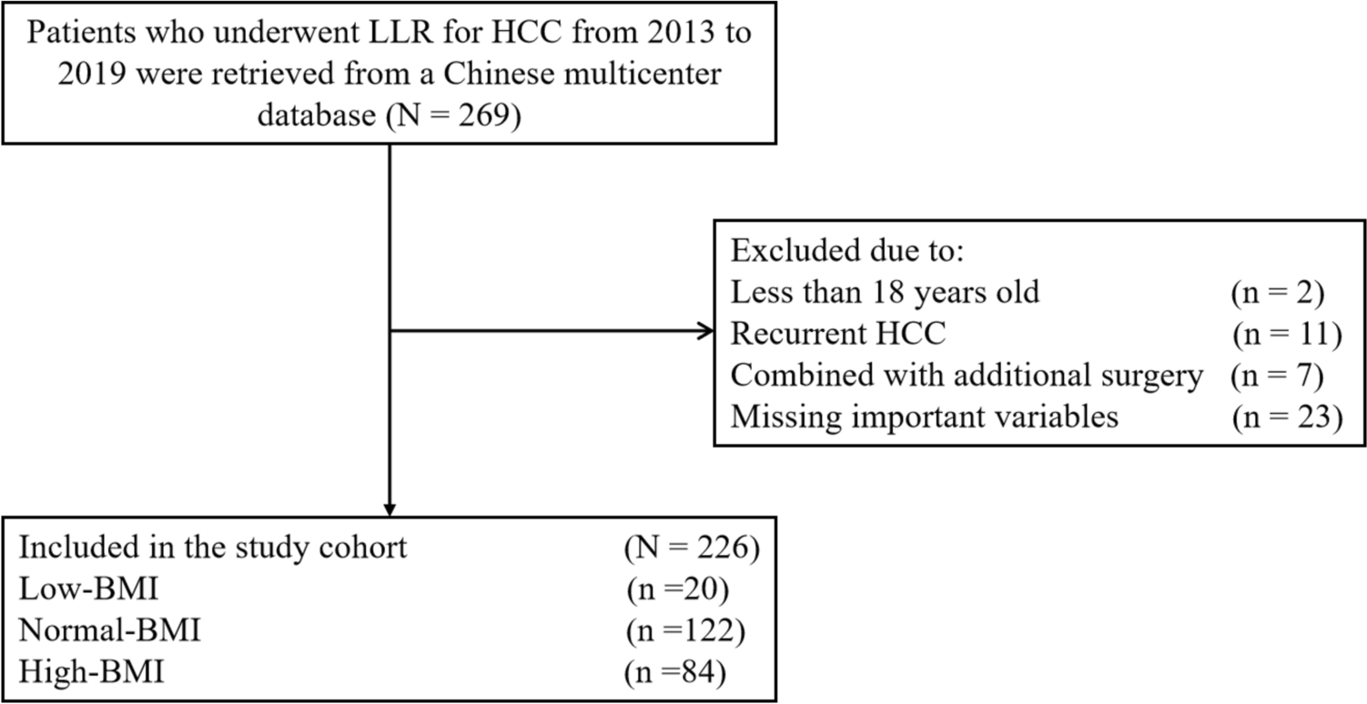
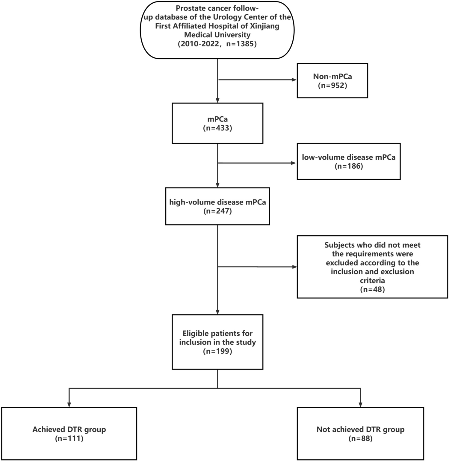
Metastatic burden plays a crucial role in determining the prognosis of patients with mPCa. Patients with a high-volume disease tend to have a shorter time to progression to CRPC and overall survival compared to those with a low-volume disease. A multicenter study revealed that the median progression-free survival time for patients with low-volume disease was 44.5 months, while for those with high-volume disease, it was 16.1 months. Similarly, the median survival time for patients with low-volume disease was 103.2 months, whereas for those with high-volume disease, it was 62.7 months. Various factors such as PSA levels, Gleason score, T stage, N stage, and treatment methods are closely associated with the progression-free survival time and overall survival of patients with high-volume disease mPCa (Shiota et al. 2021). Although CAB treatment can enhance progression-free survival time and overall survival in mPCa patients with high-volume disease, most patients will progress to the CRPC stage within 16–24 months of treatment (Nagumo et al. 2022; Achard et al. 2022). Some studies have found that serum testosterone levels during ADT are associated with the overall survival of mPCa patients. However, there is insufficient clinical evidence to support the relationship between the traditional castration cutoff point and the prognosis of PCa patients. Additionally, the clinically significant castration level has not yet been determined (Perachino et al. 2010). This study aimed to assess the correlation between serum testosterone levels below 0.7 nmol/l after one month of treatment and the prognosis in patients with high-volume disease mPCa treated with CAB. The findings of this study may provide valuable insights for adjusting therapeutic strategies in patients with high-volume disease mPCa.
As early as 10 years ago, experts emphasized the importance of monitoring serum testosterone levels to verify the response to ADT treatment. However, testosterone monitoring has not been given due attention for many years. Currently, it is believed that DTR may become a new standard (Schulman et al. 2010). Our study demonstrates a significant difference in PSA and PSA decline rate at 6 months of treatment between the group that achieved DTR in serum testosterone after one month of treatment and the group that did not achieve DTR. The group that achieved DTR had a higher proportion of PSA < 0.2 ng/ml and a greater PSA decline rate, indicating that patients with low testosterone after treatment show a better PSA response. Morote et al. (Morote et al. 2007) established a direct correlation between testosterone increase and the progression of PCa. They were the first to report that a serum testosterone level of less than 32 ng/dl after treatment can help prolong the progression-free survival of patients. Dason et al. (Dason et al. 2013) also discovered that patients with a testosterone level of less than 32 ng/dl at 9 months of treatment had a significantly longer time to progress to CRPC (P = 0.001). The median progression-free survival time was 33.1 months for those with testosterone levels below 32 ng/dl and 12.5 months for those with levels above 32 ng/dl. However, their study did not find that a testosterone level below 20 ng/dl predicts the time to develop CRPC. Nevertheless, several studies have shown that a testosterone level below 20 ng/dl after ADT treatment is important for improving patient prognosis. In a study conducted by Ding et al. (Ding* M et al. 2019), it was found that patients with testosterone levels below 20 ng/dl at 6 months of treatment experienced a significantly longer time before progressing to CRPC. The median CRPC-free survival times were 48 months for patients with testosterone levels below 20 ng/dl and 24 months for those with levels above 20 ng/dl. Kamada et al. (Kamada et al. 2015) investigated the relationship between testosterone and prognosis in PCa patients who received CAB treatment. They discovered that patients with testosterone levels below 20 ng/dl after 6 months of treatment and the lowest value of testosterone below 20 ng/dl after treatment had a significantly prolonged overall survival. Similarly, Yamamoto et al. (Yamamoto et al. 2017) conducted studies that demonstrated how a post-treatment testosterone nadir below 20 ng/dl or a testosterone reduction of at least 480 ng/dl can lead to a longer progression-free survival and OS in Japanese male patients with advanced PCa. In our study, we found that patients with serum testosterone < 0.7 nmol/l at 1 month of treatment had significantly prolonged time to progression to CRPC (17.93 ± 6.68 months vs. 13.43 ± 6.12 months) and OS compared to those with ≥ 0.7 nmol/l. The median progression-free survival time in the two groups was 18 and 12 months, respectively, and the median survival time was 57 and 32 months. The results of the our study were consistent with the previous studies, however, the results of the our study showed shorter progression-free survival time and OS than the previous studies, which may be attributed to the inclusion of a population with high-volume disease mPCa. The results of the our study further support the idea that a strict control of testosterone < 20 ng/dl will result in a longer progression-free survival and OS. Studies have shown that when testosterone levels are below 20 ng/dl after ADT treatment, there is a more effective killing effect on PCa cells. Additionally, under these conditions, residual tumor cells are more likely to proliferate into hormone-sensitive PCa cells, leading to a longer progression to CRPC. Moreover, a lower testosterone levels after treatment is associated with a reduced incidence of testosterone escape, which may contribute to a better prognosis. This finding supports the prediction that testosterone levels below 20 ng/dl after ADT treatment are indicative of a favorable outcome (Klotz and Toren 2012). Some studies have indicated that an increase in testosterone levels above the target threshold or testosterone escape after the initial month of ADT could result in poorer clinical outcomes. Tan et al. reached similar conclusions in their cohort study on the population with CRPC. The ‘bounce’ phenomenon in testosterone levels is defined as a 10% increase in testosterone levels after initiating new anti-androgen therapy compared to levels before starting the therapy. Tan et al.’s research has shown that this ‘bounce’ phenomenon is linked to poorer PSA response, shorter PSA progression-free survival, and shorter overall survival (Tan et al. 2021; Saad et al. 2020). Tan et al.’s analysis of the testosterone ‘bounce’ phenomenon underscores the importance of testosterone as a predictive factor for prostate cancer treatment response, and provides valuable insights for the clinical monitoring of testosterone levels. The underlying mechanism of the testosterone ‘bounce’ phenomenon is currently unclear. Lower testosterone levels post-treatment are associated with a decreased incidence of testosterone escape. Additionally, lower testosterone levels post-treatment may help mitigate this 'bounce' phenomenon of testosterone, which could have a direct impact on the treatment and prognosis of patients with prostate cancer. Our study aimed to identify early predictors, therefore we evaluated testosterone levels after 1 month of treatment instead of after half a year of treatment or the nadir value of testosterone. This study further analyzed the differences in progression time to CRPC and overall survival time among patients who entered DTR at different time periods. The study revealed that the median progression-free survival time for patients who achieved DTR within 1 month, reached DTR more than 1 month but within 1 year, and those who did not achieve DTR within 1 year were 18, 15, and 10 months, respectively. Similarly, the median survival time for these groups were 57, 45, and 26 months, respectively. There was a correlation between patients’ progression-free survival time and overall survival with the time taken to reach DTR. As the time to reach DTR increased, both progression-free survival time and overall survival decreased. The relationship between the time of testosterone decline and the prognosis of PCa patients has shown varying results in previous studies. Previous studies by Kamada et al. (Kamada et al. 2015) suggest that the key factor influencing the prognosis of PCa is not the rapid decline of testosterone, but rather whether the lowest testosterone level is below 20 ng/dl. However, some studies suggest that a rapid reduction in testosterone during endocrine therapy may have a positive impact on prognosis.
Wang et al. (Wang et al. 2017) conducted a multi-factor analysis and discovered that testosterone levels below 50 ng/dl after the first month of CAB treatment are not indicative of the effective hormone treatment duration. However, they observed that testosterone levels equal to or less than 25 ng/dl after the first month of treatment are significantly linked to a reduced risk of progression to CRPC (HR = 1.46, 95%CI 1.08–1.96, P = 0.013). Our study identified several independent risk factors for progression to CRPC in patients with high-volume disease mPCa. These risk factors include T stage > 3, Gleason score ≥ 9, initial PSA > 200 ng/ml, and testosterone ≥ 0.7 nmol/l at 1 month of treatment. Additionally, smoking and testosterone ≥ 0.7 nmol/l at 1 month of treatment were found to be independent risk factors for the prognosis of mPCa patients with high-volume disease. Smoking patients had a 1.707 times higher risk of death compared to non-smoking patients, while patients with testosterone ≥ 0.7 nmol/l at 1 month of treatment had a 2.087 times higher risk of death compared to those with < 0.7 nmol/l. These findings align with previous research conducted by Wang et al. and are supported by other studies (Bertaglia et al. 2013; Perachino et al. 2010; Kamada et al. 2015; Yamamoto et al. 2017). It is evident that maintaining low testosterone levels after treatment can significantly improve patient prognosis. According to another study, it was found that testosterone levels below 20 ng/dl can be used to predict the recovery of testosterone to castration levels in PCa patients undergoing external radiotherapy treated with ADT (HR = 0.35, 95%CI 0.14–0.79, P = 0.0112) (Takei et al. 2018). Previous studies have indicated that a level of 25 ng/dl can be used to differentiate patients who will progress to CRPC from those who will not progress in the short term (AUC = 0.59, 95%CI 0.51–0.66), with a sensitivity of 56% and specificity of 59%. On the other hand, a level of 30 ng/dl demonstrated the highest sensitivity (80%) and specificity (55%) in predicting patient survival (AUC = 0.69, 95%CI 0.58–0.79) (Bertaglia et al. 2013; Wang et al. 2017). Based on the results of COX multifactor analysis, this study developed a nomogram prediction model to forecast the progression and prognosis of patients with high-volume disease mPCa. This model demonstrates good predictive ability in assessing the progression and prognosis of patients with high-volume disease mPCa. This study will serve as a valuable reference point for evaluating patient progress and prognosis. It will play a crucial role in guiding clinicians to tailor treatment plans to individual patients, ultimately leading to improved patient outcomes and enhanced quality of life.
As the understanding of PCa advances, clinical practice is increasingly focusing on comprehensive management of endocrine therapy for PCa, with testosterone monitoring being a crucial component. Despite this, clinical emphasis still largely remains on monitoring PSA, with insufficient attention given to testosterone monitoring. It is worth noting that PSA monitoring alone has its limitations, as a small number of patients may still experience clinical progression despite no increase in PSA levels. There is a growing body of evidence indicating that a lower testosterone level during treatment is associated with a better prognosis for patients. Therefore, it is crucial to focus on monitoring and managing testosterone levels. This study discovered that patients who achieved DTR within 1 month had a more favorable prognosis. If the results of the current study can be replicated and confirmed, clinicians may want to consider implementing routine testing of serum testosterone at 1 month of treatment. By focusing on this early monitoring indicator, clinicians can optimize the effectiveness of castration treatment, make more precise decisions regarding treatment plans, decrease the likelihood of treatment failure, extend the patient's survival time effectively, and ultimately maximize the benefits for the patient. When monitoring patients with prostate cancer undergoing treatment, it may be observed that serum testosterone and PSA levels are not always ‘synchronized’. Patients who have achieved DTR may exhibit higher PSA levels, while patients who have not reached DTR may show lower PSA levels. The exact relationship between testosterone and PSA remains unclear at present, but it can be explored from the following perspectives. First of all, endocrine therapy drugs have a direct impact on testosterone levels and demonstrate good specificity. PSA is linked to PCa cells, and its gene transcription is regulated by androgens. This can result in an 'out-sync' between testosterone levels and PSA levels after treatment. Secondly, various pathological types of prostate cancer, as well as differing Gleason scores and individual characteristics, can result in varying sensitivities to treatment. These heterogeneities in prostate cancer can also lead to ‘out-sync’ between testosterone and PSA levels. Finally, variations in baseline levels of serum testosterone and PSA, the selection of treatment medications, and the patient's overall health condition could contribute to the lack of ‘synchronization’ between testosterone and PSA levels. Additionally, differences in the clearance rates of serum testosterone and PSA within the body should be taken into account (Zhang 2004; Saxena et al. 2012). The author's explanations for the lack of 'synchronization' between serum testosterone and PSA levels post-treatment are speculative. There is currently a lack of research establishing a clear link between the two factors and the underlying reasons for their 'non-synchronization'. The 'unsynchronized' phenomenon of testosterone and PSA highlights the complex relationship between the two in PCa. To effectively evaluate treatment outcomes and adjust strategies promptly, synchronous management of PSA and testosterone is crucial. Both biomarkers are essential for monitoring PCa patients.


留言 (0)