Herbs and extraction
Decoction piece of EHL, the dried whole grass of EHL, was purchased from Jiangzhong Traditional Chinese Medicine Piece Co., Ltd. (Jiangxi, China). Decoction piece of EHL was identified by the chief pharmacist Chunhua Gao in our hospital. The EHL was first milled and then extracted with 70% ethanol for three cycles using 500 mL per cycle. The extract was filtered and concentrated by a rotary evaporator (Hei-VAP Expert Control ML G3 XL, Heidolph, Germany) to obtain the crude extract. The crude extract was suspended in water and then re-extracted using ethyl acetate. The re-extract was concentrated using a rotary evaporator to obtain the final extract. Finally, the extracts were prepared to specific concentrations using 1% dimethyl sulfoxide (DMSO) and cell culture medium for subsequent experimental applications.
Cell culture and transfection
The human cell lines containing an integrated hepatitis B virus genome were used in this study, including Hep 3B2.1–7 (ATCC, USA) and HepG2.2.15 (BioVector NTCC, China). Eagle's Minimum Essential Medium (Gibco, USA) added fetal bovine serum (FBS, 10%) (Gibco, USA) was used to culture Hep 3B2.1–7 cell line, while RPMI1640 + 10%FBS (both Gibco, USA) was used for HepG2.2.15.
The overexpression plasmids for RAC-alpha serine/threonine-protein kinase (AKT1) (overAKT1), as well as the corresponding negative control (overNC), were synthesized by Shanghai GenePharma Co. Then the cells were transfected with overAKT1 or corresponding negative control by mixing with Lipo2000.
Determination of half maximal inhibitory concentration (IC50)
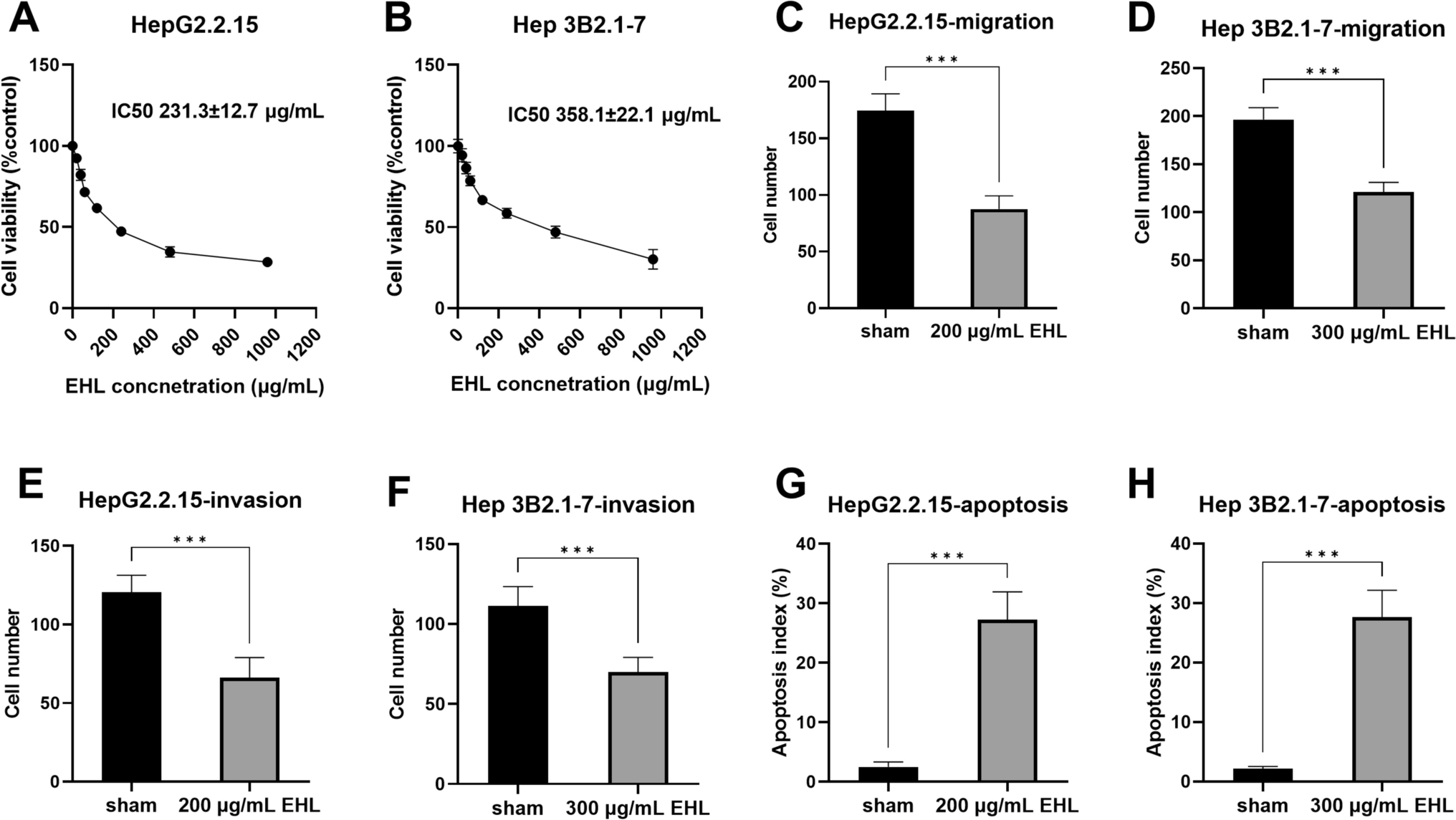
Each kind of cell was divided into an experimental group and a negative control group, both with five parallel samples. Cells in the logarithmic growth phase were collected and then subjected to trypsin digestion. The cell density was adjusted to 2 × 10⁴ per mL and inoculated in 96-well culture plates at 200 μL per well. After the cells were adhered to the wall, the culture medium was changed: the experimental group was added with different concentrations of EHL extract, at final concentrations of 20, 40, 60, 120, 240, 480, and 960 μg/mL; the control group was added with an equal amount of cell culture medium containing 5% DMSO. Cells were incubated in a CO2 (5%) incubator at 37℃ for 48 h. The cell culture plate was removed and MTT Cell Promotion Assay Kit (Sangon Biotech, China) was added to each well for further cultivation for 2 h. At the end of incubation, the culture medium is removed and the reaction is terminated. The value of each well at 490 nm was determined by a fully automated enzyme immunoassay detector. The average value of 5 wells was taken and the cell growth inhibition rate was calculated.
Transwell chamber for detecting cell migration/invasiveness
Cell migration and invasiveness were detected using 24-well chambers with Transwell® permeable supports (8 µm) (Corning, USA) and BD BioCoat™ Matrigel™ Invasion Chambers in two 24-well plates (8.0 µm) (BD Biosciences), respectively. Cells in the sham group were cultured by adding cell culture solution. Cells in the experimental group were cultured by adding EHL extracts, with 200 μg/mL for HepG2.2.15 and 300 μg/mL for Hep 3B2.1–7. Cells were collected and added to a serum-free culture medium to make a cell suspension. The upper chamber was added with pretreated cell suspension and the lower chamber was added with a culture medium containing 15% FBS. After 24 h of incubation, the unpermeabilized cells on the upper chamber surface of the filter membrane were removed, and the permeabilized cells were fixed, stained, and counted under a microscope.
Apoptosis assay
HepG2.2.15 and Hep 3B2.1–7 cells were digested with 0.25% Trypsin Solution without EDTA + 2% Bovine Serum Albumin (Beyotime, China). After washing with PBS, cells were resuspended and centrifuged for 5 min. After centrifugation, the cell precipitate seed was added with Annexin V-FITC conjugate from Annexin V-FITC Apoptosis Assay Kit (Beyotime, China) to gently resuspend the cells, and then 10 μL of propidium iodide staining solution. The cell suspension was incubated for 20 min away from light, followed by immediate detection using flow cytometry.
Network pharmacologyConstruction of "herbal candidate target-disease-related gene" interaction network
HERB database (http://herb.ac.cn/) and Traditional Chinese Medicine Systems Pharmacology (TCMSP) analysis platform (https://old.tcmsp-e.com/tcmsp.php) were used to predict the components in EHL and screen potential active compounds based on the restriction of oral bioavailability > 30% and drug-likeness > 0.18. The potential targets of active ingredients were predicted by TCMSP and SwissTargetPrediction platforms (http://www.swisstargetprediction.ch/). HBV-related human genes were retrieved through the ViRBase v3.0 database and ncRNAs were removed. The summary of gene-disease associations under "Liver carcinoma" in DisGeNET database (https://www.disgenet.org/dbinfo) was downloaded to collect the disease genes. The intersection of the EHL candidate targets with disease-related genes and HBV-related genes was taken using Venny 2.1 (https://bioinfogp.cnb.csic.es/tools/venny/index.html) mapping to obtain the targets of EHL action in HBV-HCC.
Interaction-based screening of key genes
The targets were entered into the STRING (https://cn.string-db.org/) database, and the protein–protein interaction (PPI) network was generated with the screening condition of "minimum required interaction score ≥ 0.7”; Cytoscape 3.7.1 software was used to calculate the degree value and construct the target interaction network. The target genes enriched in the hsa05161 Hepatitis B pathway were predicted using the OECloud tools (https://cloud.oebiotech.com). In addition, the top 10 hub genes were identified by Cytohubba in Cytoscape 3.7.1 software; the key subnetwork of the targets was evaluated by MCODE in Cytoscape 3.7.1 software. The common targets among Hepatitis B pathway, the top 10 hub genes, and the key subnetwork were identified as the key targets.
Prognostic value analysis of key targets
Expression differences of key target genes in Liver hepatocellular carcinoma were queried in the TCGA database and the Human Protein Atlas (HPA) database. Survival analysis based on the key targets was queried in the TCGA database. Genes with significant expression differences and significant in survival analysis were entered into subsequent molecular docking and cellular experiments for validation.
Molecular docking
The 3D structures of the compounds were downloaded from PubChem database (https://pubchem.ncbi.nlm.nih.gov/) and energy-optimized using Chem3D2019 software, stored in sdf format. 3D crystal structures of target proteins were downloaded from PDB (https://www.rcsb.org/) database and subjected to dehydrogenation and removal of small molecules and water molecules in PyMOL22.5.2 software, stored in pdb format. CB-Dock2 platform was used for auto blinding docking, which included detecting cavities on proteins based on clustering of solvent-accessible surfaces, and docking at the detected candidate pockets with AutoDock Vina. The smaller the Vina Score, the better the binding activity between the protein and the compound.
Western blotting
To estimate the effect of EHL on the expression of key target proteins, HepG2.2.15 and Hep 3B2.1–7 cells were treated with EHL for 24 h. The expression of relevant proteins, such as RAC-alpha serine/threonine-protein kinase (akt1), caspase‑3, and cleaved-caspase‑3, was analyzed by Western blotting. In brief, cells were collected and subjected to lysis, which was performed in the presence of Halt Protease and Phosphatase Inhibitor Cocktail (Thermo Scientific, USA). DNA interference was removed from lysed samples by sonication. The protein concentration was determined using a Pierce BCA Protein Assay Kit (Thermo Scientific, USA). Equal amounts of proteins were loaded on the electrophoresis apparatus, allowing different proteins to separate. Protein blots were transferred to PVDF membranes, which were blocked with 5% skimmed milk for 2 h. PVDF membranes were then incubated with diluted primary antibody, including anti-Akt1 (#2938; 1:1000; Cell Signaling Technology, USA), caspase-3 antibody (#9662; 1:1000; Cell Signaling Technology, USA), and Cleaved Caspase-3 (Asp175) Antibody (#9661; 1:1000; Cell Signaling Technology, USA), overnight and then with secondary anti-rabbit IgG antibody conjugated to HRP (#7074; Cell Signaling Technology, USA) for 2 h. PVDF membranes were developed and exposed using image Lab. The intensity of each band was quantified using ImageJ software and normalized to GAPDH.
Analysis the activity of caspase‑3
HepG2.2.15 and Hep 3B2.1–7 cells were treated with EHL or caspase-3 inhibitor (Z-DEVD-fmk) (MedChemExpress, USA) for 24 h. Caspase-3 activity was measured using APOPCYTO Caspase-3 Colorimetric Assay Kit (Medical and Biological Laboratories, Japan).
Statistical analysis
Statistical analysis was performed with Graphpad Prism software. The average data from triple experiments was used for statistical analysis. Statistical differences between groups were determined using Student’s t-test, one-way or two-way Analysis of Variance analyses. A p value less than 0.05 was considered to be statistically significant.


留言 (0)