
記住我

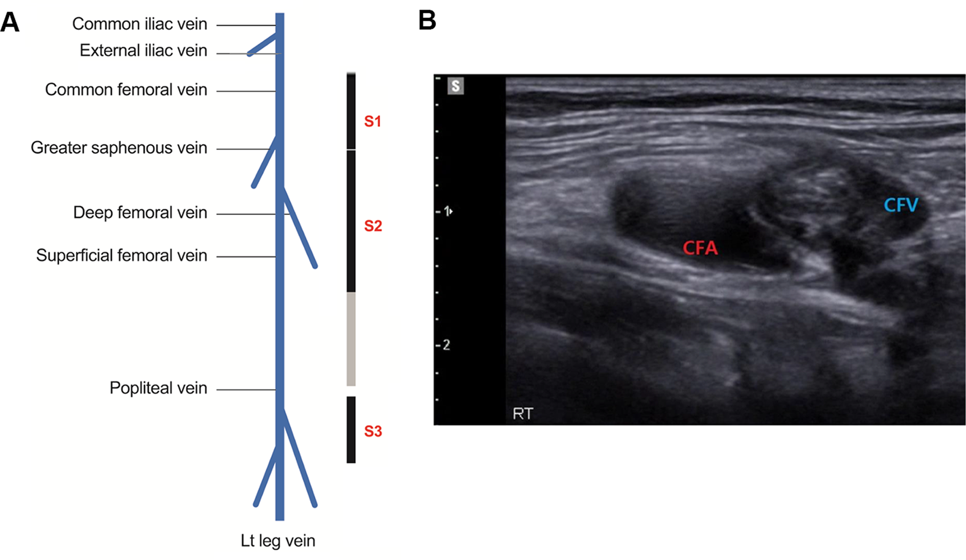
In an Australian setting, meaningful survival to good neurological outcome following out-of-hospital cardiac arrest (OHCA) is approximately 10% [1,2,3,4]. Refractory cardiac arrest (RCA) has a very poor prognosis (< 2% survival) and is often defined as those patients without sustained return of spontaneous circulation (ROSC) following 10–15 min of best-practice resuscitation or after 3 defibrillations for shockable rhythms [5,6,7]. Extracorporeal membrane oxygenation implemented during cardiopulmonary resuscitation (ECPR) provides end-organ perfusion to patients whilst the underlying cause of arrest can be treated, or recovery occurs (Fig. 1). Despite substantial increase in the use of ECPR, outcomes vary widely. A recent systematic review and meta-analysis of ECPR use for selected OHCA cases demonstrated a survival to hospital discharge of 24% of patients with 18.5% having a favourable neurological outcome.[8] Other systematic reviews and meta-analyses have also demonstrated a survival benefit for cardiac arrest patients receiving ECPR compared to conventional CPR [5, 9, 10].
Fig. 1
A schematic of an ECPR setup for cardiac arrest. Blood is taken out via the venous ‘drainage’ catheter which enters via the femoral vein with the tip located at the level of the right atrium. Oxygenated blood is pumped back to the patient via an arterial ‘return’ cannula located in the descending aorta. Picture obtained from LearnECMO.com with permission [11]
A key prognostic variable for ECPR is minimising the time from cardiac arrest to the commencement of ECMO flow, a period referred to as the low flow time. Evidence demonstrates that patients who are placed on ECPR beyond 60 min have a very low likelihood of meaningful survival [12,13,14,15]. Therefore, prehospital teams and emergency departments (ED) must incorporate systems to facilitate rapid assessment, transport, and ECMO cannulation to achieve the best outcome for carefully selected patients in RCA.
The aim of this brief narrative review is to describe the collective system improvements and implementation strategies that incorporate ECPR into prehospital and ED RCA algorithms in Sydney, Australia.
SettingIn 2020 the population of greater Sydney in New South Wales (NSW), Australia was just over 5.3 million living in a total area of 12,368.2 Sq km [16]. Currently three main hospitals provide ECPR to greater Sydney, two of which are located in central Sydney. Through shared experience and knowledge, the three tertiary-level hospitals have each established protocols and processes for ECPR within Sydney.
Our experienceWe have previously published our experience of ECPR during the 2CHEER trial and demonstrated a 44% (11/25) survival to hospital discharge with favourable neurological outcome [17]. Since the publication of this data from 2020 to 2023 we have had 51 ECPR cases (Table 1) with 16 survivors to hospital discharge (31%), of these patients 26 were OHCA with 9 survivors (35%). The majority of patients who survived to hospital discharge had good neurological function with the mean CPC score or 1.4 (STD 0.8). This fall in survival rate may reflect real-world experience outside of a clinical trial as well the challenges of managing cardiac arrests during the Covid pandemic.
Table 1 Outcome data for ECPR patients from 2020–2023 at the 3 ECPR centres in SydneyActivation processA robust and protocol driven prehospital system should facilitate the early activation of key staff and equipment preparation at the receiving hospital in potential ECPR candidates. This requires an awareness that optimising prehospital resuscitation is a critical step and there is a balance between achieving the highest likelihood of ROSC with good prehospital resuscitation whilst also minimising delays to transfer to hospital for ECPR [18]. For patients in RCA meeting criteria for ECPR small delays in transport may be associated with poorer outcomes and so it is critical to minimise the low flow time prior to ECMO initiation [19]. Our prehospital transport times and times to ECMO flows are demonstrated in Table 2.
Table 2 Key timepoints of patients with OHCA including prehospital times and times to ECMO flowsThe standard process for prehospital care in the Sydney system involves the dispatch of an ambulance unit that often comprises of two paramedics with standard ALS skills with the average time of arrival of 8 min [20]. The ALS algorithm is followed with early defibrillation, basic airway management and IV access as well as advanced airway management (supraglottic or intubation) and placement of a MCPR device (LUCAS) [21].
Once the prehospital notification has been received at the ECPR centre, the relevant team members are activated via a dedicated ECMO/ECPR page, and the resuscitation bay is prepared (Fig. 2). The ECPR team is available at all sites in standard working hours. Occasionally the ECPR team may be activated after working hours, but these are usually on a case-by-case basis and dependent on the availability of the team and the anticipated prehospital transport times.
Fig. 2
Layout of the emergency department resuscitation bay for ECPR. US = Ultrasound, TOE = Transoesophageal Echocardiography
The criteria to activate the ECPR teams differs slightly between institutions. However, the principles are similar with a balance struck between sensitivity and specificity of the activation criteria. These ECPR activation criteria use weighted factors associated with improved probabilities of survival based on the best available evidence or recommendations [12, 22,23,24,25,26] and include:
Estimated age 18–70 years.
Witnessed OHCA.
Initial rhythm VT/VF or automatic external defibrillator shock delivered or PEA arrest
Bystander CPR started within < 5 min and ongoing on ambulance arrival with no history suggestive of a prolonged period without CPR
Figure 3 shows an example of the criteria used for activation within one ECPR centre. The same criteria and activation process are used to mobilise the ECPR team for patients who suffer RCA in the ED.
Fig. 3
Inclusion and exclusion criteria utilised to activate the ECPR team (RPA hospital)
It is recommended that an institution commencing an ECPR program adheres to strict inclusion/exclusion criteria to avoid including patients who are unlikely to benefit from the intervention and therefore worsen survival rates. Inclusion "creep” is a challenge for many clinicians, especially when managing younger patients. An agreement of predefined criteria from the key stakeholders as well as a local governance structure within the hospital prior to commencement of an ECPR program improves compliance and facilitates efficient management within the ED.
RolesIt is prudent that the resuscitation team roles are pre-allocated and that key tasks are outlined (Fig. 4 and Appendix 1) to achieve efficient workflow. To maximise the productivity of the members, the team is separated into small sub-teams each with delineated roles and tasks. Further, the use of a nursing team leader to manage operational ALS concerns, and/or a second medical team leader for "event management” (see below) is often pertinent because the cognitive load on a single leader is likely to be overwhelming. Over a series of 7 ECPR calls at one of our sites there was an average peak attendance of 24 staff members (range 16–30) emphasising the challenge of managing both allocated roles and interested staff bystanders.
Fig. 4
A schematic of staff positions and roles including differentiation into various sub-groups. Any staff members not directly involved in the resuscitation are advised to remain behind a visible red line in the resus bay
On arrival to ED the team members should don the appropriate personal protective equipment (PPE) and lanyards that identifies the persons role within the team (Appendix 1). The majority of the ECPR centres utilise a cardiac anaesthetist to perform a transoesophageal echocardiogram (TOE) during the arrest. The TOE becomes essential for: 1. Identifying a potential cause of arrest i.e., pericardial tamponade, visualisation of embolism, aortic dissection 2. Confirming correct wire/cannula placement for ECMO and 3. Ensuring effective left ventricle compression via MCPR [27,28,29].
The cannulation team are often Intensive Care specialists or Cardiothoracic surgeons and consist of one cannulator plus an assistant positioned to right side of the patient to access the right femoral artery and vein.
A nurse is assigned the role of CPR to ensure that chest compressions are adequate, that the MCPR device is secured in the correct location, and to take over the role of chest compressions if the MCPR device fails.
ECPR cases may require duplication of various roles due to the workload required for various tasks (e.g. medication preparation listed in appendix 2) and attract bystander interest that are in addition to the list active roles (Fig. 4). Therefore, as with any major resuscitation in the ED, crowd control and communication to team members is crucial. Strategies that actively mitigate the latent threat of large numbers of responders and role redundancy are a key consideration in ECPR cases. Having a dedicated team member(s) to manage crowd control, noise levels and perform regular summaries may be efficacious but have not been studied in clinical studies.
ResuscitationThe resuscitation of ECPR patients is divided into four main phases (Fig. 5). These phases of resuscitation have been adapted from the 'Learn-ECMO’ team and allows for team members to smoothly transition through a complicated resuscitation [11]. Dividing this complex and stressful resuscitation into different phases with key task priorities ensures that all staff members are focused on manageable goals that can be achieved in a timely manner [30]. The team leader utilises a checklist with these tasks for each phase to reduce cognitive burden during the resuscitation.
Fig. 5
A flowchart of the ECPR patient journey and task priorities for each phase of the resuscitation. Phase 1 = Arrival and decision. Phase 2 = Proceed with cannulation. Phase 3 = Preparation for disposition. MCPR = Mechanical CPR; CT = Computerised tomography; ICU = Intensive care unit; US = Ultrasound; TOE = Transoesophageal Echocardiogram; ETCO2 = End-tidal Carbon Dioxide; SpO2 = Oxygen saturations; NG = Nasogastric; CXR = Chest x-ray
Pre-arrival phaseOnce the notification has been received at the ECPR centre and the team is activated, the priority is to ensure the staff, drugs (Appendix 2) and equipment are ready to receive the patient. All members of the team don standard resuscitation PPE including eye protection, gowns, and gloves. The cannulation team will prepare for the procedure using standard surgical aseptic technique. The resuscitation bay needs to be large enough (suggested a minimum 25m2) to allow for the ECPR equipment and 360-degree access to the patient by nursing and medical staff. Non-essential equipment should be removed and equipment that is essential for cannulation moved into the correct positions (Fig. 2). One head of bed US is used to perform TOE and one or two additional adjacent US machines are to assist with vessel cannulation.
Phase 1 Arrival and decisionPhase 1 of the resuscitation begins when the patient arrives in ED and ends when drapes are placed. The patient is rapidly transferred to a resuscitation bed and an abbreviated handover that focuses on the ECPR inclusion/exclusion criteria by the paramedics. The ED nursing team leader will continue the cardiac arrest algorithm as per standard ALS recommendations. During this time the ED team leader and ECMO cannulators perform a rapid assessment of whether the patient is suitable using the criteria mentioned previously. Once the patient is deemed appropriate then a list of tasks is utilised by the ED team leader to prepare the patient for cannulation. If not already performed in the field, then an endotracheal tube is placed and secured by the most senior ED doctor, facilitating the ability to perform a TOE.
Phase 2 – Proceed with cannulationThe commencement of Phase 2 is indicated once the sterile drapes are placed for cannulation. The medical team leader must notify staff that the team is no longer performing standard ALS management as defibrillation and adrenaline dosing are paused in preference for successful cannulation. MCPR is performed continuously with only very brief interruptions if cannulation is challenging. Ideally an arterial line is placed by the ED or ICU 'circulation’ doctor. Intravenous heparin 5000units is given as the guidewires are placed.
Phase 3 – Preparation for dispositionOnce ECMO flows are established a myriad of challenges and complexities are possible that are outside the scope of this article or standard practice for the emergency physician. Some of these acute challenges include haemodynamic instability, coagulation management and circuit complications. A list of key priorities to mitigate these challenges are described in Fig. 5. In our experience we have found that once adequate ECMO flow is established then patients frequently revert to malignant reperfusion rhythms including VF. Approximately one minute of CPR helps to decompress the distended right ventricle that frequently occurs during cardiac arrest followed by defibrillation if in a shockable rhythm.
Often whilst in ED awaiting transfer to either the cardiac angiography suite, CT, or ICU a distal perfusion cannula is placed in the superficial femoral artery ipsilateral to the arterial return cannula. The distal perfusion cannula is again placed under US guidance and ensures adequate antegrade perfusion to the lower leg as flow distal to the common femoral artery is limited or absent due to the size of the arterial return cannula.
A nasogastric tube is placed in ED so that antiplatelet medications (Aspirin and Clopidogrel or Ticagrelor) can be commenced for patients with a suspected coronary artery cause. Further anti-coagulation management is completed in ICU and under the direction of the perfusion or ICU teams and may be directed by activated clotting time (ACT), APTT/anti-Xa and/or visco-elastic testing. Once the patient is ready the medical team leader hands over care of the patient to the cardiac anaesthetist for transfer.
Post- ED careActivation of the cardiac angiography suite occurs routinely on the notification of a potential ECPR patient arriving with OHCA. Immediate coronary angiography is performed after establishment of ECMO flow. However, if during the resuscitation the ED team leader in communication with the resuscitation team feels that another cause is more likely, then the angiography can be deferred. Whilst controversy remains regarding the utility of empiric angiography for patients post cardiac arrest of unknown cause or without ST Elevation, many of the recent trials did not include haemodynamically unstable patients and patients without ROSC or ECPR [31,32,33]. Following cardiac arrest the presence of acute coronary lesions in patients with non – ST elevation cardiac arrests vary by 40–70%, however, this prevalence is increased in refractory cardiac arrest patients [31, 32, 34]. In our experience, 61% (n = 20) of ECPR patients who had a coronary angiogram (n = 33) required stenting of a coronary lesion indicating that there are a significant proportion of ECPR patients that may benefit from cardiac angiography.
Prior to ICU admission a computerised tomography (CT) of the head, and chest to knees is performed to identify any evidence of intracranial pathology (e.g. intracranial haemorrhage) or sequelae of chest compressions (e.g. traumatic pneumothorax, rib fracture, solid organ trauma), and cannulation injuries and configuration. In approximately 40% of CT pan scans a pathology will be identified that will change the management of patients [35].
留言 (0)