Cell culture, treatments, and SARS-CoV-2 infectionCell cultures
CaLu-3 cells (ATCC, HTB-55™, human lung adenocarcinoma) were grown in DMEM supplemented with 10% Fetal Bovine Serum (FBS), 1% non-essential aminoacids (NEAA), and 1% penicillin–streptomycin (PS)/L-glutamine. A549-hACE2 epithelial cells (NR-53522, BEI Resources, NIAID, NIH, human lung adenocarcinoma cells expressing Human Angiotensin-Converting Enzyme 2), and VERO-E6 cells (ATCC, African green monkey epithelial kidney cells) were grown in DMEM supplemented with 10% FBS, and 1% PS/L-glutamine. Human Umbilical Vein Endothelial Cells (HUVECs, Lonza, Walkersville, USA) were grown in EMG-2 medium (Lonza, Walkersville, USA) containing 2% FBS.
SARS-CoV-2 infection and treatments
SARS-CoV-2 (strain 2019-nCoV/Italy-INMI1) was expanded in VERO-E6 cells and viral titers were assessed through the TCID50 endpoint dilution assay in CaLu-3 and A549-hACE2 epithelial cells. Given the absence of cytopathic effects, viral titers from cell supernatants were determined in order to assess TCID50 through quantitative, real-time, Reverse Transcriptase-Polymerase Chain Reaction (RT-qPCR), as previously described [63, 64]. The TCID50 values were used to calculate the Multiplicity of Infection (MOI). CaLu-3, and A549-hACE2 cells were seeded in 24-well plates (1.25 × 10⁵, and 0.8 × 10⁵ cells/well, respectively, in a final volume of 500 μL) for 24 h. HUVECs were seeded into a 24-well cell culture plate (0.5 × 10⁵ cells/well), containing 0.5% gelatinized cover slips, and grown to confluence until they reached the cobblestone morphology (7 days).
Type-I IFN (human recombinant IFN-β, 500 IU/ml, BEI resources) and human recombinant α-syn monomers (1 μM, Merck-Sigma), were exogenously pre-administered to cells, both alone and in combination, at 24 h pre-infection. CaLu-3 and A549-hACE2 cells were Mock-infected or infected with SARS-CoV-2 at a MOI of 0.05 for 1 h, while HUVECs were Mock-infected or SARS-CoV-2-infected at a MOI of 1 for 1 h. Cells were washed in PBS to remove unbound virus and cultured in fresh medium. Supernatants and/or cells were collected at different time intervals post-infection (24 and 48 h for CaLu-3, and A549-hACE2; 24, 48, and 72 h for HUVECs), and processed accordingly for different methodological assays.
In a different set of experiments, CaLu-3, and A549-hACE2 cells were seeded at a low density (1.25 × 10⁵, and 0.8 × 10⁵ cells/well) in 12-well plates in a final volume of 1 mL growth medium. Cells were cultured for 24 h and then infected with SARS-CoV-2 at three different MOIs (0.01, 0.005, 0.001) for 1 h. CaLu-3, and A549-hACE2 were washed in PBS, replenished with 1 mL fresh medium, and cultured up to 3 and 5d, respectively. At these time-points, cells were harvested for transcriptional analysis aimed at investigating correlations between the mRNA expression of SNCA and SARS-CoV-2 Nucleocapsid (N) gene.
Eventually, a co-culture system of PBMCs and CaLu-3 was set up as previously described [39]. Briefly, 1.25 × 10⁵ Calu-3 cells were cultured in the lower chamber of a 12 well plate with 0.4 µm pore polycarbonate membrane inserts (Costar, Corning Incorporated, Corning, NY, USA) in 1 mL medium with 10% FBS. The same day, 2 × 10⁶ PBMCs isolated from healthy, SARS-CoV-2-negative volunteers, were cultured in 1 mL RPMI medium with 10% FBS and 15 ng/mL of IL-2 in the upper chamber of the insert membranes placed on the pre-seeded Calu-3 cells. After 24 h, cells were either Mock-infected or challenged with SARS-CoV-2 (2019-nCoV strain 2019-nCoV/Italy-INMI1, Rome, Italy) at a MOI of 0.01 for 1 h. Cells were than thoroughly washed with PBS and refilled with proper growth medium. At 3d post-infection, both PBMCs and Calu-3 cells and supernatants were collected for RNA extraction, and evaluation of SARS-CoV-2 replication rate by using the viral RNA extraction method and the SARS-CoV-2-N mRNA detection protocol described below. Ethical clearance was obtained from the University of Milan Ethics Committee (number 14/22). Written informed consent was obtained after receiving information about use of their biological samples. The biological material was anonymized. All the experiments with SARS-CoV-2 virus were performed in the BSL3 facility.
Quantification of viral replication from cell supernatants
For assessment of SARS-CoV-2 replication, RNA from cell culture supernatants (volume = 100 μl from 24-well plates; 200 μl from 12-well plates) was extracted through the Maxwell® RSC Instrument (Promega, Fitchburg, WI, USA), and quantified through real time, RT-qPCR by means of well-validated primers for SARS-CoV-2 N gene (2019-nCoV_N2, 2019-nCoV_N2, Forward Primer 5ʹ-TTA CAA ACA TTG GCC GCA AA-3ʹ, Reverse Primer 5ʹ-GCG CGA CAT TCC GAA GAA-3ʹ), as previously described [64]. RT-qPCR was performed through the CFX96 instrument (Bio-Rad). Melting curves besides Cq values were analyzed for primer and reaction specificity. Absolute viral copy number quantification was performed by referring to a standard curve from the quantified 2019-nCoV_N-positive Plasmid Controls (IDT, USA). Before sample analysis outside the BSL3 area, the virus was inactivated according to institutional safety guidelines.
Cell viability
CaLu-3 and A549-hACE2 cells (3 × 10⁴, and 2 × 10⁴ cells/well, respectively) were seeded in 96-well plates for 24 h and then treated with Type-I IFN (human recombinant IFN-β, 500 IU/ml, BEI resources) and/or human recombinant α-syn monomers (1 μM, S7820, Merk-Sigma). Cells were then either mock-infected or infected with SARS-CoV-2 as detailed above. After 48 h, cell viability was assessed by 3-(4,5-dimethylthiazol-2-yl)-2,5-diphenyltetrazolium bromide (MTT) method. Briefly, 30 μL of MTT (final concentration, 0.5 mg/mL) were added to each well under sterile conditions, and the 96-well plates were incubated for 4 h at 37 °C. Supernatants were removed, and dimethyl sulfoxide (100 µL/well) was added. The plates were then agitated on a plate shaker for 5 min. The absorbance of each well was measured at 490 nm with a Bio-Rad automated EIA analyzer (Bio-Rad Laboratories, Hercules, CA, USA). The viability of Control cells (Mock-infected, vehicle-treated) was considered 100%, while the other conditions were expressed as percentages of control.
For Trypan Blue exclusion assay, cells in 24-well plates were incubated in Cell Dissociation Buffer 1X (Merck-Sigma, Milan, Italy) for 10 min at 37 °C. Then, an equal volume of DMEM was added to the wells to stop the dissociation reaction. Ten μL of cell suspension were mixed and briefly incubated with 10 μL of 0.4% Trypan Blue (Merck-Sigma, Milan, Italy) in 96-well plates. Ten microliters of the mix were loaded on chamber slides and counted with the T20 Automated Cell Counter (Bio-Rad Laboratories, Hercules, CA, USA). Results are expressed as the mean ± SEM from n = 3 independent experiments.
SNCA RNA interference
To silence SNCA expression, specific small interfering RNA oligonucleotides (SNCA-siRNA, AM16708, Thermo Scientific, Waltham, MA, USA) were used:
Sense 5ʹ-GGGUAUCAAGACUACGAACtt-3ʹ.
Antisense 5ʹ-GUUCGUAGUCUUGAUACCCtt-3ʹ.
A549-hACE2 cells were transfected with 10 nM siRNA, either Non-Targeting negative control (siRNA AM4613 Negative Control No. 2, Thermo Scientific, Waltham, MA, USA) or against SNCA, using Lipofectamine 2000 according to the manufacturer’s instructions (Thermo Scientific, Waltham, MA, USA). Transfected cells were cultured for 24 h before infection with SARS-CoV-2 and harvested/fixed at 48 h post-infection (72 h post-transfection), for cell viability assessment through Trypan Blue exclusion assay, transcriptional analysis through qPCR, or immunofluorescence analysis.
Immunofluorescence
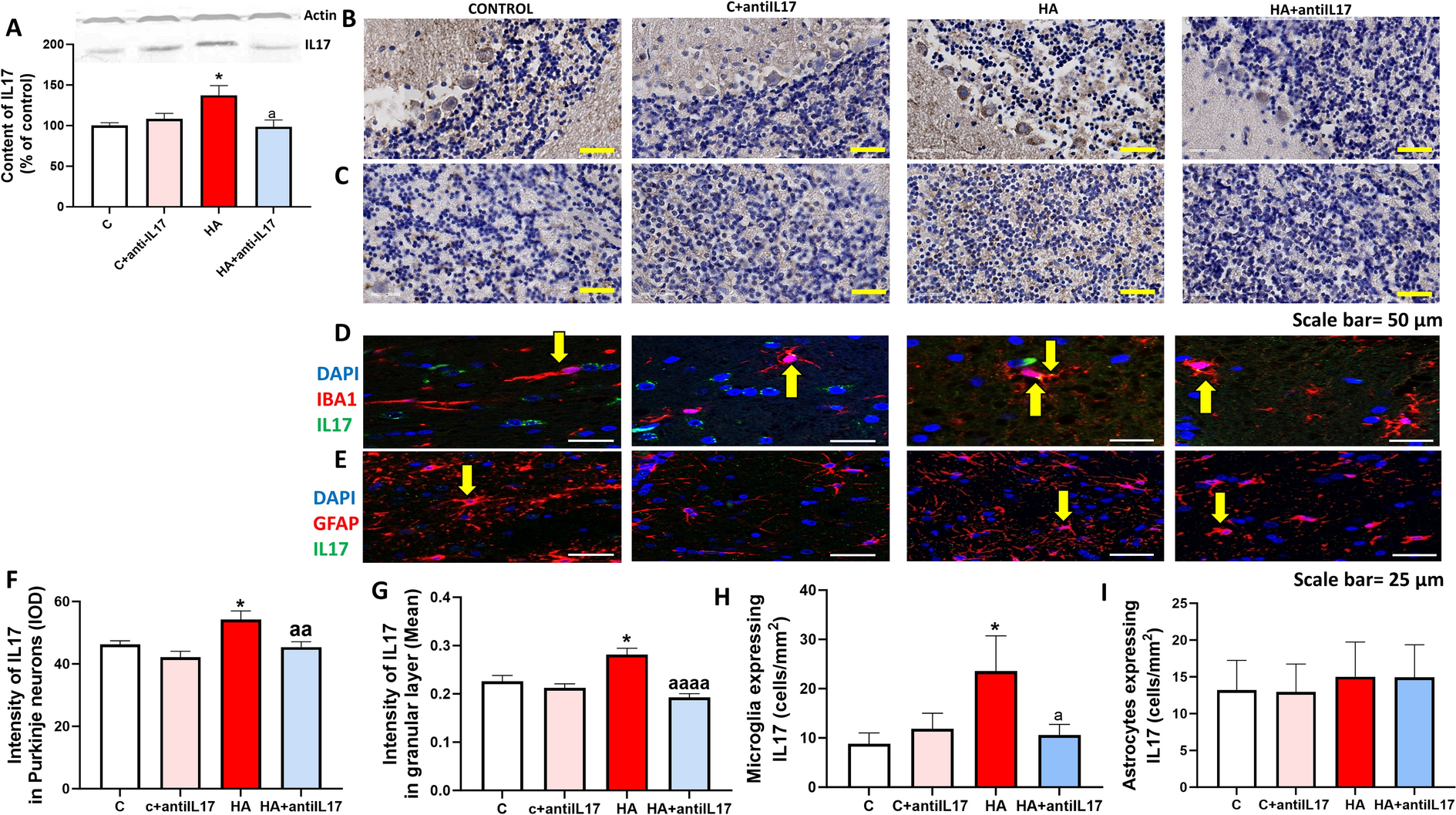
For immunofluorescence, cells were seeded on Poly-Lysine-coated glass coverslips placed on 24-well plates. At the indicated times post-infection, cells were fixed and permeabilized with different approaches slightly modified from [31] to provide better sensitivity for the detection of total α-syn, monomeric or multimeric α-syn species. In detail, for experiments shown in Figs. 1, 2, and 6, cells were fixed for 15 min in 4% Formaldehyde solution (containing methanol as a stabilizing agent), and then permeabilized with 0.1% Triton 100X for 15 min. This procedure is expected to moderately permeabilize cells allowing rough detection of both α-syn oligomers and monomers. For experiments in Figs. 4, and 5, fixation and permeabilization procedure was modified for better visualization of α-syn monomers vs oligomers/multimers. In detail, for preservation and better visualization of α-syn monomers, cells were fixed in 4% paraformaldehyde (PFA) for 15 min and briefly permeabilized with 0.1% Triton 100X for 10 min. For better visualization of α-syn oligomers/multimers, cells were fixed for 15 min in 4% Formaldehyde solution (containing methanol as a stabilizing agent) and then further permeabilized with 0.3% Triton 100X for 15 min. After thorough washing, cells were incubated in a blocking solution of 5% BSA for 1 h, and then with primary antibodies against α-syn (rabbit mAb, 1:200, BSM-54277R 3H12, Bioss, USA), and SARS-CoV-2 Spike (mouse mAb, 1:1000, 1A9, Genetex, Cat. No. GTX632604, Prodotti Gianni, Milan, Italy) or Nucleocapsid (mouse mAb, 1:1000, E8R1L, #33717, Cell Siganling, Euroclone, Milan, Italy) prepared in 1% BSA for 1 h at RT. Cells were washed thrice in PBS and incubated with Alexa-Fluor-conjugated secondary antibodies raised against the host species of the primary antibodies, namely Goat anti-mouse Alexa Fluor 488 (abcam, ab150113) or 647 (abcam, ab150115), or Goat anti-rabbit Alexa Fluor 488 (abcam, ab150077) or 647 (abcam, ab150079), 1:500 prepared in 1% BSA-PBS. Negative controls were performed by omitting primary antibodies. After 3 × 5 min washes in PBS, coverslips were mounted on Superfrost glass slides using a mounting medium with DAPI (Enzo Life Sciences, Milan, Italy).
Thioflavin-S-α-syn co-staining
After the incubation with the secondary antibodies, and following 3 × 5 min washes in PBS, CaLu-3 cells were incubated for 15 min at RT in the dark with a solution of 0.05% w/v Thioflavin-S (T1892, Merck-Sigma, Milan, Italy), which was freshly prepared in 50% ethanol/water and 0.22 μm filtered. Cells were washed twice with 50% ethanol for 10 min each, and then washed once with 80% ethanol for 20 min. Eventually, cells were washed in PBS, briefly rinsed with water, and coverslips were mounted on Superfrost glass slides using a mounting medium with DAPI (Enzo Life Sciences, Milan, Italy). Confocal images were acquired on a TCS SP8 System equipped with a DMi8 inverted microscope and a HC PL APO 40 × /1.30 Oil CS2 (Leica Microsystems, Wetzlar, Germany) at a resolution of 1024 × 1024 pixels.
Quantification of SARS-CoV-2 Nucleocapsid (N)-positive cells
SARS-CoV-2-infected cells were quantified by counting N-positive cells/total cells per microscopy field by using Image J software (NIH, Bethesda, MD, USA). Counts per performed from at least n = 3 microscopy fields per experimental group, and per each independent experiment. At least 300 total cells were counted per each experimental group. Values were expressed as percent N-positive cells ± SD.
RNA extraction and transcriptional analyses through real-time qPCR
For transcriptional analyses, cells were collected in collected in RNAzol® (TEL-TEST Inc., Friendswood, TX, USA) and RNA extraction was performed through the phenol–chloroform method, as previously described [63]. RNA was quantified by the Nanodrop 2000 Instrument (Thermo Scientific, Waltham, MA, USA). One μg of RNA was purified from genomic DNA with RNase-free DNase (RQ1 DNase; Promega) and reverse transcribed into cDNA with Moloney murine leukemia virus reverse transcriptase along with random hexanucleotide primers, oligo dT, and dNTPs (Promega, Fitchburg, WI, USA). cDNA (25 ng) was amplified and quantified by real-time qPCR (CFX96 connect, Bio-Rad, Hercules, CA, USA) through SYBR Green PCR mix (Promega, Fitchburg, WI, USA). Negative controls (distilled water), as well as positive controls (human cDNA), were included in each run. Results for gene expression analyses were calculated by the 2−ΔΔCt equation. Melting curves besides Cq values were analyzed for primer and reaction specificity. Results are presented as the percent mean ± SEM of the relative expression units to an internal reference sample, and normalized to the Glyceraldehyde-3-Phosphate Dehydrogenase (GAPDH) housekeeping gene. Results show the quantifications from at least n = 3 independent experiments. The following genes were analyzed: synuclein alpha (SNCA), Interferon beta (IFNB), SARS-CoV-2 Nucleocapsid (N2), Signal transducer and activator of transcription 1 (STAT1), Myxovirus Resistance Protein 1 (MX1), Myxovirus Resistance Protein 2 (MX2), 2'-5'-oligoadenylate synthetase 1 (OAS1), retinoic acid-inducible gene I (RIG-I), tumor necrosis factor alpha (TNFA), tumor necrosis factor receptor superfamily 1A (TNFRSF1A), Toll-like receptor 8 (TLR8), Toll-like receptor 9 (TLR9). Primers for STAT1, and TNFRSF1A were purchased as already optimized (PrimePCR, Bio-Rad). The sequences of the remaining primers are listed in Additional file 1: Table S1.
Western blot
Cells were collected in ice-cold lysis buffer (RIPA buffer with 1% Triton X-100, and 0.1% SDS) supplemented with a cocktail of protease and phosphatase inhibitors (cOmplete and PhosSTOP; Roche Applied Science, Mannheim, Germany). Samples were incubated on ice for 30 min on a platform rotator, sonicated, and then centrifuged at maximum speed for 30 min at 4 °C. Protein concentration was determined through BCA protein assay kit (Pierce, USA). 50 μg proteins per sample were prepared by combining the appropriate volume with 5 × Laemmli SDS sample buffer (final 1 ×) and Milli-Q H2O. Samples were boiled for 5 min and loaded on 4–12% SDS-PAGE gel (Bio-Rad), which was run at 150 V for 1–1.5 h. Proteins were transferred on PVDF membrane through The Trans-Blot Turbo Transfer System TM and Transfer packTM (Bio-Rad). To preserve α-syn protein, membrane fixation with 0.4% PFA for 30 min at RT was performed [31]. Prior to the blocking step, the membrane was imaged as a loading control for normalization to total protein. The membrane was blocked in 1 × TBS with 5% non-fat milk for 1 h at RT, washed in TBS-0.2% Tween, and incubated overnight at 4 °C with rabbit anti- α-syn antibody (1:800, BSM-54277R 3H12, Bioss, USA) in blocking buffer with 2.5% non-fat dry milk. The day after, the blot was washed 3 × 5 min with 1 × TBS-0.2% Tween and incubated for 1 h with HRP-conjugated goat anti-rabbit secondary antibody (1:5000, STAR208P, Bio-Rad) in blocking buffer with 2.5% non-fat dry milk. The blot was washed 3 × 5 min with 1 × TBS-0.2% Tween and then incubated for 5 min with Clarity Western ECL substrate and visualized with a ChemiDoc MP imaging system (Bio-Rad). Results were analyzed using the Image Lab software (Bio-Rad). For each experiment, a representative blot is shown, and the graphs show to the mean ± SEM of raw normalized values obtained from indicated (n) independent experiments.
Statistical analysis
The GraphPad Prism software package (GraphPad Software, San Diego, CA, USA) was used to generate all the graphs. Data normality was assessed through the Shapiro–Wilk test. In case of normal distribution, the statistical significance was evaluated using the unpaired Student’s t test (single comparisons), or one-way, or two-way ANOVA (as appropriate, for multiple comparisons), followed by multiple testing correction by false discovery rate (FDR) through the Two-stage linear step-up procedure of Benjamini, Krieger and Yekutieli. When data normality was non confirmed, non-parametric Wilcoxon ranked or Kruskal Wallis tests were applied. Obtained q values corresponding to adjusted p values were shown in the graphs with statistical significance set as *p < 0.05, **p < 0.01, ***p < 0.001, ****p < 0.0001. Pearson’s r coefficient was calculated for correlation analyses followed by two-way, paired Student’s t test. To avoid graphs overcrowding, p values were shown for statistically significant groups of interest only. Results are expressed as mean ± SEM or SD of the indicated n values, as specified in the figure legends.


留言 (0)