1 Background
One amongst the major issues cardiologists are tending is risk stratification in ACS patients to spot the seriousness and complexity of coronary heart disease (CHD). For this reason, many scoring scales and laboratory parameters are employed in clinical practice.1
Flowers et al. within the 1960s illustrated that fragmented QRS (f-QRS) complexes (high-frequency components) are more common among patients with preceding myocardial infarction as well as amongst patients with either right or left ventricular enlargement.2,3
Das et al provided practical definitions for f-QRS in 2006, followed later in 2008 by f-QRS definition in the wide QRS complex.4,5
Territorial f-QRS designs signify the presence of a prominent corresponding focal localized myocardial scar on stress MPI.6 The f-QRS appeared to be an independent indicator of adverse cardiac events in CAD patients on long-term follow-up.7 The number of f-QRS leads, especially the existence of ≥3 electrocardiogram (EKG) leads with f-QRS, is an independent predictor of cardiac fatality or hospitalization for heart failure in patients with prior MI.8,9
2 Methods
A retrospective observational review search of medical records. The study recruited eighty-four acute coronary syndrome (ACS) patients with age range (29–80 years). Patients were admitted to the Intensive Care Medicine Department within the Faculty of Medicine. Our retrospective non-interventional non-invasive study was done after approval and informed consent of all patients or their relatives and all personal patients’ data are hidden carefully.
The study protocol was approved by the Critical Care Department; Faculty of medicine Cairo University ethical committee followed by the approval of the faculty of medicine Kasr Al-Ainy College and Cairo University Council in 2015; Cairo University, Egypt.
They were separated according to 12 leads surface electrocardiogram within 2 days of admission into two groups. The group A (cases group) counting forty six patients with f-QRS and the group B (controls group) counting thirty eight patients without f-QRS. Patients with previous myocardial infarction (ST elevation myocardial infarction/Non-ST-elevation myocardial infarction), cardiomyopathies, Interventional procedures (PCI/CABG) or permanent AF were excluded.
We reviewed carefully the accessible medical records. The patients’ data included thorough medical record, clinical examination counting vital signs, the key risk factors of CHD, laboratory investigations counting complete blood picture, liver and kidney function tests, cardiac enzymes, cTn and trans-thoracic echocardiography.
Within the initial 48 hours, we inspect ECGs for first the recognition of f-QRS according to Das and associates definition of f-QRS in 2006 and 2008. In 2006 Das et al characterized (f-QRS) being the presence of [R′] a further R wave or indenting within the lowest point of the S wave, or the existence of >1 R′ (fragmentation) in two adjoining leads, corresponding to a chief coronary arteria domain. Two years later, Das et al characterized the f-QRS within wide QRS complex, ie ≥120 ms (Bundle Branch Block and paced rhythms) as different RSR′ designs with/without a Q wave, with >2 R waves (R′) or >2 indenting within the R wave, or >2 indents within the down-or-up-stroke of the S wave, in two adjoining EKG leads corresponding to a chief coronary artery supply domain in fragmented bundle branch block and also the existence of >2 R′ or >2 notches within the S waves in two adjoining fragmented paced QRS leads.4,5 represents one of the present research ECGs.
Second we recorded the location of the f-QRS complex. Anterior f-QRS was characterized by the occurrence of f-QRS in two adjoining anterior chest leads (V1–V5). Lateral location of f-QRS was characterized as the existence of f-QRS in two adjoining lateral leads (I, aVL, and V6) while the inferior f-QRS location was distinct by the occurrence of f-QRS in two adjacent inferior leads (II, III, and aVF).10 Third the f-QRS number of leads was tallied (at the minimum 2 adjacent leads relating to a chief coronary territory).
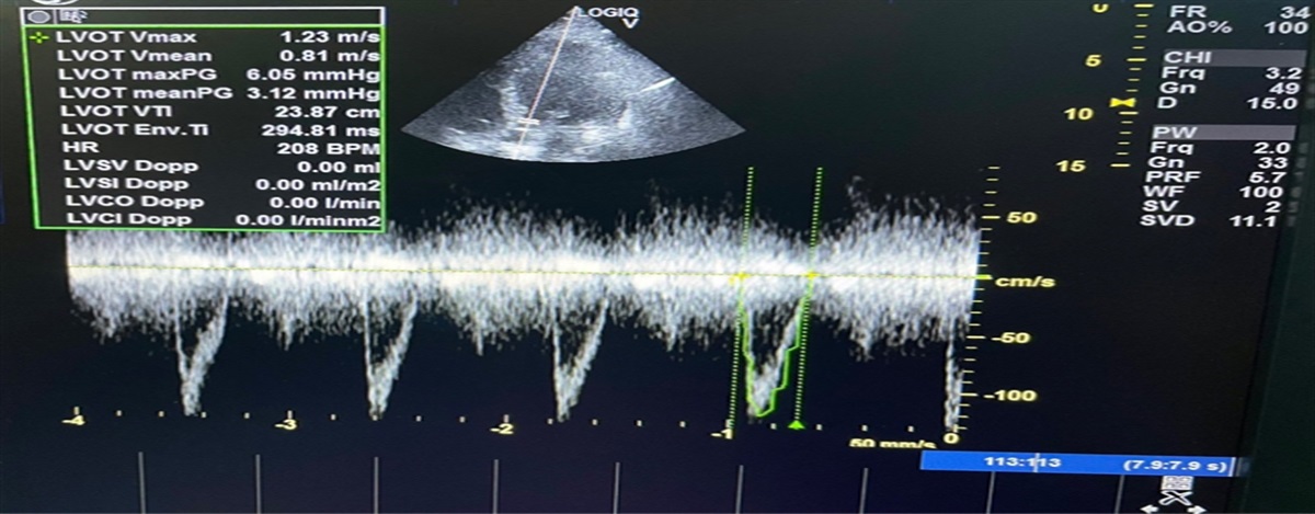
Echocardiography was employed for measurement of left ventricle systolic function and segmental wall motion abnormality (SWMA). Wall motion score index (WMSI) was calculated in accordance to the European Association of Echocardiography (EAE) in conjunction with American Society of Echocardiography (ASE) employing a 16 segments model tool.11 Each segment was visually scored employing a semi-quantitative rating framework where 1 = normal, 2 = hypokinesia, 3 = akinesia, and 4 = dyskinesia. A global WMSI was calculated as the summation of the scores of each segment divided by the number of the visualized segments.
Coronary angiography (CA) was subjected to estimation of severity and extent of coronary arteria disease using Gensini score, in which diminution within the lumen diameter of 25%, 50%, 75%, 90%, 99%, and complete obstruction are given Gensini scores of 1, 2, 4, 8, 16, and 32, respectively. Each principal vascular segment is then allocated a multiplier in accordance with the functional importance of the myocardial region supplied by that segment: the left main coronary arteria × 5; the proximal segment of left anterior descending (LAD) × 2.5; the proximal segment of the circumflex arteria × 2.5; the mid-segment of the LAD × 1.5; the distal segment of the LAD, the posterolateral arteria, the right coronary arteria, and the obtuse marginal arteria × 1; and others × 0.5.12,13.
2.1 Statistical data
Quantitative data was presented in terms of mean ± SD if normally distributed or median and range if not normally distributed while qualitative data was presented in terms of frequencies and percentages. Assessment of mathematical factors was performed between the research groups employing Student t test for independent samples in comparing two groups of normally distributed data and/or large groups and Mann Whitney U test for independent samples for comparing non normal data and whenever appropriate. Chi-square (χ2) test was performed when comparing categorical data. When the expected frequency is <5, Exact test was used instead. P values less than .05 was considered statistically significant. All statistical calculations were done using Microsoft Office Excel 2007, 2010, and Microsoft Excel 365, the statistical package SPSS version 22, MedCalc Statistical Software versions 16.8 and 16.8.4 (MedCalc Software bvba, Ostend, Belgium; https://www.medcalc.org;206), Social Science Statistics website (http://www.socscistatistics.com) and computer program SPSS (Statistical Package for the Social Science; SPSS Inc., Chicago, IL, USA) release 15 for Microsoft Windows (2006).
3 Results
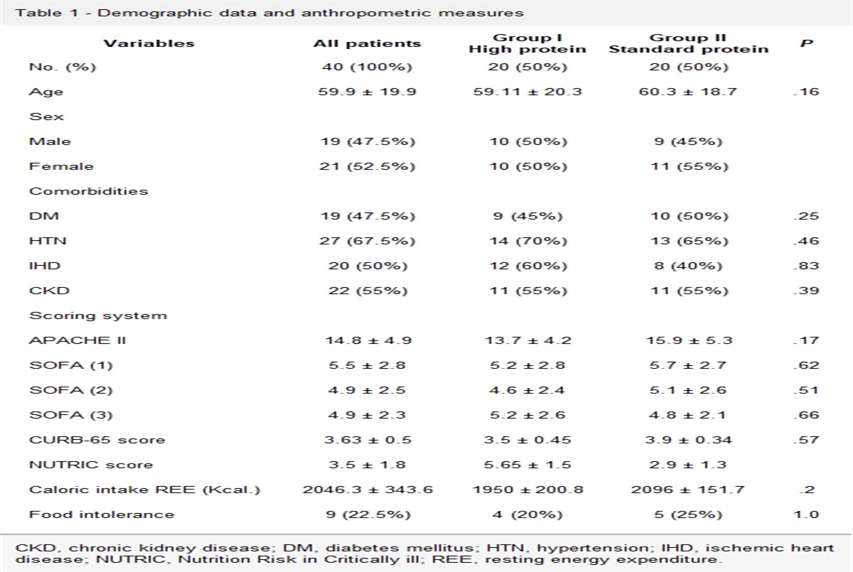
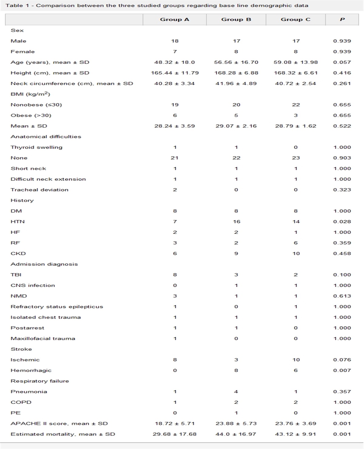
We analyzed 84 patients’ information. Forty-six patients had f-QRS (group A/cases) whereas 38 patients lack f-QRS (controls/group B). The study showed insignificant discrepancy between the two groups as regards age and gender, coronary heart diseases risk factors. Moreover, in term of LVEF, WMSI and Gensini score, no statistical significant difference was depicted between the cases and controls (Tables 1 and 2).
Table 1 -
Demographic information, chief risk factors of heart diseases
f-QRS Mean ± SD
Non f-QRS Mean ± SD
P-value
Age, y
52.52 ± 10.04
54.29 ± 12.8
.480
Gender, male/female
40/6
27/11
.071
Body weight, kg
80.22 ± 11.78
79.42 ± 8.94
.736
Smoking status
36/46
24/38
.127
Hyperlipidemia
16/46
14/38
.481
Hypertension
18/46
16/38
.782
Diabetes mellitus
16/46
11/38
.569
Table 2 -
Echocardiography and coronary angiography data
f-QRS
Non f-QRS
P-value
EF, (%)
53.78 ± 12.016
55.67 ± 11.962
.493
WMSI
1.36 ± 0.311
1.26 ± 0.279
.154
Gensini score
64.88 ± 41.84
48.67 ± 32.65
.103
EF, ejection fraction; f-QRS, fragmented QRS; SWMA, segmental wall motion abnormalities; WMSI, wall motion score index.
Presence of f-QRS did not infer worse myocardial systolic function or performance index. Also, presence of f-QRS was not correlated to anatomical complexity through angiographic examination (Table 2).
Eighteen patients (39%) of group A had f-QRS in two leads; 12 patients (26% of group A) had f-QRS in three leads whereas the rest of patients in this group had f-QRS in four to eight leads.
3.1 Impact of number and position of f-QRS
We additionally sub-grouped the group A into two subgroups counting to the numbers of f-QRS leads. subgroup (A1) counting patients with more than three f-QRS leads and subgroup (A2) tallying patients with 3 or less f-QRS leads. Subgroup (A1) showed significant difference compared to subgroup (A2) in the form of higher WMSI (1.55 ± 0.33 versus 1.27 ± 0.27, P = .007) and a higher Gensini score (86.12 ± 47.2 versus 55.08 ± 35.97, P = .030). We can conclude that Number of f-QRS showed positive correlation with decline in systolic functions and Gensini score (
Table 3).
Anterior f-QRS position occurred in 20/46 patients (43.5%) of the cases. While inferior f-QRS location was experienced in 76.1% of patients in group (A), the lateral f-QRS position was the least (21.7%) of patients.
In our study, f-QRS location additionally impacted Gensini score and myocardial performance assessed by WMSI. Anterior f-QRS showed considerable differences compared to non-anterior f-QRS; with a lower SBP (P = .006), a higher HR (P = .040), a lower LVEF (P = .039), a higher Gensini score (P = .016) and a higher WMSI (P = .004) (
Table 4).
Table 3 -
The important results of the two subgroups (A1) and (A2)
Subgroup (A1) > 3f-QRS
Subgroup (A2) < 3f-QRS
P-value
EF (%)
48.08 ± 13.07
56.14 ± 10.92
.049
WMSI
1.55 ± 0.33
1.27 ± 0.27
.007
Gensini score
86.12 ± 47.2
55.08 ± 35.97
.030
EF, ejection fraction; f-QRS, fragmented QRS; WMSI, wall motion score index.
Table 4 -
Relations of different locations of f-QRS to important variables
Anterior
P (1–2)
Lateral
P (2–3)
Inferior
P (1–3)
EF (%)
48.7 ± 12.03
.039
49.4 ± 14.82
.253
54.7 ± 13.08
.369
WMSI
1.55 ± 0.33
.004
1.44 ± 0.35
.374
1.32 ± 0.3
.146
Gensini score
85.5 ± 46.18
.016
66.43 ± 36.6
.735
60.28 ± 38.8
.42
EF, ejection fraction; f-QRS, fragmented QRS; WMSI, wall motion score index.
4 Discussion
Daszyk and partners accredited that the discerning esteem of f-QRS within the surface EKG in ACS patients continues to be questionable. Research meted out in outsized patients’ groupings displayed contradictory results. Consequently, a necessity to hold out a variety of meta-analysis is important so as to depict the correct conclusions.14
We aimed to research whether or not the presence of f-QRS in surface EKG leads may predict the extent of cardiac muscles injury by means of LVEF and WMSI measured by echocardiography or the severity of CAD using coronary angiographic score (Gensini score) in ACS patients.
Our conclusion is that the quantity and the site of f-QRS leads are what actually impact the outcome and not just the simple presence of f-QRS per se.
LVEF perceived to be lower in f-QRS group without statistical significance. Lorgis and associates confirmed that the f-QRS is not a reliable predictor of ejection fraction or arrythmias.10 Cheema and colleagues in 2010 studied f-QRS and mortality risk in 842 patients with left sided cardiac dysfunction, the presence or absence of f-QRS failed to impact the EF.15
WMSI has been antecedently valid in several echocardiographic studies. WMSI correlated well with LVEF as reported by Lebeau et al in 2012.16 Additionally M⊘ller et al in 2006 demonstrated that LVEF and WMSI offer powerful prognostic information after acute cardiac muscle infarction; however, the predictive power of WMSI is larger.17 In our study, WMSI as well as Gensini angiographic score failed to provide any important distinction between the f-QRS and the non-f-QRS groups. In contrast Bekler et al.1 found a significantly higher Gensini score amongst the patients with f-QRS taking into consideration that they incorporated patients with previous MI however we did not.
Subgroup (A1) showed lower LVEF than subgroup (A2) (P-value = 0.049). Yildirim and colleagues found that lower EF was related to a higher number of fragmented derivations.18 Interestingly within the cases group we found that anterior f-QRS was associated considerably with lower EF than non-anterior f-QRS. WMSI was drastically higher in subgroup (A1) than subgroup (A2), P-value = 0.007. The different locations of f-QRS had important impacts on vital signs and the development of cardiogenic shock and LVEF8 Uslu et al. in 2015 explicit that WMSI was notably related to the existence of the f-QRS, which reflects the affiliation between regional left ventricular pump dysfunction and the occurrence of severe cardiac muscle damage in STEMI.19 Furthermore anterior f-QRS showed notably higher WMSI than non-anterior f-QRS, P-value = .004. Finally in term of Gensini score, subgroup (A2) showed notably lower Gensini score than subgroup (A1) (P-value = .03). Likewise Bekler et al. in 2015 concluded that the severity and complexness of CAD in ACS patients is related to the number of f-QRS leads on admission 12-lead surface EKG.1 Ma et al. in 2016 showed also the important relation of higher number of f-QRS leads to Gensini score.20 Anterior f-QRS in our study was associated drastically with higher Gensini score (P-value = .016) than non-anterior f-QRS.
4.1 Research limitations
The sample size of 84 patients enclosed in the research is moderately small. An even bigger sample size could validate the statistical results.
A retrospective study could be restricted by the poor management over covariates and possible confounders.
We did not enclose in our research analysis the f-QRS in right EKG chest leads. To the foremost of our information, f-QRS within the right chest leads aVR, V3r & V4r leads was not enclosed previously in any study.
The finest low pass filter accustomed to perceive f-QRS is 100–150 Hz. This could be veiled once employing a filter with inferior setting.
5 Conclusions
F-QRS showing in more than three leads as well as anterior f-QRS location, when compared to non-anterior is associated with a considerably greater impact on myocardial performance as assessed by EF and WMSI, and associated with a higher Gensini angiographic score.
References
[1]. Bekler A, Barutçu A, Tenekecioglu E, et al. The relationship between fragmented QRS complexes and SYNTAX and Gensini scores in patients with acute coronary syndrome. Kardiol Pol 2015;73(4):246–254.
[2]. Flowers NC, Horan LG, Thomas JR, Tolleson WJ. The anatomic basis for high-frequency components in the electrocardiogram. Circulation 1996;39:531–539.
[3]. Pietrasik G, Zaręba W. QRS fragmentation: diagnostic and prognostic significance. Cardiol J 2012;19(2):114–121.
[4]. Das MK, Khan B, Jacob S, Kumar A, Mahenthiran J. Significance of a fragmented QRS complex vs. a Q wave in patients with coronary artery disease. Circulation 2006;113:2495–2501.
[5]. Das MK, Suradi H, Maskoun W, et al. Fragmented wide QRS on a 12-lead ECG: a sign of myocardial scar and poor prognosis. Circ Arrhythm Electrophysiol 2008;1:258–268.
[6]. Mahenthiran J, Khan BR, Sawada SG, Das MK. Fragmented QRS complexes not typical of a bundle branch block: a marker of greater myocardial perfusion tomography abnormalities in coronary artery disease. J Nucl Cardiol 2007;14(3):347–353.
[7]. Das MK, Saha C, El Masry H, et al. Fragmented QRS on a 12-lead ECG: a predictor of mortality and cardiac events in patients with coronary artery disease. Heart Rhythm 2007;4(11):1385–1392.
[8]. Torigoe K, Tamura A, Kawano Y, Shinozaki K, Kotoku M, Kadota J. The number of leads with fragmented QRS is independently associated with cardiac death or hospitalization for heart failure in patients with prior myocardial infarction. J Cardiol 2012;59(1):36–41.
[9]. Younis AS, ElHallag MI, ElBAdry MA, Abbas NI. Fragmented QRS complex frequency and location as predictor of cardiogenic shock and mortality following acute coronary syndrome. Egyptian Heart J 2020;72:43.
[10]. Lorgis L, Jourda F, Hachet O, et al. RICO Survey Working Group. Prognostic value of fragmented QRS on a 12-lead ECG in patients with acute myocardial infarction. Heart Lung 2013;42(5):326–331.
[11]. Lang RM, Bierig M, Devereux RB, et al. Recommendations for chamber quantification: a report from the American Society of Echocardiography's Guidelines and Standards Committee and the Chamber Quantification Writing Group, developed in conjunction with the European Association of Echocardiography, a branch of the European Society of Cardiology. J Am Soc Echocardiogr 2005;18(12):1440–1463.
[12]. Gensini GG. A more meaningful scoring system for determining the severity of coronary heart disease. Am J Cardiol 1983;5(3):606.
[13]. Du LJ, Dong PS, Jia JJ, et al. Association between left ventricular end-diastolic pressure and coronary artery disease as well as its extent and severity. Int J Clin Exp Med 2015;8(10):18673–18680.
[14]. Daszyk AM, Zygmund K, Mitręga KA, Cebula S, Kalarus Z, Średniawa B. Fragmentation of the QRS complex in patients with acute coronary syndrome treated invasively. Kardiol Pol 2016;74(7):644–649.
[15]. Cheema A, Khalid A, Wimmer A, et al. Fragmented QRS and mortality risk in patients with left ventricular dysfunction. Circ Arrhythm Electrophysiol 2010;3(4):339–344.
[16]. Lebeau R, Serri K, Morice MC, et al. Assessment of left ventricular ejection fraction using the wall motion score index in cardiac magnetic resonance imaging. Arch Cardiovasc Dis 2012;105(2):91–98.
[17]. M⊘ller JE, Hillis GS, Oh JK, Reeder GS, Gersh BJ, Pellikka PA. Wall motion score index and ejection fraction for risk stratification after acute myocardial infarction. Am Heart J 2006;151(2):419–425.
[18]. Yildirim E, Karaçimen D, Özcan KS, et al. The relationship between fragmentation on electrocardiography and in-hospital prognosis of patients with acute myocardial infarction. Med Sci Monit 2014;20:913–919.
[19]. Uslu N, Gul M, Cakmak HA, et al. The assessment of relationship between fragmented QRS complex and left ventricular wall motion score index in patients with ST elevation myocardial infarction who underwent primary percutaneous coronary intervention. Ann Noninvasive Electrocardiol 2015;20(2):148–157.
[20]. Ma X, Duan W, Poudel P, Ma J, Sharma D, Xu Y. Fragmented QRS complexes have predictive value of imperfect ST-segment resolution in patients with STEMI after primary percutaneous coronary intervention. Am J Emerg Med 2016;34(3):398–402.

留言 (0)