
記住我

A 23-year-old Japanese female was referred to our department for fever and polyarthritis. Approximately 8 years prior to this presentation, the patient had undergone four cycles of rituximab for glucocorticoid-resistant nephrotic syndrome. Two months prior, she had been hospitalized for empyema and underwent surgical debridement; however, the pathological and microbiological etiologies remained unclear. Subsequently, she developed fever and polyarthritis affecting the elbow, shoulder, hip, wrist, and ankle joints. At the previous hospital, ceftriaxone and 30 mg/day of prednisolone were initiated for suspected septic and reactive arthritis; however, her symptoms did not improve, and she became bedridden because of severe joint pain.
Upon admission, she was alert and her vital signs were stable, except for fever. Physical examination revealed severe tenderness of the left wrist, left sternoclavicular joint, left ankle joint, and both hip joints. Laboratory testing demonstrated an elevation in white blood cell count (20,920/µL) and serum C-reactive protein (CRP, 23.36 mg/dL). Serum levels of IgG, IgA, and IgM decreased to 243.3 mg/dL (normal range, 861.0–1747 mg/dL), 20.4 mg/dL (normal range, 93.0–393.0 mg/dL), and < 2.0 mg/dL (normal range, 50.0–269.0 mg/dL), respectively, leading to a diagnosis of hypogammaglobulinemia, for which we promptly initiated immunoglobulin replacement therapy. Both antinuclear and antineutrophil cytoplasmic antibodies tested negative. Immunoglobulin levels were confirmed to be within normal limits before receiving rituximab.
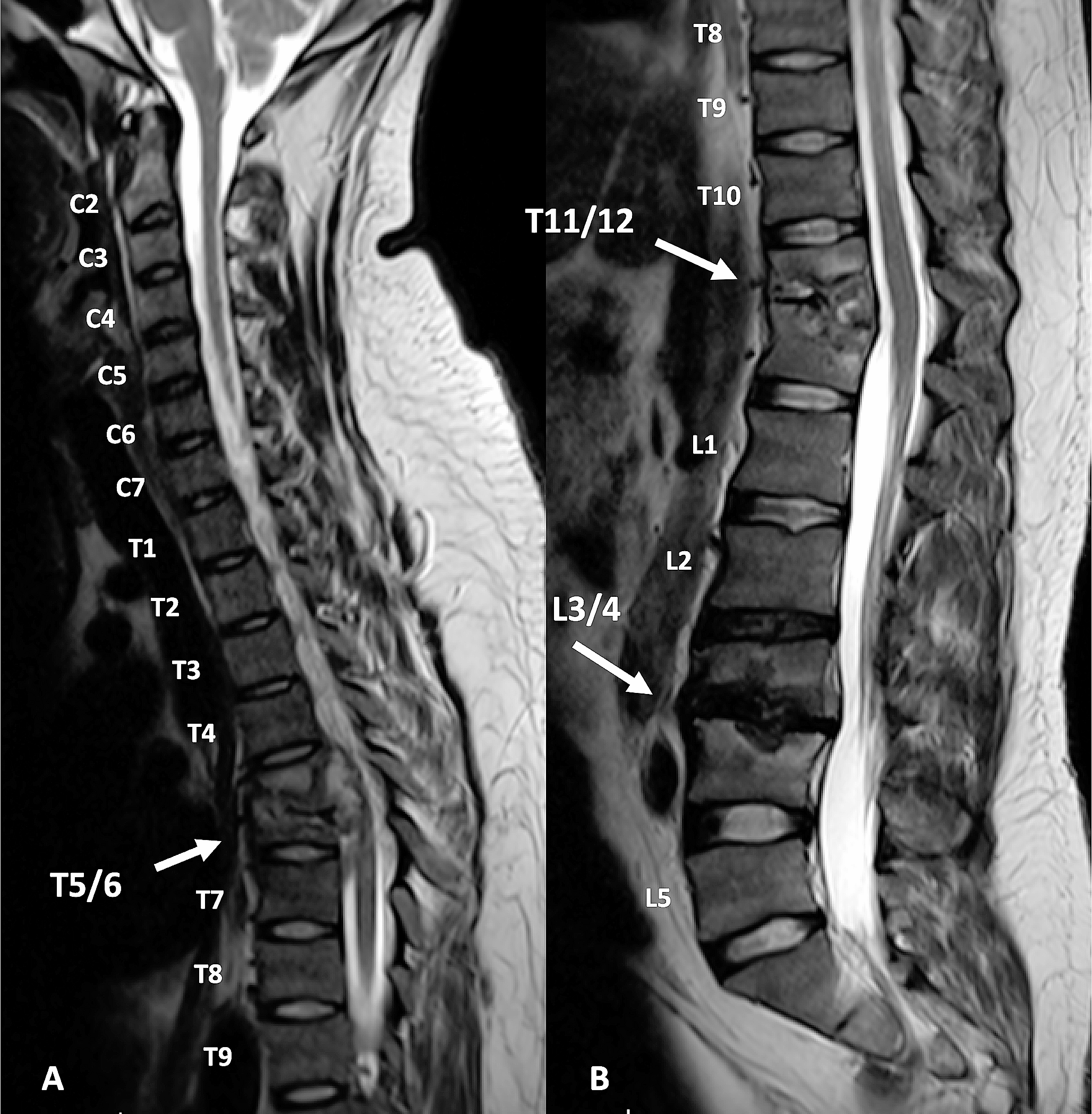
Contrast-enhanced computed tomography (CT) revealed effusions in the left sternoclavicular and bilateral hip joints (Fig. 1A, B). Gallium-67 scintigraphy revealed uptake in the left sternoclavicular, hip, wrist, knee, and ankle joints (Fig. 1C). CT-guided drainage of the left hip joint detected yellowish, cloudy joint fluid, with an increased cell count (60,000/µL) showing a predominance of polynuclear cells (99%) over mononuclear cells (1%). Despite these findings, Gram staining and bacterial culture of the joint fluid did not reveal any pathogens. While, we observed a positive testing result for alpha-defensin lateral flow tests (Synovasure® lateral flow test; Zimmer Biomet, IN, USA) in synovial fluid of both hips.
Fig. 1
Radiological findings A, B Contrast-enhanced computed tomography showing effusions in the left sternoclavicular and both hip joints. C Gallium-67 scintigraphy demonstrating uptakes in bilateral wrist, knee, ankle joints, and large joints. D Magnetic resonance imaging of the hips detecting bone marrow edema along with fluid retention bilaterally
Suspecting septic arthritis caused by atypical organisms, treatment with 200 mg doxycycline (DOXY) daily was initiated. Following antibiotic therapy, the joint symptoms and serum C-reactive protein (CRP) levels improved (Fig. 2). Although clinically effective, DOXY therapy was switched to 500 mg/day levofloxacin (LVFX) because of DOXY-induced gastritis, as confirmed using gastroendoscopy. Soon after, the serum CRP level was elevated during LVFX therapy, accompanied by severe joint tenderness, suggesting LVFX resistance. We then switched to oral DOXY therapy. Gastric symptoms recurred, and DOXY was converted to intravenous minocycline.
Fig. 2
The clinical course of the patient Serum C-reactive protein levels decreased following DOXY therapy and resurged after switching to LVFX. Combination therapy with DOXY (MINO) and AZM was effective, and the patient was discharged with AZM monotherapy. PSL prednisolone; CRP C-reactive protein; IVIG intravenous immunoglobulin; DOXY doxycycline; MINO minocycline; LVFX levofloxacin; AZM azithromycin
Around that time, we performed an in-house two-step PCR for the 16S rRNA gene to accurately identify the causative organism. DNA was isolated from hip joint fluid using the DNeasy® PowerSoil Pro Kit (QIAGEN). The 16S rRNA gene was amplified with 8UA and 1485B primers (forward primer: 5′-AGAGTTTGATCMTGGCTCAG-3′; reverse primer: 5′-TACGGTTACCTTGTTACGAC-3′). PCR was performed using the following regimen: 98 °C for 3 min followed by 45 cycles at 98 °C for 4 s, 60 °C for 30 s, 72 °C for 1 min, and a final extension at 72 °C for 5 min (ramp rate = 1 °C/s). The second PCR was carried out with 341A and 519B primers (forward primer: 5′-CTACGGGAGGCAGCAGTGGG-3′, and reverse primer: 5′-ATTACCGCGGCKGCTG-3′) under a following amplification process: 96 °C for 1 min, followed by 25 cycles at 96 °C for 10 s, 50 °C for 5 s, 60 °C for 1 min (ramp rate = 1 °C/s). The sequence data of the PCR products were analyzed using the Basic Local Alignment Search Tool (BLAST), and the isolate was identified as U. urealyticum with a 100% concordance rate with the reference strain (GenBank accession number: NR_041710.1).
Under the diagnosis of disseminated arthritis caused by U. urealyticum, we added oral azithromycin (500 mg for 3 days, followed by 250 mg/day) as combination therapy to prevent the emergence of antimicrobial resistance. Magnetic resonance imaging revealed bilateral joint effusions, bone marrow edema in both hip joints (Fig. 1D) and fluid retention in the ankle joints. Thereafter, arthroscopic irrigation and debridement of both hip joints were performed 63 days after admission. During hospitalization, the patient remained bedridden because of severe joint tenderness, even after intravenous fentanyl therapy. Combination therapy with antibiotics and surgical intervention led to an improvement in her fever and polyarthritis. Intravenous minocycline was discontinued and the patient was discharged in good condition on oral azithromycin, with plans to continue antibiotic treatment for several months.
留言 (0)