
記住我
Molecular diagnostics are increasingly recognized as a crucial complement to traditional phenotypic antibiotic susceptibility testing to address the time-consuming limitations inherent to bacterial culture. In China, the burden of TB prevention and control predominantly lies on numerous county-level secondary hospitals which often operate under resource constraints but lacking adequate facilities for M. tuberculosis culture and gold standard DST. Our collaborating Chaoshan Hospital of Jinan University First Affiliated Hospital represents such hospitals facing constraints associated with biosafety-compliant bacterial culture laboratories and costly MGIT™ 960 liquid culture/DST system. However, amidst the endeavor to curb local outbreaks of the severe acute respiratory syndrome coronavirus 2 (SARS-CoV-2) virus and the resulting coronavirus disease 2019 (COVID-19) pandemic, fluorescence PCR instruments have gained significant popularity in China’s primary care [35]. This serendipitously positions Chaoshan Hospital, equipped with multi-channel fluorescence PCR instruments, as a conducive setting for deploying our multiplex PCR-MPMA assay to fully exploit the potential of fluorescence real-time PCR technology for rapid molecular diagnostics, enabling frontline healthcare facilities to perform follow-up testing to sputum smear microscopy. Additionally, this assay, by monitoring drug resistance in patients receiving specific medications, facilitates timely adjustments of therapeutic regimens, aligning with the objective of the WHO 2021TPP.
M. tuberculosis acquires drug resistance through specific genetic alterations, including point mutations, deletions or insertions [14]. The WHO recommends routine testing of all TB patients for resistance to RIF and INH, the classic first-line drugs [29]. Extensive studies have illumined the genetic mechanisms of resistance. RIF resistance is predominantly attributed to mutations in the 81-bp fragment RRDR of the rpoB gene, which accounts for about 95% of cases [36]. However, it is worth noting that certain studies have reported rare mutations outside this region, such as I491F, I59T, V146F and I572F, which potentially confer RIF resistance but are not currently targeted by commercially available assays [37, 38]. Specifically, I572F and six other located within the RRDR-namely, L511P, D516Y, H526L, H526N, H526C, and L533P-are often considered “disputed” or “occult”, which can mask MDR-TB when phenotypically assessed by methods such as the MGIT™ 960, and can lead to potential treatment failure or relapse if not detected early [38]. In contrast, INH resistance unravels through a complex genetic narrative, with previous studies reporting over 80% of INH-resistant M. tuberculosis strains harbouring mutations at codon 315 of katG, alongside positions -15 and -8 of the inhA promoter and positions -6 to -47 of the ahpC promoter [39,40,41]. Noteworthily, while some previous reports suggest that the ahpC gene serves only to compensate for the loss of peroxidase function caused by katG mutations in many INH-resistant isolates, two meta-analyses have jointly reported that mutations in 11 loci within a 106-bp intergenic region between the pseudogene oxyR and the ahpC gene are associated with INH resistance [42, 43]. One of the two studies also observed a global prevalence of 1.84%, with regional prevalence reaching 2.71% in Asia, a phenomenon transcending the mere compensatory mechanism of katG mutations [43]. Despite the generally acknowledged clinical relevance of the fabG1 gene for INH resistance detection, it was not specially investigated in this study. The decision was steered by a recent study employing whole-genome sequencing to analyze mutations associated with INH resistance in 188 MDR-TB and single INH-resistant clinical isolates collected in a Chinese national survey. Remarkably, no mutations in the fabG1 gene were detected, whereas the same study reported high mutation frequencies in the katG (86.2%), inhA promoter (19.6%), and oxyR-ahpC intergenic regions (18.6%) [19]. As described in the Methods section, our strategy for selecting variants to detect, including rpoB RRDR coding and deletion mutations, katG coding mutations, ahpC coding and promoter mutations, and inhA promoter mutations, was informed by the WHO guideline, the listed commercial assays and the literature review. Furthermore, in recent years, very few PCR-HRMA studies demonstrating a mutation-resistance association have directly tested sputum samples from TB patients, with one exception, but that study also did not include the inhA gene [44]. Consequently, this study aims to bridge this gap by evaluating diagnostic performance using clinical sputum samples, thereby providing valuable insights for researchers in this field.
Our multiplex PCR-MPMA assay utilizes an inventive combination of fluorescence and melting temperature labelling to detect the selected mutant sequences in a high-throughput, multicolour melting curve analysis framework. Consequently, by meticulously scrutinizing a unique melting peak plus a Tm value in each mutation as identifiable markers for the discrimination of 4 WT from a total number of 40 mutant sequences, our assay possess the diagnostic potential to analyse the genetic information of resistance to RIF and INH. Careful optimization improved assay’s performance, including optimized nucleic acid extraction to obtain high-quality template DNA, intensive Molecular Beacon probe design to increase the specificity of melting analysis, and fragment length adjustment to avoid primer dimerization. A salient feature of our assay is its ability to discriminate between insertion and deletion mutations, whereas we observed that base substitution mutations can lead to very diverse differentiation effects, depending on the difference in Tm values between the mutant and WT targets. For example, mutation types G/A, G/T, C/A and C/T were readily distinguished (Tm difference > 0.5 °C), while G/C (Tm difference < 0.4 °C) and A/T (Tm difference < 0.2 °C) were much more difficult to distinguish. To address this problem, our application of Molecular Beacon probes significantly bolsters the specificity of experiments. This was demonstrated that the shorter double-stranded portion formed by the binding of the probes to their complementary regions, combined with the Touch-down PCR reaction cycling program, worked better to identify selected mutant sequences. In addition, the application of asymmetric PCR as a replacement for standard PCR was crucial because, after the completion of asymmetric PCR, the probes hybridized to a large number of specific sites on the single strands with increasing temperature. As a result, this combination enabled at least 2.4 °C Tm shifts between mutant and WT samples, as shown by single-plex PCR evaluation data (Supplemental Table S5). These visible differences allowed us to effectively discriminate between drug-susceptible and drug-resistant clinical specimens for the diagnosis of TB patients. However, this sensitivity leads to false positives from synonymous mutations. Our data from the detection of plasmids containing the five synonymous mutations, Q510Q, F514F, Q513Q, D516D and L533L reported RIF resistance. Despite the remarkable sensitivity of the probe being advantageous in identifying minor variations, the detection of synonymous mutations may not necessarily be pertinent for cases pertaining to RIF resistance in certain clinical scenarios. This fundamental drawback is inherent in the melting curve assay methodology. In this regard, our assay did not prove superior to Cepheid’s Xpert MTB/RIF, which uses a similar methodology that is nested PCR combined with melting curve analysis, in the detection of synonymous mutations to avoid false positives [25].
The multiplex PCR-MPMA assay was evaluated for its analytical performance, validating 40 different mutations within rpoB (18 codon mutations), katG (4 codon mutations), ahpC (3 codon mutations and 13 promotor region mutations) and inhA (2 promotor region mutations) using the relevant commercially synthesized plasmids (Supplemental Table S2). The ddPCR technique was used for precise quantification of the nucleic acid molecules in the template plasmids, aiding in determining the assay’s analytical sensitivity. All 40 mutations plus the corresponding WT from each gene were simultaneously correctly detected by the four-tube, multiplex PCR-MPMA assay. Unlike other fluorescence PCR assays that use amplification curves and Ct values as detection limit indicators, the LOD of our assay was determined using a proportional mixture of WT and mutant artificial plasmids as templates to test for qualitative detection of characteristic melting peaks and Tm values representative of WT and mutant samples under serial dilution. The assay LOD was defined as the lowest concentration at which characteristic melting peaks/ Tm values were reliably detectable, based on achieving 100% success rate in 10 replicates. Therefore, our assay was determined to achieve a LOD of 5000 copies/ml, equivalent to 125 copies/reaction, but it must be clarified that these outcomes pertain solely to the input DNA samples. Given the increasing prevalence of infection with mixed susceptibility and resistance to a drug in the MDR population as evidenced by recent studies [45, 46], an alternative LOD validation experiment was performed to investigate the extent to which our assay could unequivocally identify hetero-resistant mutants mixed with WT drug-sensitive strains. Our data corroborated the assay’s analytical rigor, with its capacity to distinguish as little as 40% of the desired hetero-resistant mutants mixed with WT being particularly noteworthy. This suggests that our assay’s performance for detecting hetero-resistance is consistent with a study using laboratory-modified Xpert MTB/RIF assay to achieve comparable results [47]. Intra- and inter-assay CVs below 0.5% demonstrated robust and reproducible Tm values, thereby reinforcing the consistency of our assay’s performance and affirming its stability and precision.
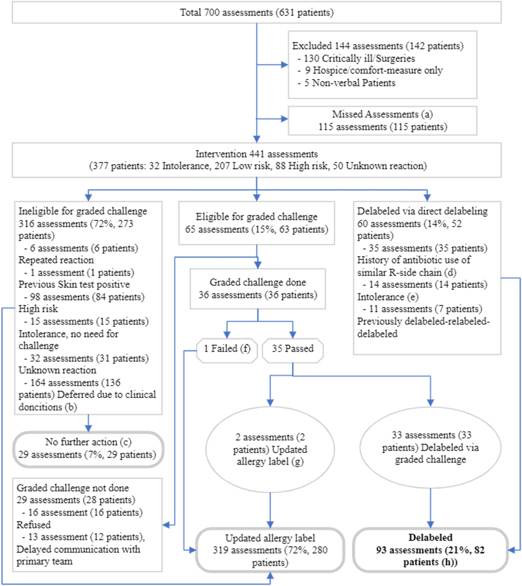
The latter phase of this study transitioned from assay development to a critical evaluation of its diagnostic performance, involved in collaboration with clinical patients. We designed a protocol to consecutively recruit a compact cohort of confirmed TB patients, facilitating a comparative analysis of our assay against DNA sequencing data obtained during the same period and retrospectively, against reference standards like liquid culture, phenotypic DST and the WHO recommended Xpert MTB/RIF assay. The authors candidly admit that, at the inception of this study, challenges in establishing a collaboration with a centralized diagnostic laboratory precluded the synchronous implementation of all designated control tests during the clinical evaluation phase of our assay. Initially, we intended to partner with a specialist hospital and Chaoshan Hospital to conduct all tests concurrently, thereby validating our assay’s efficacy in the intended application scenario. However, unforeseen circumstances led to a year’s delay in performing the gold standard tests and comparator test post clinical sample collection, necessitating a shift to retrospective analyses. This timing misalignment forced us to conduct the assay’s field test suboptimally. A significant limitation is that solely detecting resistance mutations from smear- positive clinical sputum samples doesn’t provide a comprehensive assessment of our assay, as data from smear- negative samples are essential for a balanced evaluation. Understandably, enrolling suspected TB patients without bacterial confirmation of M. tuberculosis infection during the participant screening phase would introduce bias. Additionally, the inability to utilize advanced diagnostic equipment not available in county-level hospitals, such as bronchofiberscopes, results in another notable limitation of this study-specifically, the lack of comprehensive evaluation of diagnostic performance across other types of respiratory specimens, including bronchoalveolar lavage fluid.
Nevertheless, after the retrospective analysis, this study aligns the minimal criteria for the evaluation of innovative products for molecular TB-DR detection products defined by the WHO 2021TPP at the level of experimental protocol design and implementation, encompassing the requisite reference standards and comparative tests. Given the potential of this technique for TB diagnosis, the selection of the rpoB gene as the target within the Mycobacterium genomic region is a noteworthy step. However, extensive clinical trials are requisite to validate the application of our assay as a primary screening test for M. tuberculosis. A pertinent illustration of a comparable diagnostic approach is Cepheid’s Xpert MTB/RIF. Much like our focus on the rpoB gene, the Xpert MTB/RIF, a WHO-approved molecular rapid diagnostic (mWRD) test, facilitates the initial diagnosis of M. tuberculosis rather than serving as a reflex test to a TB-positive result [22, 24, 36]. The inclusion of DNA sequencing in this study was founded on the premise of phenotypic DST being the gold standard reference used in retrospective validation. Selecting DNA sequencing as a reference tool to furnish genotypic information for the multiplex PCR-MPMA assay outcomes was buttressed by a WHO report. This document elucidated that the accuracy of sequencing in predicting INH, RIF, FQ and SLI resistance compared to phenotypic DST results, coupled with its sufficient sensitivity in appraising the prevalence of drug resistance in TB surveillance, substantiates its choice as a reference tool [48].
The focus of this study was to develop a novel multiplex PCR-MPMA assay for ascertaining drug resistance status, for which we conducted verification experiments under defined scenarios mirroring the assay’s intended application. The empirical data obtained from these experiments supplied substantial evidence of the assay’s efficacy when deployed on samples from the target patient population. Our findings underscored a sensitivity of 93.33% and 95.24% for detecting RIF and INH resistance, respectively, with a specificity of 100% against phenotypic DST. These findings were consistent with the previously published study validating their PCR-HRMA assay via sputum samples, which reported HRMA sensitivity of 90.5%, 86.4% and 100% for pinpointing mutations in the rpoB, katG and inhA genes, respectively [44]. In our assay, we observed a notable discrepancy: one missing sample detection each for the rpoB and katG probes. This was primarily due to the presence of mutations in the patient samples that fell outside the predefined mutation detection range of our designed probes, leading to reduced diagnostic sensitivity. In comparison, the retrospective analysis of the Xpert MTB/RIF assay revealed three false-negative samples with a sensitivity of 80%. These false-negative results were attributed to the inability of the manufacturer’s probes to capture the specific mutation sites carried by these patients. The AUC values, delineated in Fig. 5 F, evinced a comparable accuracy among our assay, Xpert MTB/RIF and DNA sequencing in detecting RIF resistance within the examined cohort, with respective AUC values of 0.967, 0.900 and 1.000. Similarly, in detecting INH resistance, our assay and DNA sequencing exhibited a tight diagnostic concordance, as illustrated in Fig. 5G, with AUC values of 0.976 and 1.000, respectively. The diagnostic performance of our assay, juxtaposed against both the WHO-approved reference test and the Xpert MTB/RIF assay, showcased comparable efficacy. Overall, despite the fact that the phenotypic DST results used as reference standards were obtained from retrospective analyses, the measured sensitivities of 93.33% for RIF resistance and 95.24% for INH resistance stand in parallel with the pooled sensitivities of 94% for RIF and 93% for INH elucidated in a 2021 systematic review and meta-analysis encompassing 47 HRMA-based studies [49]. It’s necessary to emphasize that, unlike the meta-analysis which primarily analyzed data from clinical isolates of M. tuberculosis derived from 47 studies, our investigation uniquely utilized sputum samples, enhancing the clinical feasibility of our findings.
Our multiplex PCR-MPMA assay identified resistant samples, a finding corroborated by DNA sequencing. These samples harboured a total of 17 mutated loci, including well-documented mutation hotspots such as codons 531, 526 and 516 within the RRDR of the rpoB gene responsible for RIF resistance, alongside locus 315 of katG, locus -15 of inhA and locus -8 of inhA, the three most frequently mutated loci related to INH resistance. Moreover, our assay detected the disputed mutations at codons 511 and 533, specifically L511P and L533P, in two samples, providing compelling evidence of our assay’s capability to identify mutations that contribute to low-level DR. DNA sequencing additionally identified two uncommon loci, locus 561 of rpoB and locus 337 of katG. These mutations have been consistently reported in numerous studies employing commercial testing protocols or laboratory-developed assays [21, 44, 47, 50, 51]. Among the 50 samples analyzed, DNA sequencing authenticated 11 mutations across 15 samples associated with RIF resistance, revealing no significant variance in their distribution. Although the high-frequency mutation S531L in rpoB (TCG → TTG) manifested in merely 3 samples, three mutations at another commonly reported locus, rpoB526 (CAC → GAC, CAC → TAC and CAC → CGC), were found across 4 samples cumulatively. Similarly, this clinical validation confirmed that our assay, in alignment with DNA sequencing, accurately identified the mutation S315T (AGC → ACC) in the katG gene in 9 (18%) samples, thereby corroborating with literature that this mutation type responsible for INH resistance is predominant. Interestingly, the mutations rpoB I561V (ATC → GTC) and katG Y337C (TAC → TGC), overlooked by our assay yet recognized by DNA sequencing, have been delineated in merely two preceding publications, hence are deemed rare [52, 53]. In addition, regarding rpoB mutation detection, our experiments using the Xpert MTB/RIF assay failed to detect samples carrying rpoB A516G (GAC → GGC) and rpoB del 510–512, alongside rpoB I561V, likely due to these mutations transcending the probe target range as per Cepheid’s disclosed information. Should ensuing studies persist in discovering these two rare mutations in larger cohorts, their sequences could be integrated into newly developed probes within an upgraded assay. Overall, 28 out of 50 TB patients (56%) were found to harbor mono-resistance to RIF (7, 14%) or INH (13, 26%), or MDR (8, 16%) to both drugs, as ascertained by our assay coupled with DNA sequencing. This substantial proportion of drug resistance, encapsulating MDR-TB, as demonstrated by the findings of this study, represents a fundamental challenge confronting global TB control, highlighting its marked significance in public health and direct clinical relevance.
Similar to other molecular tests, our assay is specifically designed to detect certain resistant mutations, inherently introducing a risk of misdiagnosis due to its inability to cover all potential mutations. For instance, our study demonstrated sensitivities of 93.33% for RIF and 95.24% for INH, slightly lower than those reported in a prior study, which claimed a 100% HRMA sensitivity in smear-positive sputum samples [44]. The discrepancy can be attributed to the non-detection of a single mono-resistant sample for each drug, a limitation stemming from our probe design. Nonetheless, the inherent flexibility of our assay facilitates swift adaptation to newly reported mutations. By incorporating or modifying probes based on prevalent types discovered through pathogenic and epidemiological studies, we can effectively reduce the misdiagnosis risk. Addressing our decision against selecting the fabG1 gene as a detection target, our probe design strategy has appropriately considered this factor. It is noteworthy that mutations in the inhA promoter, including those upstream of fabG1, overlap with the inhA gene and serve as promoters for the entire inhA-fabG1 operon [43]. Our assay targets specific sites like C-15T and T-8C within the promoter region, which are well-documented and globally recognised as drivers of INH resistance [43]. Additionally, as mentioned earlier, rpoB mutations outside the 81-bp RRDR, including I491F, I59T, V146F and I572F, are infrequent and remained undetected by our current probes. We remain committed to monitoring emerging literature to determine if adding probes for these mutations enhances RR-TB detection sensitivity. Pertaining to the potential false positives associated with the identification of synonymous mutations in the RRDR within the rpoB gene, we are planning rigorous clinical validations to assess the incidence of such mutations. Upon validation through sequencing, we can refine our probes and modulate fluorescent moiety to classify these mutations as WT, effectively minimizing false positive impact.
The updated WHO 2021TPP outlines both minimum and optimal criteria for technical and operational specifications of next generation TB DST protocols, guiding the development of innovative molecular diagnostics intended for use in primary healthcare centres [9, 10]. Our multiplex PCR-MPMA assay aligns with the 2021TPP’s four technical specifications: target selection, analytical performance, operational and infrastructural requirements, and pricing. This ensures an affordable, efficient TB DR screening tool tailored for peripheral laboratories with specific needs. We retrospectively conducted phenotypic DST on liquid culture, using it as the gold standard, and compared our assay’s performance with Cepheid’s Xpert MTB/RIF during the clinical validation phase. This comprehensive evaluation contextualized our assay within the 2021TPP framework. Furthermore, we benchmarked technical specifications of our assay against the six commercially available assays, utilizing WHO published documents as our data resource [22,23,24, 36] (detailed in Supplementary Material Figure S4). Our assay excels in drug and mutation site coverage and offers competitive diagnostic performance (Table S1). For RIF resistance diagnosis, our 93.33% combined sensitivity is slightly lower than Cepheid’s Xpert MTB/RIF (94.4%), BD’s Max MDR-TB (99.1%), Bruker/Hain’s FluoroType MTBDR (97%), and Abbott’s RealTime MTB RIF/INH (94%), but outperforms Roche’s cobas MTB-RIF/INH (91%) and Molbio’s Truenat MTB-RIF Dx (84%). Moreover, our 95.24% combined sensitivity for detecting INH resistance is consistent with competitors.
Uniquely, unlike our assay necessitates minimal infrastructural investment compared to the six proprietary commercial systems. It is fully compatible with various renowned real-time fluorescence PCR instruments, thereby eliminating the necessity to modify existing molecular pathogen detection protocols, making its compatibility beneficial in diverse laboratory environments. Our assay workflow comprises three key steps: sputum liquefaction, decontamination and centrifugal concentration, nucleic acid extraction, and PCR amplification and melting curve analysis, aligning with the 2021TPP’s five-step maximum. All six competitors also require sputum liquefaction, albeit with varying incubation times. Notably, protocols from Cepheid, Roche, and BD offer integrated nucleic acid extraction and testing, while others, including ours, necessitate a separate nucleic acid extraction and PCR amplification detection process. Thus, despite its significantly lower initial infrastructure costs, our assay is marginally inferior in terms of biosafety compared to the aforementioned fully integrated systems, owing to the necessity for manual sample transfers between distinct procedural stages. This manual handling, despite using closed tube operations to reduce exposure to infectious agents, introduces potential biosafety challenges that are inherently mitigated in more seamless, integrated assay configurations. Our assay’s time efficiency stands out: 40 min for sputum specimen pre-processing, 40 min for nucleic extraction, 10 min for PCR manipulation, and 2 h 40 min for PCR amplification and melting curve analysis. Consequently, our assay can analyse up to 22 patient samples (including negative and positive quality control references) within approximately 4.2 h using a standard 96-well fluorescence PCR instrument. In contrast, Cepheid’s Xpert MTB/RIF requires 1.5 h for M. tuberculosis and RIF resistance detection, followed by an additional 1.5 h for INH resistance detection. Molbio’s protocol entails 20 min for DNA extraction, 1 h for M. tuberculosis detection and 1 h for RIF resistance detection. It is worth noting that the total protocol time for the other four platforms to complete both drug resistance detections exceeds that of our assay. Considering the expenses associated with fluorescent probes, molecular diagnostic enzymes, and other necessary reagents for assay development, we have set an ex-factory price at $10 per test. In contrast, Cepheid offers negotiated mass availability rates of $9.90 for Xpert MTB/RIF and $19.80 for Xpert MTB/XDR, implying an ex-factory cost of $29.70 for the required RIF and INH resistance tests. The authors acknowledge that the fee of $29.70 levied by Cepheid, represents a costlier yet comprehensive test for suspected MDR-TB cases, covering not only the detection of resistance to RIF and INH but also extending to FQ, second-line injectable drugs (such as amikacin, kanamycin, and capreomycin), and ethionamide [24], thereby justifying the value of Xpert MTB/XDR in terms of the extensive DR profiling it offers. Negotiations between the other companies and the WHO have yet to determine a final ex-factory price for low- and middle- income countries. Therefore, our assay demonstrates compatibility and aligns with the guiding principles outlined in the 2021TPP, promising to enhance public health in peripheral centres.
Incorporating matched sequencing data in our study was essential for identifying and interpreting mutations linked to resistance phenotypes which were retrospectively verified by bacteriological culture and phenotypic DST. To ensure a thorough evaluation of our assay, it is imperative to use bacteriological evidence and DST as reference standards. Future studies should draw from a diverse patient population, factoring in various screening and inclusion criteria, such as age, gender, socio-geographical characteristics, clinical symptoms, diagnosis/treatment history, and culture outcome. This holistic approach facilitates robust analytical modelling. It is also advisable for subsequent research to categorize specimens, encompassing sputum smear-negative specimens, clinical culture isolates and bronchoalveolar lavage fluid. Analyzing varied sample types offers a richer diagnostic performance evaluation. Once thoroughly evaluated, our assay promises several advantages: solid analytical results, extensive coverage, and the flexibility to update mutation targets, positioning it highly effectively for first-line M. tuberculosis drug resistance screening and epidemiological studies. Finally, the rigorous validation and comprehensive reporting conducted for our assay adheres to the key criteria outlined in the Standards for Reporting Diagnostic Accuracy (STARD) 2015 guidelines [54], including appropriate use of reference standards and transparent discussion of limitations and further investigation, ensuring a trustworthy and accurate evaluation of this novel diagnostic tool.
留言 (0)