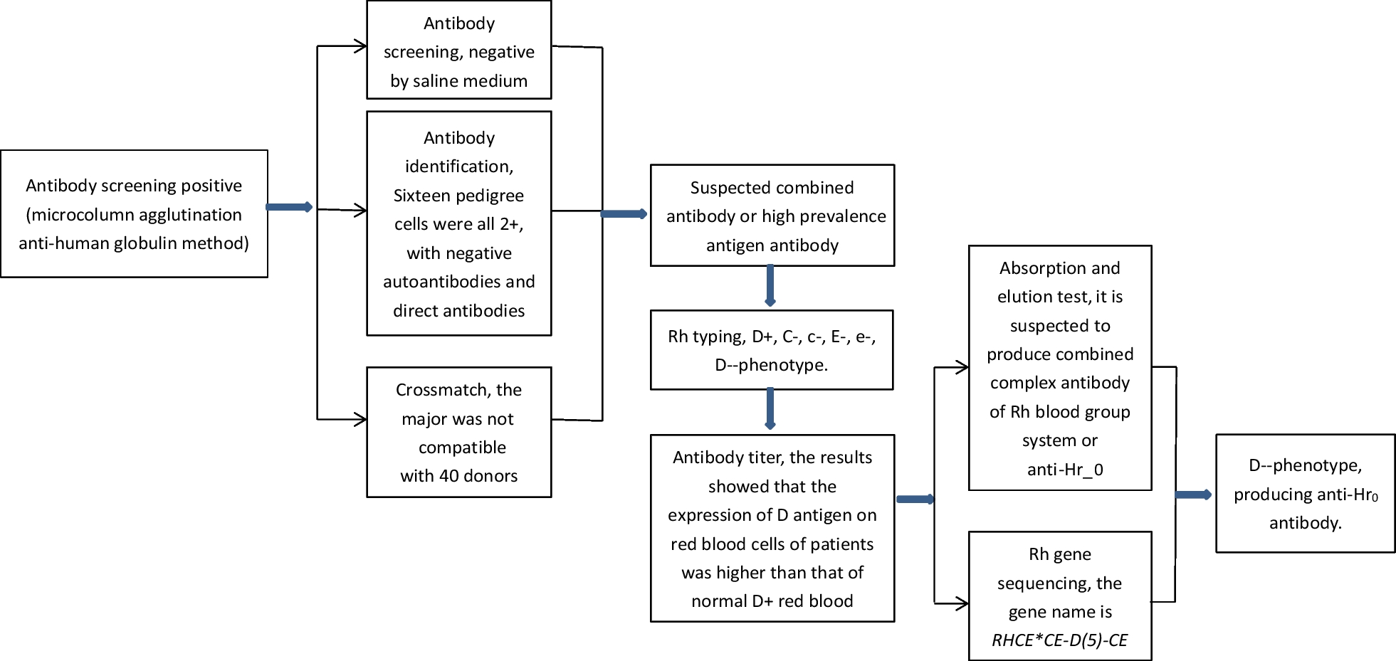
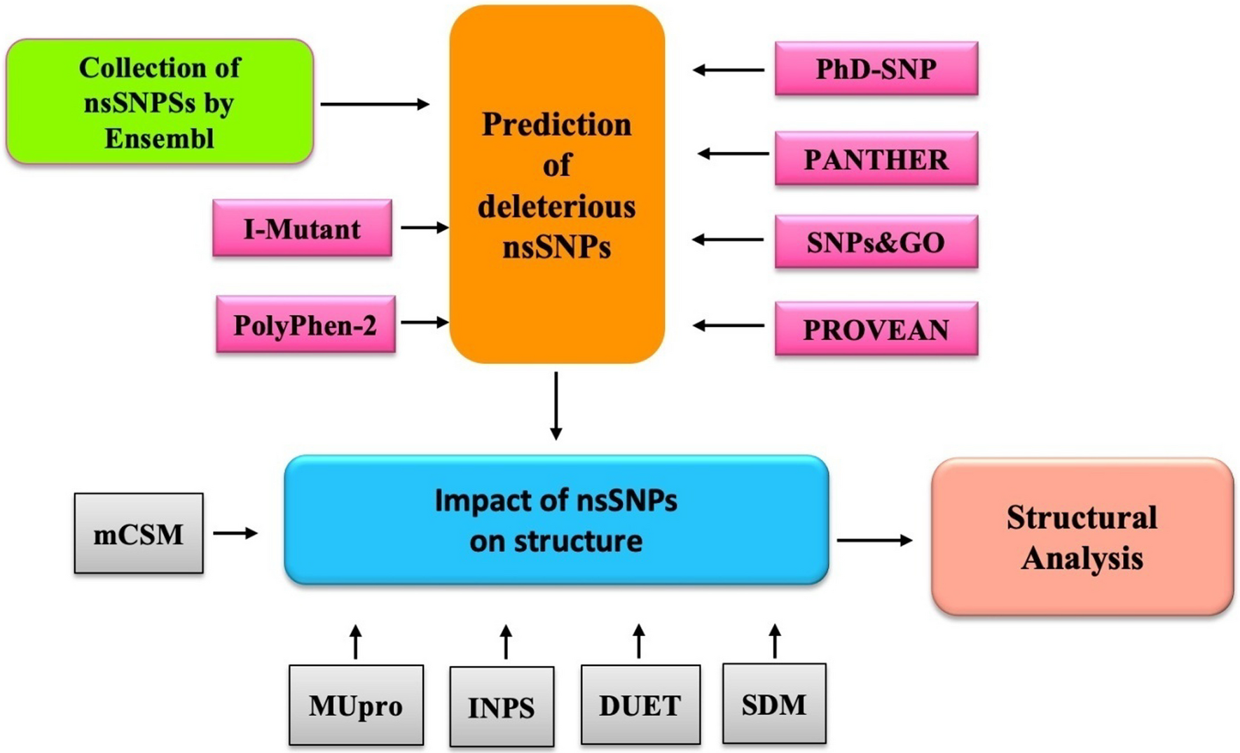
With the advances in genomics research, the bioinformatics-based approach is one of the potential approaches to proposing biomarkers for various diseases. In our study, we employed this approach to identify potential genetic-driven biomarkers for liver fibrosis, marking the first of its kind in liver fibrosis research. Utilizing advanced bioinformatic tools, we delved into the intricate molecular mechanisms associated with the disease and identified promising biomarker candidates. Herein, we utilized DisGeNET as a valuable platform for exploring genes and variations associated with human diseases, including liver fibrosis [16]. Our study employed DisGeNET, in conjunction with GO enrichment studies, KEGG pathway enrichment analyses, and PPI analysis, to conduct a comprehensive bioinformatics analysis.
Liver fibrosis is characterized by the excessive accumulation of extracellular matrix (ECM) in the subendothelial compartment. The ECM can be broken down by matrix metalloproteinases (MMPs), while tissue inhibitor matrix metalloproteinases (TIMPs) promote ECM formation and prevent its breakdown [17]. Under normal physiological conditions, there is a balanced regulation of MMPs and TIMPs to maintain ECM homeostasis. However, in liver fibrosis, this balance is disrupted [18]. Hepatic stellate cells (HSCs) are the primary source of ECM in the liver. Normally, HSCs are responsible for vitamin A storage, but liver injury triggers their activation [19]. This HSC activation is crucial in the early stages of liver fibrosis. Activated HSCs produce collagen-1 (Col-1), a major constituent of the ECM. This study identified ten potential biomarkers for liver fibrosis: TGF β-1, MMP-2, CTNNB-1, FGF-2, IL-6, LOX, CTGF, SMAD-3, ALB, and VEGFA. Among these biomarkers, TGF β-1 and MMP-2 stood out due to their high systemic scores in the CytoHubba MCC algorithm, suggesting their potential as useful biomarkers for liver fibrosis [20].
Transforming growth factor beta (TGFβ) plays a central role in the development of tissue fibrosis, particularly in conjunction with Smad signaling, which leads to the activation of myofibroblasts and subsequent extracellular matrix transformation (ECMT) [21]. TGF-β1, known for its profibrogenic and immunosuppressive properties, is released during liver injury by Kupffer cells, sinusoidal endothelial cells, and other inflammatory cells, contributing to its activation. This cytokine serves as a master profibrogenic agent, activating hepatic stellate cells (HSCs) via the TGFβ/Smad3 signaling pathway [22]. Consequently, TGF-β1 not only increases HSC activation but also influences the expression of MMPs and TIMPs. Inhibition of TGF-β1 has been demonstrated to suppress HSC activation both in vivo and in vitro [23]. The TGF-β1/Smad pathway plays a role in ECM deposition by enhancing TIMP1 expression and inhibiting MMP2 expression. Additionally, serum levels of TGF-β1 have been associated with the severity of inflammation and stages of liver fibrosis. TGF-β1 shows promise as a serum biomarker for the progression of liver inflammation and fibrosis, particularly in chronic HCV infection [23,24,25].
Matrix metalloproteinases (MMPs) are enzymes that degrade components of the extracellular matrix (ECM), maintaining ECM integrity and composition, while also participating in ECM-mediated signaling. MMP-2, also known as gelatinase A, is predominantly expressed by hepatocytes, particularly hepatic stellate cells (HSCs) and Kupffer cells (KCs). It is one of the extensively studied enzymes in liver fibrosis and is involved in maintaining vascular homeostasis in the liver’s vascular region. Studies have demonstrated a correlation between MMP-2 expression and fibrosis progression, irrespective of the underlying etiology, suggesting its profibrogenic properties. Elevated MMP-2 expression has been associated with various liver conditions, including chronic hepatitis, liver fibrosis, alcoholic cirrhosis, ischemia–reperfusion injury (IRI), biliary atresia (BA), and hepatocellular carcinoma (HCC) [26]. MMP-2 activity has shown potential as a serum marker for disease severity in alcoholic liver disease [27].
The utilization of DisGeNET and bioinformatic approaches enabled the identification of potential genetic-driven biomarkers for liver fibrosis. The dysregulation of MMPs and TIMPs, along with the involvement of TGF-β1 and MMP-2, underscores their significant contributions to the pathophysiology of liver fibrosis. TGF-β1 activates hepatic stellate cells (HSCs) and promotes the deposition of extracellular matrix (ECM) through the TGFβ/Smad3 signaling pathway, whereas MMP-2 plays a role in ECM remodeling. These findings offer valuable insights into the underlying genetic mechanisms of liver fibrosis and present potential candidates for future clinical investigations and early detection of the disease.
However, it is essential to acknowledge certain limitations that require careful consideration. The presented results are based on current information obtained from DisGeNET, and future updates or new data may influence these findings. Moreover, the analysis conducted in this study is primarily exploratory, and further confirmation through functional studies is necessary to validate the results.

留言 (0)