
記住我
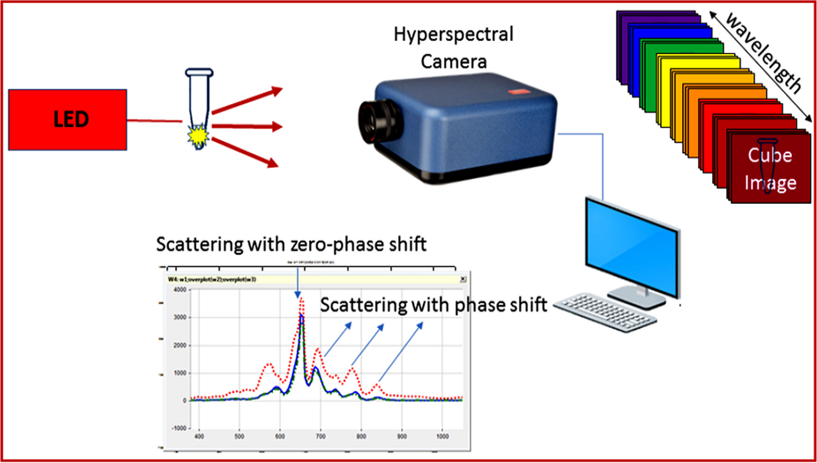
The field of care testing toward the analysis of blood lack nowadays raped and simple test techniques for biomarkers. In this study, we have developed a novel DNA/RNA genetic signature via nonlinear polarization with hyperspectral imaging for helping hepatocellular carcinoma early diagnosis.
Table 1 presents the participant's clinical and demographic details. According to a prior study, the higher baseline platelet level and lower baseline hemoglobin may be associated to possible prognostic factors for cancer.
Table 1 Clinical and demographic data characteristicsCharacterizing the nonpolarized signature of DNA/RNA controlThe most prevalent malignant primary liver tumor is hepatocellular carcinoma (HCC) that usually develops from the progression of cirrhosis. In terms of the pathological observation, many pathological stain-based diagnosis studies are performed on tissue specimens and cells. Staining is time-consuming. With the help of hyperspectral imaging by light polarization signature of DNA/RNA in specimen, it is possible to observe specimens without staining or fixation.
A study by Hashimoto et al. [27] conducted a computer-aided diagnosis on liver pathological samples which were hematoxylin and eosin (H & E) stained. Applying hyperspectral imaging (HSI) to the same samples, the accuracy improvement has reached to 24% for fibers and 5% for cytoplasm. So, DNA/RNA genetic signature via nonlinear polarization with hyperspectral imaging is possible help to hepatocellular carcinoma early diagnosis.
Total DNA and RNA under polarized light can provide information regarding chemical structure links and dielectric characteristics. Light emission and absorption can be used to assess the dielectric characteristics of macromolecules. Due to mutations in macromolecule (DNA and RNA) chemical structure, the harmonic oscillation signature could offer a distinctive signature [28]. The scattered and re-emitted fingerprint for total DNA and RNA (control) is demonstrated in Fig. 2 (mean of 5 samples).
Fig. 2
Resonance harmonic oscillations of whole DNA and RNA (control) samples irradiated by a laser source at 656 nm (scattered and returned energy)
In an attempt to conclude spectral oscillation signature, normalized (scattered and re-emitted) signatures were calculated to investigate polarized light intensity (Fig. 3).
Fig. 3
Normalized harmonic oscillations (scattered and re-emitted radiation) of total DNA and RNA (control) after being irradiated with laser source at 656 nm
Light polarization signature of DNA/RNA (control)Nonpolarized signature of total DNA and RNA could be employed to offer platform for determining the spectral fingerprint signature of both total DNA and RNA. Characteristic resonance frequency for each sample is represented in Table 2.
Table 2 Charactersitic light polarization signature of total DNA/RNA (control)Total DNA demonstrated characteristic resonance frequencies that differ from total RNA at 2nd, 3rd, 4th, 5th harmonic. Variable polarization states can be noticed by varying the total DNA and RNA structural arrangement with variable light absorption (attenuation). Signal intensity of total DNA and RNA signature is demonstrated in Table 3. Nonpolarized signature attenuation offered another path to differentiate between total DNA and RNA spectral signatures.
Table 3 Attenuation of transmitted light polarization of total DNA and RNA samplesFrequency changes and phase differences of DNA/RNA (control)In an effort to distinguish between the spectral fingerprint characteristics of total DNA and RNA control samples via change in frequency response, harmonic oscillations (scattered and re-emitted) radiations were processed using rapid Fourier transform (Fig. 4).
Fig. 4
Frequency change of total DNA and RNA (control)
Briefly, for the first time, blood analysis by nonlinear polarization with hyperspectral imaging has demonstrated the potential use of DNA/RNA genetic signature in blood spectral biomarkers as a novel alternative for hepatocellular carcinoma diagnosis, which could be used for screening purposes.
Total DNA demonstrated characteristic frequency response that could be discriminated from total RNA. DNA demonstrated spectrum band starting from 0.0044 to 0.0118 GHz compared with RNA spectrum lines. In an attempt to explicitly discriminate between total DNA and RNA, phase change was investigated. Change in frequency signal phase can be considered as a potent tool to discriminate the spectral signatures of both total DNA and RNA (Fig. 5). DNA can be efficiently distinguished from RNA via phase shift. While DNA demonstrated phase shift dominates at 0.088 GHz, RNA dominates at 0.106 GHz (Fig. 5). These stage shifts could be attributed to DNA's distinct chemical composition and dielectric features from RNA.
Fig. 5
Phase difference between DNA and RNA
Impact of HCC tumor on RNA nonlinear polarization signatureThe optoelectronic and dielectric characteristics of RNA may change along with HCC-related changes. Re-emitted light radiation at resonance frequencies could provide a fingerprint sign for chemical composition and physical characteristics. The nonlinear response of RNA (control) was investigated to RNA (HCC) for early HCC diagnosis (Fig. 6).
Fig. 6
Scattered and re-emitted radiation light of RNA (control) and RNA (HCC) after being irradiated with laser source at 656 nm
To counteract the impact of the intensity of polarized light, normalized signatures (scattered and re-emitted radiations) were determined (Fig. 7).
Fig. 7
Normalized scattered and re-emitted radiation light of RNA (control) and RNA (HCC) after being irradiated with laser source at 656 nm
The re-emitted signature could be distinguished due to changes in molecular-electronic configuration and absorber size in the order of (nm). The resonance frequency for RNA (control) to RNA (HCC) is shown in Table 4.
Table 4 Characteristic light polarization signature of RNA (control) and RNA (HCC)Various polarization states produced by various structural configurations can exhibit variable light attenuation. Table 5 lists the attenuation of the investigated samples.
Table 5 Attenuation of transmitted light polarization (scattered and re-emitted) of RNA (Control) and RNA (HCC)Impact of HCC tumor on RNA frequency changes and phase differencesTo distinguish between RNA (control) and RNA (HCC), nonlinear signature was processed using a quick Fourier transform. RNA (HCC) demonstrated characteristic frequency signals at 0.006, 0,014, and 0,022 MHz. These signals could offer good discrimination between RNA (HCC) and RNA (control) (Fig. 8).
Fig. 8
Determination of RNA resonance frequency change due to HCC
Phase difference between RNA (control) and RNA (HCC) was investigated. Whereas RNA (control) dominates at 0.014 and 0.072 MHz, RNA (HCC) dominates at 0.021 and 0.032 MHz (Fig. 9). Phase shift could offer good discrimination between RNA (control) and RNA (HCC). Nonlinear polarization could secure facile HCC diagnosis.
Fig. 9
Phase difference between RNA (control) and RNA (HCC)
Ten signal measurements of the DNA (control), RNA (control), and RNA (HCC) responses were calculated into a histogram for the frequency distribution. The DNA (control), RNA (control), and RNA (HCC) all had standard deviations of 0.164, 0.155, and 0.146, respectively. The RNA (HCC), DNA (control), and RNA mean values are 0.091, 0.08, and 0.123, respectively. We notice that the mean values shift to right when the patient suffering from HCC cancer (Fig. 10).
Fig. 10
The calculated histogram for RNA, DNA, and RNA (HCC)
留言 (0)