
記住我


Tumors with peritoneal dissemination were long considered to be terminal and incurable, therefore, treatment was focused on palliation; however, in the last decades, cytoreductive surgery associated with hyperthermic intraperitoneal chemotherapy (HIPEC) has shown itself to be a strategy associated with palliative care with relapse-free periods higher than 87% at 20 years, in comparison with curative therapy. Currently, precise indications exist for HIPEC for patients with peritoneal metastatic disease1. Recent studies have shown higher survival rates in selected patients with peritoneal carcinomatosis, and it is currently considered the treatment of choice for pseudomyxoma peritonei and patients diagnosed with peritoneal mesothelioma2.
It is also the recommended treatment in selected cases of patients with gastric, colon, and ovarian primary carcinomatosis.
The adverse effects of intraperitoneal chemotherapy in patients with peritoneal carcinomatosis are limited and generally due to poor blood supply and low penetration of chemotherapy through the peritoneum2. Therefore, the mainstay of treatment starts with cytoreductive surgery to eradicate the macroscopic tumor by peritonectomy and multivisceral resections, followed by intraperitoneal chemotherapy to eliminate the microscopic disease to produce synergism between cytotoxic drugs and hyperthermia which allows an increase in the permeability of tumor cells leading to apoptosis3.
This report presents an unusual case of a patient with peritoneal pseudomyxoma secondary to mucinous adenocarcinoma of the urachus, who underwent complete macroscopic cytoreduction and intraoperative HIPEC.
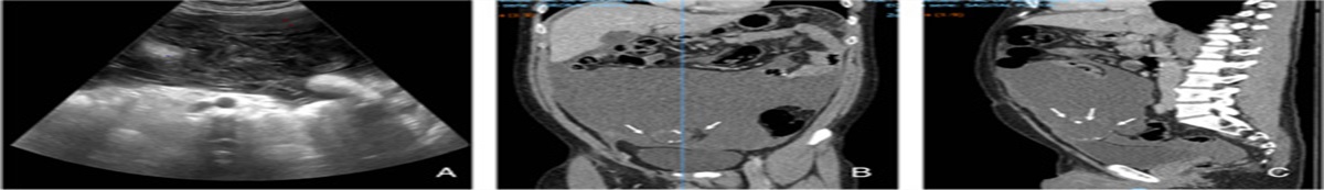
Case descriptionA 43-year-old male patient with no past medical history had been experiencing symptoms over a period of 8 months characterized by abdominal pain associated with intermittent distension and unintentional weight loss. He went to the emergency department where initial imaging studies were performed through abdominal ultrasound (Fig. 1A). These found a predominantly infraumbilical lesion towards the anterior abdominal wall, with a liquid appearance and multiple septa in its interior of ∼110×288 mm that displaced adjacent structures. For a better characterization of the lesion, a total abdomen computed tomography (CT) scan was performed in which a lesion of complex cystic aspects was found, with calcified areas and septa that enhanced after contrast administration. It measured ∼84×72×52 mm with a volume of 157 ml (Figs. 1B and C) obtained through a normal chest CT scan. This led to a paracentesis with 20 ml of thick mucinous content being extracted. The patient was referred to our peritoneal oncologic surgery unit for suspected peritoneal pseudomyxoma. Laparoscopy was considered to classify the extent of peritoneal disease and to evaluate the possibility of complete resection and biopsy.
 Figure 1:
 (A) Ultrasound of the abdomen: showing an infraumbilical lesion with septa inside ∼110×299 mm. Contrast abdominal computed tomography scan: coronal section (B) and sagittal section (C). Complex cystic lesion, with calcific areas and septa with contrast medium, ∼84×72×52 mm, and volume of 157 ml.
A laparoscopy was performed and given the findings, it was later converted to a laparotomy with 6 l of mucinous ascites. The carcinomatosis index was calculated at 20 and the disease was considered resectable. Histopathological analysis revealed mucinous adenocarcinoma with a colloid component of the urachus, these results were presented to a multidisciplinary meeting where it was determined to take the patient to complete cytoreduction and HIPEC.
En bloc resection was performed, including the previous surgical scar and umbilicus involving the posterior sheath of the rectum bilaterally following the path of the urachus to the bladder dome, proceeding to perform a partial cystectomy. Note the orifice that communicates the mucinous tumor with the cupula through which mucus comes out (Fig. 2A).
 Figure 2:
 (A) Xiphopubic incision, en bloc resection of the umbilicus and previous scar, caudal dissection involving the posterior sheath of the rectus abdominis, bilaterally. (B and C) Dissection to delimit the urachus trajectory, identifying its healthy borders and involvement of the bladder dome. White arrows, bladder dome. Asterisk, urachus trajectory.
Cytoreduction procedures were performed to achieve complete macroscopic cytoreduction (Figs. 2B and C).
Cytoreduction was performed achieving complete macroscopic cytoreduction. HIPEC was performed by open technique (Fig. 3A), for 90 min with oxaliplatin at an average temperature of 42°C. The patient was taken to the intensive care unit for postoperative monitoring as part of our protocol.
 Figure 3:
 (A) Partial cystectomy of the bladder done. (B) Cytoreduction, with peritonectomy and multivisceral resections. (C) HIPEC with open technique – colisthesis.
After 72 h of being monitored in the intensive care unit, the patient was transferred to a room with multidisciplinary management. A liquid diet was given to the patient on the fifth day, with adequate tolerance, modulation of abdominal pain, and presence of intestinal transit. Hospital discharge was given on the 14th postoperative day, and ambulatory follow-up was provided by our unit.
The pathology report confirmed the suspected diagnosis of mucinous adenocarcinoma of the urachus, with infiltration of the bladder dome, with the presence of focal mucin without infiltration of the muscular wall nor evidence of dysplastic epithelium (Fig. 4).
 Figure 4:
 Mucinous adenocarcinoma of the urachus, with the presence of fibrous tissue capsule with calcifications and foreign body type gigantic cellular reaction, with acellular mucin extravasation on the surface of the capsule. Negative for dysplastic epithelium (A-D).
Immunohistochemistry stains evidenced morphologic and immunohistochemical features that correlated with the image data, confirming the presence of a mucinous cystadenocarcinoma of urachal origin (Figs. 4A–D).
Posttherapeutic follow-ups were performed with peritoneal magnetic resonance and with the evaluation of carcinoembryonic antigen, carbohydrate antigen 19-9, and cancer antigen 125 markers; this follow-up was performed every 6 months for 3 years, then every year for 3 years, and finally every 2 years until 15 years after initial care were completed. In this patient, there was no evidence of tumor relapse at 48 months of follow-up after surgery.
DiscussionPseudomyxoma peritonei due to urachal adenocarcinoma is a rare pathology, representing 0.5–2% of malignant tumors related to the bladder and 0.01% of all cancers in adults. It affects patients between 40 and 70 years of age, with a 12:1 male predilection4,5.
It originates from the embryological remnant of the urachus, resulting from the involution of the allantoic duct and the ventral cloaca that connects the bladder dome to the umbilicus, the urachus remnant may persist in up to 30% of cases and may persist as a tubular or cystic structure6. The urachus is composed of three layers of tissue: an epithelial canal lined by epithelium, submucosal connective tissue, and an outer layer of smooth muscle. Urachal neoplasms can arise from any of these layers and can be epithelial (from which mucinous adenocarcinoma of the urachus originates) or mesenchymal7.
Approximately 70% of urachal carcinomas are mucin-producers. With the progression of the disease, the mucin produced by the neoplastic cells accumulates in the urachal tract, subsequently progresses to the rupture of the urachal tumor mass and spreads, giving access of the mucinous neoplastic cells to the peritoneal cavity, which subsequently produces ascites with mucinous characteristics (pseudomyxoma peritonei)8.
The clinical presentation of this entity may begin with nonspecific symptoms which can make very challenging the correct and early diagnosis: it may debut with abdominopelvic pain, associated with abdominal distension, palpable mass, weight loss, intermittent bowel obstruction, and urological symptoms such as hematuria, dysuria, and mucosuria; the latter originated by the persistent connection between the urachal remnant and the bladder is very distinctive of this disease9.
Imaging studies for the diagnostic approach range from the ultrasound that can visualize a heterogeneous mass located in the midline of the abdomen or free fluid; CT or MRI are used to evaluate the tumor (staging, visceral involvement, lymphadenopathy, or distant metastasis) and can characterize the lesion as an infraumbilical midline cystic mass attached to the abdominal wall and the upper aspect of the bladder, which should make us suspect an adenocarcinoma of the urachus2,10; The stage of the disease allows us to guide the treatment and oncologic prognosis.
Surgery is the standard treatment for localized tumors and involves resection of the urachal remnant and en bloc resection of the bladder dome and umbilicus. According to MD Anderson Cancer Center data, they achieved a 5-year disease-free survival rate of 44% with this procedure6. Perioperative chemotherapy has a limited role.
Cytoreductive surgery plus HIPEC was developed as a treatment for peritoneal tumor disease3. It has been associated with improved survival rates, disease-free time, and better quality of life compared with systemic chemotherapy2,11.
In patients diagnosed with urachal adenocarcinoma, the tumor biology has been related to colorectal adenocarcinomas, considering the similarity of histological findings of urachal and enteric adenocarcinomas. Therefore, in patients with metastatic involvement limited to the peritoneal cavity, cytoreductive surgery, and HIPEC have been considered as the treatment of choice, phase II and III trials have shown benefits over systemic chemotherapy5,11–15.
Mertens et al.3 evaluated the 5-year oncologic outcomes in patients with urachal adenocarcinoma and peritoneal pseudomyxoma undergoing cytoreduction plus HIPEC with evidence of recidivism in 50% (N=5) of cases, a 2-year survival rate of 66.7% and a 5-year survival rate of 55.6%.
Liu and colleagues8,16,17 performed cytoreductive surgery/HIPEC in nine patients with pseudomyxoma peritonei rising from the urachus. Only one recurrence occurred; the median survival was 27.5 months (range: 6–61 months). In the case of our patient, complete cytoreduction was performed and oxaliplatin (a chemotherapeutic agent with a high molecular weight that allows for maintaining a high intraperitoneal concentration) was administrated.
MRI was an essential complement for the surveillance and monitoring of patients with PMP and was very helpful for carrying out exhaustive lesion mapping. For low-graded tumors peritoneal MRI and evaluation of carcinoembryogenic antigen, carbohydrate antigen 19-9, and cancer antigen 125 markers were used as follow-up techniques, these were performed every 6 months for 3 years, then every year for 3 years, and finally every 2 years until 15 years were completed from initial care.
For high-grade tumors, contrasted thoracic–abdomen–pelvic CT scan (without digestive opacification) plus the evaluation of markers (previously mentioned) were indicated for follow-up, this was performed every 4 months for 3 years, then every 6 months for 2 years and finally every year until 15 years were completed from initial care18. Peritoneal MRI was performed in case of doubt about the CT scan results or if serum markers increased in absence of an abnormal CT scan.
Therapeutic management is still a matter of debate, considering that it is a rare clinical entity, studies to standardize management strategy are lacking.
ConclusionsUrachal adenocarcinoma is a rare entity with a poor prognosis usually due to the advanced condition or metastatic disease at the time of diagnosis. The most frequent symptoms are abdominal pain, the presence of a palpable mass in the hypogastrium, and mucosuria. The surgical treatment represents the standard therapy, systemic chemotherapy has a limited role. Cytoreductive surgery associated with HIPEC can be considered a treatment strategy for mucinous urachal neoplasms presenting with peritoneal pseudomyxoma.
MethodsThis case report was written and corrected in line with SCARE 2020 guidelines19.
Ethical approvalThe patient was informed of the intention to publish this case report and accompanying images and a written informed consent was obtained. A copy of the written consent is available on request.
Sources of fundingNo funding was received for this paper.
Author contributionG.J.S. and R.G.: conception and revision. A.C.R.B.: conception, writing, revision, and submission. M.A.H.: conception, writing, and revision. R.D. and A.B.: writing and revision.
Conflicts of interest disclosureThe authors declare that they have no financial conflict of interest with regard to the content of this report.
Research registration unique identifying number (UIN)None.
GuarantorNone.
AcknowledgmentsThe authors thank Mariana Ospina for her contribution in the translation of the article.
References 1. Helderman RFCPA, Löke DR, Kok HP, et al. Variation in clinical application of hyperthermic intraperitoneal chemotherapy: a review. Cancers 2019;11:78. 2. Mah M, Mack LA, Hurton S, et al. Cytoreductive surgery and heated intraperitoneal chemotherapy for peritoneal carcinomatosis from rare etiologies. Am J Surg 2019;217:923–927. 3. Mertens LS, Behrendt MA, Mehta AM, et al. Long-term survival after cytoreductive surgery and hyperthermic intraperitoneal chemotherapy (HIPEC) for patients with peritoneal metastases of urachal cancer. Eur J Surg Oncol 2019;45:1740–1744. 4. Jia Z, Chang X, Li X, et al. Urachal carcinoma: are lymphadenectomy and umbilectomy necessary? Med Sci Monit 2020;26:e927913. 5. Li S, Meng X, Liang P, et al. Clinical and radiological features of urachal carcinoma and infection. Front Oncol 2021;11:702116. 6. Siefker-Radtke AO, Gee J, Shen Y, et al. Multimodality management of urachal carcinoma: the MD Anderson Cancer Center experience. J Urol 2003;169:1295–1298. 7. Gopalan A, Sharp DS, Fine SW, et al. Urachal carcinoma: a clinicopathologic analysis of 24 cases with outcome correlation. Am J Surg Pathol 2009;33:659–668. 8. Liu Y, Ishibashi H, Hirano M, et al. Cytoreductive surgery plus hyperthermic intraperitoneal chemotherapy for pseudomyxoma peritonei arising from urachus. Ann Surg Oncol 2015;22:2799–2805. 9. Ponzini F, Kowal L, Ghafoor M, et al. Rare occurrence of pseudomyxoma peritonei (PMP) syndrome arising from a malignant transformed ovarian primary mature cystic teratoma treated by cytoreductive surgery and HIPEC: a case report. World J Surg Oncol 2022;20:78. 10. Sugarbaker PH, Verghese M, Yan TD, et al. Management of mucinous urachal neoplasm presenting as pseudomyxoma peritonei. Tumori 2008;94:732–736. 11. Yang XJ, Huang CQ, Suo T, et al. Cytoreductive surgery and hyperthermic intraperitoneal chemotherapy improves survival of patients with peritoneal carcinomatosis from gastric cancer: final results of a phase III randomized clinical trial. Ann Surg Oncol 2011;18:1575–1581. 12. Krane LS, Kader AK, Levine EA. Cytoreductive surgery with hyperthermic intraperitoneal chemotherapy for patients with peritoneal carcinomatosis secondary to urachal adenocarcinoma. J Surg Oncol 2012;105:258–260. 13. Mercier F, Passot G, Villeneuve L, et al. BIG-RENAPE Working Group. Peritoneal carcinomatosis of urachus origin treated by cytoreductive surgery and hyperthermic intraperitoneal chemotherapy (HIPEC): an international registry of 36 patients. Ann Surg Oncol 2018;25:1094–1100. 14. Verwaal VJ, van Ruth S, de Bree E, et al. Randomized trial of cytoreduction and hyperthermic intraperitoneal chemotherapy versus systemic chemotherapy and palliative surgery in patients with peritoneal carcinomatosis of colorectal cancer. J Clin Oncol 2003;21:3737–3743. 15. Agrawal AK, Bobiński P, Grzebieniak Z, et al. Pseudomyxoma peritonei originating from urachus-case report and review of the literature. Curr Oncol 2014;21:e155–e165. 16. Siefker-Radtke A. Urachal carcinoma: surgical and chemotherapeutic options. Expert Rev Anticancer Ther 2006;6:1715–1721. 17. Agha RA, Fowler AJ, Saeta A, et al. SCARE Group. The SCARE Statement: Consensus-based surgical case report guidelines. Int J Surg 2016;34:180–186. 18. Delhorme JB, Villeneuve L, Bouché O, et al. Appendiceal tumors and pseudomyxoma peritonei: French Intergroup Clinical Practice Guidelines for diagnosis, treatments and follow-up (RENAPE, RENAPATH, SNFGE, FFCD, GERCOR, UNICANCER, SFCD, SFED, SFRO, ACHBT, SFR). Dig Liver Dis 2022;54:30–39. 19. Agha RA, Franchi T, Sohrabi C, et al. for the SCARE Group. The SCARE 2020 Guideline: Updating Consensus Surgical CAse REport (SCARE) Guidelines. Int J Surg 2020;84:226–230.
留言 (0)