
記住我


Breast cancer is the world’s most prevalent and the second leading cause of cancer-associated mortality among women in the world1,2 with 2.3 million diagnosed cases and 685 000 deaths at the end of 20203. CRC in turn is the third most common cancer in males and the second most common malignancy diagnosed in females, with over 1.9 million new cases and ~940 000 deaths reported in 20204. The current therapeutic strategies for breast cancer, include surgery, chemotherapy and radiotherapy, but they may lack efficacy due to a high risk of relapse, poor patient response and the emergence of drug resistance5. The prognosis for CRC varies according to the stage of the cancer, with nearly half of the patients that undergo curative surgery and another 20–25% who receive post-surgical adjuvant chemotherapy, experience cancer relapse, metastasis and eventual death6–8, emphesizing the inadequacy of the state of modern treatment options for this fatal malignancy at the present time9. There is therefore, a growing interest in identifying natural compounds that are safe and affordable as adjunctive treatments to the conventional therapy currently offered for these patients. Curcumin from the roots of the Curcuma longa L., is one such compound that has become one of the leading and most studied natural medicines for its role in cancer prevention. Curcumin is a polyphenolic phytochemical 1,7-bis(4-hydroxy-3-methoxyphenyl)-1,6-heptadiene-3,5-dione10,11 which interacts with numerous biological targets, including inflammatory mediators, growth factors, enzymes, carrier proteins, metal ions, tumour suppressors, transcription factors, oncoproteins and cellular nucleic acids12–14. In Asian countries curcumin has been consumed for more than 2000 years, due to its various medicinal properties against human diseases, including cancer and auto-immune diseases15–23. However, in spite of the potential for cancer prevention and treatment being supported by plenty of preclinical studies and even clinical studies, there is concern regarding the selectivity and bioavailability of curcumin following oral ingestion. The poor bioavailability of curcumin is attributed in part to its chemical instability24, poor absorption, rapid metabolism and systemic elimination, hampering its application as a therapeutic agent25,26. To overcome these practical limitations of curcumin bioavailability, numerous approaches have been adopted to enhance its systemic absorption27,28. Extensive efforts have also been devoted to the synthesis of new curcumin derivatives29,30, to optimize the beneficial properties of curcumin against cancer and improve its pharmacokinetic profile31.
Although, there have been many curcumin analogues and their hybrid molecules synthesized and tested, as exemplified by a recent review of 220 compounds32, the report concludes with the assertion that most of the studies are incomplete and lacking SAR and clinical studies for the most potent compounds. In this our preliminary work 14 novel heterocyclic curcumin derivatives were screened using the MTT assay to assess their cytotoxicity on human colorectal cancer cell line HCT-116 and mammary gland breast adenocarcinoma MDA-MB-231 and also on a normal GM08402 (human fibroblast) cell line for the purpose of generating lead compounds with improved bioavailability and anti-cancer activity. Electrochemical oxidation potentials were also determined for selected compounds to reveal their electron-donating capacity and as a general indicator of their radical scavenging ability. Compounds with good antioxidant and free-radical quenching properties, can intercept and neutralize potent chemical carcinogens. The curcumin derivatives in this study are based on two heterocycles, the 3,4-dihydropyrimid-2(1H)-one (DHPM) and 3,4-dihydro-2(1H)-pyridone (DHPDO). DHPMs have a remarkable scope of pharmaceutical efficiency and therapeutic activities such as antibacterial, antiviral, anti-inflammatory, analgesic, antitumor, and cardiovascular activity33–35. Several DHPM curcumin derivatives have been synthesized previously36–38 and their cytotoxic and antioxidant properties reported. Significant therapeutic and biological activities, such as antibacterial, antifungal, antitumor, and HIV-1-specific reverse transcriptase inhibitors have also been reported for the analogous DHPDO core containing alkaloids39. By incorporating curcumin motifs in these heterocycles one may expect to see even more diverse or enhanced pharmacological properties.
Materials and methods Cell culturesHuman colorectal cancer cell line HCT-116 (ATCC CCL-247EMT) and mammary gland breast adenocarcinoma MDA-MB-231 (ATCC HTB-26) were obtained from the American Type Culture Collection (ATCC, LGC Standards). Human fibroblast GM08402, apparently healthy were obtained from the (Coriell Institute for Medical Research). HCT-116 cells were subsequently cultured in Mycos 5 A medium supplemented with 10% foetal bovine serum (Sigma-Aldrich). MDA-MB-231 cells were subsequently cultured in Dulbecco’s Modified Eagle’s Medium supplemented with 10% foetal bovine serum (Sigma-Aldrich). GM08402 cells were subsequently cultured in Eagle’s Minimum Essential Medium supplemented with 15% foetal bovine serum (Sigma-Aldrich). Cells were incubated at 37°C in humidified 5% CO2.
Proliferation assayCells were seeded in 96-well plates in concentration 5000 cells per well and cultivated for 24 h, then compounds to be tested were added in concentration range 100–0.4 µM and incubated 48 h. Cell viability was measured using 3-(4,5-dimethylthiazol-2-yl)-2,5-diphenyltetrazolinium bromide (MTT). In brief, after incubating with compounds culture medium was removed and fresh medium with 0.2 mg/ml MTT was added in each well of the plate. After incubation (3 h, 37°C, 5% CO2) the MTT solution was removed and replaced with 200 µl of dimethyl sulfoxide (DMSO) and 25 µl Sorenson’s glycine buffer (glycine 0.1 M, NaCl 0.1 M, pH:10.5 with 0.1 NaOH). The optical density of the wells was determined using a plate reader Thermo Scientific Multiskan EX at test wavelength 540 nm. The half-maximal inhibitory concentration (IC50) of each compound was calculated using Graph Pad Prism 3.0, by GraphPad Software, see additional information in the Supplementary, Supplemental Digital Content 1, https://links.lww.com/IJSO/A15.
Electrochemical oxidation potentialsCyclic voltammetry experiments were carried out on a PARSTAT 2273 electrochemical system, Princeton Applied Research. A stationary glassy carbon disk electrode (d=0:5 mm) served as the working electrode, while the counterelectrode was a Pt wire. The oxidation potentials were measured relative to a Ag/Ag+ reference electrode. Acetonitrile was dried over P2O5 and distilled; the distillate was stored over CaH2 and redistilled just before use. Recrystallized tetrabutylammonium tetrafluoroborate (TBABF4) was used as a supporting electrolyte at 0.1 M concentration.
Results Antiproliferative activityThe cytotoxic effects of heterocyclic curcumin derivatives 1–14 synthesized at the Latvian Institute of Organic Synthesis, Aizkraukles 21, Riga LV-1006, Latvia (the synthesis is not part of this work, and will be or are published elsewhere40–42), were determined by MTT assay on two cancer cell lines HCT-116 and MDA-MB-231, and also on a normal cell line GM08402 (human fibroblast). In Table 1 are reported the IC50 µM concentrations of curcumin and compounds 1–14. If at 100 μM concentration the cell viability was 75% or lower then it was deemed to be greater than 100 μM; If on the other hand at 100 μM concentration the cell viability was higher than 75% then the compound was deemed as *Not cytotoxic at 100 μM concentration. Curcumin was 4 times more cytotoxic to MDA-MB-231 cancer cells and 6 times more cytotoxic to HCT-116.
Table 1 - Cell growth inhibitory effects of curcumin and compounds 1–14 evaluated after 48 h of treatment by MTT assays and oxidation potentials (volts) of selected compounds. MDA-MB-231 HCT-116 GM08402 Oxidation Potentials No Compound IC50 (µM) IC50 (µM) IC50 (µM) Eox1(V) Eox2(V) Curcumin 18.9±1.1 12.7±2.3 75.5±8.2 0.88 1.41 1 a
a
a
2
a
a
a
2
 a
a
a
1.82
3
a
a
a
1.82
3
 a
a
a
1.68
4
a
a
a
1.68
4
 a
a
a
Not solub
5
a
a
a
Not solub
5
 17.6±2.2
3.5±0.3
33.2±3.0
1.6
6
17.6±2.2
3.5±0.3
33.2±3.0
1.6
6
 7.2±0.5
1.9±0.7
95.6±5.2
1.16
1.85
7
7.2±0.5
1.9±0.7
95.6±5.2
1.16
1.85
7
 98.0±0.6
65.5±7.8
a
1.6
1.9
8
98.0±0.6
65.5±7.8
a
1.6
1.9
8
 100.6±3.8
34.0±6.4
>100
1.61
9
100.6±3.8
34.0±6.4
>100
1.61
9
 a
98.6±2.1
>100
1.44
10
a
98.6±2.1
>100
1.44
10
 9.5±1.8
31.7±8.4
1.6±0.2
1.44
11
9.5±1.8
31.7±8.4
1.6±0.2
1.44
11
 32.6±0.1
34.0±5.2
53.0±7.9
12
32.6±0.1
34.0±5.2
53.0±7.9
12
 100.0±5.2
99.2±5.7
>100
13
100.0±5.2
99.2±5.7
>100
13
 5.0±0.2
5.6±1.7
9.3±2.1
14
5.0±0.2
5.6±1.7
9.3±2.1
14
 68.0±5.7
63.3±8.5
>100
68.0±5.7
63.3±8.5
>100
At bottom of table: Data are expressed as the concentrations which inhibit 50% (IC50) cell growth and are means ± SE of at least three separate experiments.
aNot cytotoxic at 100 µM concentration.
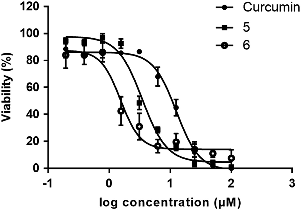
Table 1 cancer cells than to the normal GM08402 cells. Compounds 1–4 at 100 µM concentration had no toxicity on cancer cells and no toxicity to normal cells. Compounds 7, 8, 11, 12, and 14 had IC50 concentrations substantially higher than curcumin and thus they aren’t useful as anti-cancer compounds. Compounds 10 and 13 are toxic to normal cells to the same extent as to cancer cells. The antiproliferative activity of curcumin and compounds 5 and 6 are presented graphically in Figs. 1, 2, and 3 below.
 Figure 1:
 Cell growth inhibitory effect of compounds 5, 6 and curcumin on HCT-116 cell line. Average value from two independent experiment, R>0.950.
 Figure 2:
 Cell growth inhibitory effect of compounds 5, 6 and curcumin on MDA-MB-231 cell line. Average value from two independent experiment, R>0.950.
 Figure 3:
 Cell growth inhibitory effect of compounds 5, 6 and curcumin on GM08402 cell line. Average value from two independent experiment, R>0.950.
Compound 5 is 1.9 times more cytotoxic to MDA-MB-231cancer cells and 9.6 times more cytotoxic on HCT-116 cancer cells compared to the the normal GM08402 cells. Compound 6 is 13.3 times more cytotoxic to MDA-MB-231cancer cells and 50.3 times more cytotoxic on HCT-116 cancer cells compared to the the normal GM08402 cells. Thus compound 6 is superior to curcumin being more toxic on both tested cancer cell lines and less toxic than curcumin on normal cells. The best result of the previously reported DHPM curcumin derivative37 on HCT-116 cells using the MTT assay had IC50 value at 12.5 µM concentration.
Oxidation potentialsThe electrochemical oxidation of selected compounds was performed by cyclic voltammetry on a stationary glassy carbon electrode in dry acetonitrile, the data are presented in Table 1.
DiscussionAs a preliminary study the purpose of our research was to screen 14 curcumin heteterocyclic derivatives for cytotoxicity against 2 cancer cell lines and hopefully diminished cytotoxicity to normal cells. We chose the colorimetric MTT assay for this task: a yellow tetrazole dye which is reduced to a purple colour formazan dye in living cells, and is generally assumed to be dependent on NAD(P)H oxidoreductase enzymes located largely in the cytosolic compartment of the cell reflecting the number of viable cells present. A literature review revealed that several tetrazolium-based assays, such as the MTT may show interactions with many phytochemicals demonstrating intrinsic reductive potential including antioxidants and polyphenols43,44. The 3-(4,5-dimethylthiazol-2-yl)-2,5-diphenyl-2H-tetrazolium bromide dye in the MTT assay has two reduction potentials at around −0.2 and −0.6 V45. Since none of the compounds in our study have oxidation potentials in this range there should not be any interference from the MTT dye being oxidized by the sample compounds. The MTT assay screening revealed two lead compounds 5 and 6 with superior anti-cancer activity compared to curcumin, but this assay provides no details concerning the mechanism where by this activity is achieved. Curcumin itself is recognized by numerous intracellular targets, including proteins involved in antioxidant response, immune response, apoptosis, cell cycle regulation, tumour progression, and moreover, was suggested to effectively overcome chemoresistance during cancer treatment. Dimethoxy curcumin (DMC) induced cell death in breast carcinoma MCF7 cells through S-phase arrest and apoptosis or triggered a strong proteasome inhibition and high induction of CHOP to achieve potent anti-cancer effects on malignant breast cancer cells46. In human renal carcinoma Caki cells, DMC induced apoptosis, through pro-oxidant production of ROS, the release of mitochondrial Cytc, and the subsequent activation of caspase-347. Two colon cancer cells (HT-29 and SW480) and one normal human colon mucosal epithelial cell (NCM460) were studied by western blotting analysis, which indicated that cleavage of pro-caspases-3 and PARP were clearly induced by DMC to their active form, while the expression of survivin was reduced and E-cadherin was enhanced in both cells in vitro and in vivo48.
The oxidation of curcumin is an irreversible process that proceeds in two steps in 0.1 M TBABF4 in dry acetonitrile. The half-wave potential (Eox1) of the first step of curcumin oxidation, as determined by cyclic voltammetry, is 0.88 V and the half-wave potential (Eox2) of the second step is 1.41 V affording a product with two ortho-benzoquinone substituents Fig. 4.
 Figure 4: Proposed electrochemical oxidation mechanism of curcumin49.
The main activities of curcumin in normal cells can be attributed to its potent antioxidant and free-radical quenching properties, which result in the interception and neutralization of potent chemical carcinogens, such as ROS (superoxide anions, peroxyl radicals, and hydroxyl radicals) and NOS (nitric oxide and peroxynitrite compounds)49. The 4-hydroxy-3-methoxystyryl DHPDO compound 6 (red colour representing curcumin components) also undergoes electrochemical oxidation in 2 steps Fig. 5.
 Figure 5:
 Proposed electrochemical oxidation mechanism of styryl DHPDO (6).
From our previous studies on DHPDO electrochemical oxidations, we have observed that 4-arylsubstituted DHPDO derivatives have oxidation potentials in the 1.46–1.72 V range50. Thus (Eox1) of 1.16 V would correspond to the phenol oxidation forming the ortho-benzoquinone and the 1.85 V (Eox2) is required for the DHPDO oxidation to the 2-pyridone. This is confirmed by compound 5 which only has the DHPDO moiety and is oxidized to the 2-pyridone at 1.6 V. Compound 6 with a 2-methoxyphenol group has a good electron-donating capacity indicated by the low Eox1 and thus can be expected to be a good radical scavenger or antioxidant. More studies will be needed to ascertain the mechanism by which compounds 5 and 6 induce cytotoxicity in the cancer cells leading to futher optimizations.
ConclusionsThe cytotoxic effects of 14 novel heterocyclic curcumin derivatives were tested on human colorectal cancer cell line HCT-116 and mammary gland breast adenocarcinoma MDA-MB-231 and also on normal GM08402 (human fibroblast) cell line for the purpose of generating lead compounds with improved chemical stability, bioavailability, and anti-cancer activity. The curcumin derivatives are based on two heterocycles, DHPM and DHPDO. After performing MTT assays, only heterocyclic curcumin derivatives 5 and 6 displayed notable cytotoxicity against both cancer cell lines and both derivatives were from the DHPDO series. Significantly DHPDO curcumin derivative 6, compared to curcumin is 2.6 times more toxic to the breast cancer cell line and 6.7 times more toxic to the colorectal cancer cell line (IC50 at 1.9±0.7 µM) and at the same time being 1.3 times less toxic than curcumin to normal fibroblast cells. The electrochemical oxidation experiments revealed a 1.16 V (Eox1) oxidation potential for compound 6 compared to 1.41 V (Eox2) for curcumin, which indicates a good electron-donating capacity for compound 6 and possibly good free-radical scavenging properties. This outcome has established DHPDO curcumin derivative 6 as a promising lead compound for further elaboration, tests, and optimization, which are currently pursued in our labs.
Ethical approvalNot applicable.
Source of fundingThis research was funded by ERDF project No.1.1.1.2/VIAA/1/16/241 ‘Synthesis of Curcumin Heterocyclic Derivatives‘.
Author contributionR.S.: writing the manuscript, synthesis of compounds, interpretation of data analysis I.D.: cytotoxicity studies, data collection and analysis. B.T.: determined the oxidation potentials for the compounds
Conflicts of interest disclosureThere are no conflicts of interest.
Research registration unique identifying number (UIN)Not applicable.
GuarantorRufus Smits.
References 1. Garcia-Aranda M, Redondo M. Protein kinase targets in breast cancer. Int J Mol Sci 2017;18:2543. 2. Sun YS, Zhao Z, Yang ZN, et al. Risk factors and preventions of breast cancer. Int J Biol Sci 2017;13:1387–1397. 3. Sung H, Ferlay J, Siegel RL, et al. Global Cancer Statistics 2020: GLOBOCAN Estimates of Incidence and Mortality Worldwide for 36 Cancers in 185 Countries. Ca Cancer J Clin 2021;71:209–249. 4. Xi Y, Xu P. Global colorectal cancer burden in 2020 and projections to 2040. Transl Oncol 2021;14:101174. 5. Ye JC, Formenti SC. Integration of radiation and immuno-therapy in breast cancer—treatment implications. Breast 2018;38:66–74. 6. Obrand DI, Gordon PH. Incidence and patterns of recurrence following curative resection for colorectal carcinoma. Dis Colon Rectum 1997;40:15–24. 7. O’Connell MJ, Campbell ME, Goldberg RM, et al. Survival following recurrence in stage II and III colon cancer: findings from the ACCENT data set. J Clin Oncol 2008;26:2336–2341. 8. Andre T, Boni C, Navarro M, et al. Improved overall survival with oxaliplatin, fluorouracil, and leucovorin as adjuvant treatment in stage II or III colon cancer in the MOSAIC trial. J Clin Oncol 2009;27:3109–3116. 9. Weng W, Goel A. Curcumin and colorectal cancer: an update and current perspective on this natural medicine. Semin Cancer Biol 2020;80:73–86. 10. Fadus MC, Lau C, Bikhchandani J, et al. Curcumin: an age-old anti-inflammatory and anti-neoplastic agent. J Tradit Complement Med 2016;7:339–346. 11. Banik U, Parasuraman S, Adhikary AK, et al. Curcumin: the spicy modulator of breast carcinogenesis. J Exp Clin Cancer Res 2017;36:98. 12. Shishodia S, Sethi G, Aggarwal BB. Curcumin: getting back to the roots. Ann N Y Acad Sci 2005;1056:206–217. 13. Aggarwal BB, Kumar A, Bharti AC. Anticancer potential of curcumin: preclinical and clinical studies. Anticancer Res 2003;23:363–398. 14. Han X, Deng S, Wang N, et al. Inhibitory effects and molecular mechanisms of tetrahydrocurcumin against human breast cancer MCF-7 cells. Food Nutr Res 2016;60:30616. 15. Liu JL, Pan YY, Chen O, et al. Curcumin inhibits MCF-7 cells by modulating the NF-κB signaling pathway. Oncol Lett 2017;14:5581–5584. 16. Zheng J, Zhou Y, Li Y, et al. Spices for prevention and treatment of cancers. Nutrients 2016;8:E495. 17. Wang Y, Yu J, Cui R, et al. Curcumin in treating breast cancer: a review. J Lab Autom 2016;21:723–731. 18. Imran M, Ullah A, Saeed F, et al. Cucurmin, anticancer, & antitumor perspectives: a comprehensive review. Crit Rev Food Sci Nutr 2018;58:1271–1293. 19. Ko EY, Moon A. Natural products for chemoprevention of breast cancer. J Cancer Prev 2015;20:223–231. 20. Hossain DM, Bhattacharyya S, Das T, et al. Curcumin: the multi-targeted therapy for cancer regression. Front Biosci (Schol Ed) 2012;4:335–355. 21. Zhou H, Beevers CS, Huang S. The targets of curcumin. Curr Drug Targets 2011;12:332–347. 22. Huang G, Xu Z, Huang Y, et al. Curcumin protects against collagen-induced arthritis via suppression of BAFF production. J Clin Immunol 2013;33:550–557. 23. Song X, Zang M, Dai E, et al. Molecular targets of curcumin in breast cancer (Review). Mol Med Rep 2019;19:23–29. 24. Ojo1 OA, Adeyemo TR, Rotimi D, et al. Anticancer properties of curcumin against colorectal cancer: review. Front Oncol 2022;12:881641. 25. Liu W, Zhai Y, Heng X, et al. Oral bioavailability of curcumin: problems and advancements. J Drug Target 2016;24:694–702. 26. Toden S, Goel A. The holy grail of curcumin and its efficacy in various diseases: is bioavailability truly a big concern?. J Restor Med 2017;6:27–36. 27. Jayaprakasha GK, Chidambara Murthy KN, Patil BS. Enhanced colon cancer chemoprevention of curcumin by nanoencapsulation with whey protein. Eur J Pharmacol 2016;789:291–300. 28. Chaurasia S, Chaubey P, Patel RR, et al. Curcumin-polymeric nanoparticles against colon-26 tumor-bearing mice: cytotoxicity, pharmacokinetic and anticancer efficacy studies. Drug Dev Ind Pharm 2016;42:694–700. 29. Zhang J, Feng Z, Wang C, et al. Curcumin derivative WZ35 efficiently suppresses colon cancer progression through inducing ROS production and ER stress-dependent apoptosis. Am J Cancer Res 2017;7:275–288. 30. Pröhl M, Schubert US, Weigand W, et al. Metal complexes of curcumin and curcumin derivatives for molecular imaging and anticancer therapy. Coord Chem Rev 2016;307:32–41. 31. Mbese Z, Khwaza V, Aderibigbe BA. Curcumin and its derivatives as potential therapeutic agents in prostate, colon and breast cancers (Review). Molecules 2019;24:4386. 32. Noureddin SA, El-Shishtawy RM, Al-Footy KO. Curcumin analogues and their hybrid molecules as multifunctional drugs. Eur J Med Chem 2019;182:111631. 33. Ben Moussa S, Mehri A, Badraoui B. Magnesium modified calcium hydroxyapatite: An efficient and recyclable catalyst for the one-pot Biginelli condensation. J Molec Struct 2020;1200:1–7. 34. Dos Anjos JV, Srivastava RM, Costa-Silva JH, et al. Comparative computational studies of 3,4-dihydro-2,6-diaryl4-oxo-pyrimidine-5-carbonitrile derivatives as potential antinociceptive agents. Molecules 2012;17:809–819. 35. Kappe CO. Biologically active dihydropyrimidones of the Biginelli-type—a literature survey. Eur J Med Chem 2000;35:1043–1052. 36. Lal J, Gupta SK, Thavaselvam D, et al. Synthesis and pharmacological activity evaluation of curcumin derivatives. Chinese Chem Lett 2016;27:1067–1072. 37. Sahu PK. Design, structure activity relationship, cytotoxicity and evaluation of antioxidant activity of curcumin derivatives/analogues. Eur J Med Chem 2016;121:510–516. 38. Rodrigues FC, Kumar NVA, Thakur G. The potency of heterocyclic curcumin analogues: An evidence-based review. Pharmacol Res 2021;166:105489. 39. Noguez OM, Marcelino V, Rodríguez H, et al. Infrared assisted production of 3,4-dihydro-2(1H)-pyridones in solvent-free conditions. Int J Mol Sci 2011;12:2641–2649. 40. Smits R. Curcumin dihydropyridone derivatives with anti-cancer activity. WO 2021/234468 Al. 41. Smits R. Synthesis of curcumin heterocyclic derivatives. VIII EFMC International Symposium on Advances in Synthetic and Medicinal Chemistry Athens. Greece; 2019:264. 42. Smits R, Belyakov S, Petrova M, et al. Synthesis of 6-styryl dihydropyrimidines and dihydropyridones via the respective 6-alkylphosphonates. Chem Heterocycl Compd 2022;58:687–694. 43. van Tonder A, Joubert AM, Cromarty DA. Limitations of the 3-(4,5-dimethylthiazol-2-yl)-2,5-diphenyl-2H-tetrazolium bromide (MTT) assay when compared to three commonly used cell enumeration assays. BMC Res Notes 2015;8:47. 44. Ghasemi M, Turnbull T, Sebastian S, et al. The MTT assay: utility, limitations, pitfalls, and interpretation in bulk and single-cell analysis. Int J Mol Sci 2021;22:12827. 45. Li J, Dimitrov N. NTBC and MTT reduction electrochemistry: impact on cu superconformal plating for fabrication of glass interposers. J Electrochem Soc 2019;166:D3120–D3128. 46. Yoon MJ, Kang YJ, Lee JA, et al. Stronger proteasomal inhibition and higher CHOP induction are responsible for more effective induction of paraptosis by dimethoxycurcumin than curcumin. Cell Death Dis 2014;5:e1112. 47. Lee JW, Hong HM, Kwon DD, et al. Dimethoxy curcumin, a structural analogue of curcumin, induces apoptosis in human renal carcinoma caki cells through the production of reactive oxygen species, the release of cytochrome C, and the activation of caspase-3. Korean J Urol 2010;51:870–878. 48. Chen D, Dai F, Chen Z, et al. Dimethoxy curcumin induces apoptosis by suppressing survivin and inhibits invasion by enhancing E-Cadherin in colon cancer cells. Med Sci Monit 2016;22:3215–3222. 49. Maseka A, Chrzescijanska E, Zaborski M. Characteristics of curcumin using cyclic voltammetry, UV–vis, fluorescence and thermogravimetric analysis. Electrochimica Acta 2013;107:441–447. 50. Smits R, Turovska B, Belyakov S, et al. Synthesis of 5-carboxy-6-methyl-3,4-dihydro-2(1H)-pyridone derivatives and their electrochemical oxidation to 2-pyridones. Chem Phys Lett 2016;649:84–87.
留言 (0)