Study population
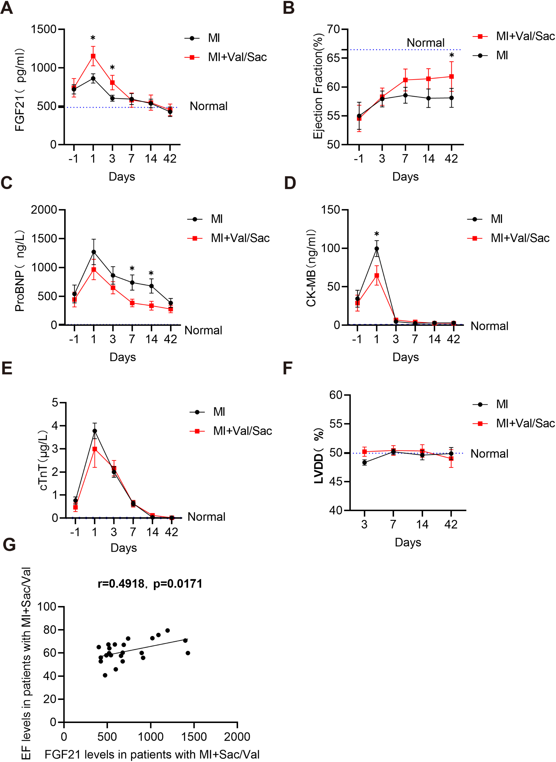
All participating subjects provided written informed consent, and the study received approval from the Human Ethics Committee of Hangzhou Xiaoshan First People’s Hospital. The prospective study cohort was compiled from the hospital’s registry of patients, adhering to strict inclusion and exclusion criteria. Inclusion criteria included: (i) patients aged between 18 and 85 years, (ii) patients with an ejection fraction of 50% or less, and (iii) patients with a ProBNP level of 600 ng/L or higher (iv) Patients with ST-segment elevation or non-ST-segment elevation acute myocardial infarction and heart failure. The exclusion criteria were as follows: (i) patients who had used Sac/Val or similar drugs within the past six months, (ii) patients with renal insufficiency, defined as a blood creatinine level of 256 µM or higher, (iii) patients with obesity, having a body mass index (BMI) of 28.0 or higher, (iv) patients with hepatic insufficiency, (v) patients with menstrual disorders, and (vi) patients with tumors, severe infections, autoimmune diseases, or other significant comorbidities. Ultimately, 83 patients met the eligibility criteria and were enrolled in the study, all of whom had myocardial infarction. Of these, 31 patients received Sac/Val treatment, 52 patients did not. We also recruited 28 normal healthy individuals and used them as negative controls. Normal healthy individuals were examined for BMI, cTnT, ProBNP, CK-MB, ejection fraction, left ventricular end-diastolic internal diameter (LVDD), hemoglobin, total cholesterol, triglycerides, fasting glucose, creatinine, and albumin, which were all within the normal range of values, and did not have a history of smoking, alcohol consumption, or a history of hypertension or diabetes. Only blood samples were collected and used for all patients, and no medical interventions were involved. The project was reviewed by the Ethics Committee of the First People’s Hospital of Xiaoshan District, Hangzhou.
Mice
The heterozygous fgf21 mice, kindly donated by Professor Aimin Xu of the University of Hong Kong, underwent heterozygous amplification through breeding with C57BL/6J wild-type mice. Subsequently, FGF21 heterozygous males and females were selectively mated for several consecutive generations to achieve a genetically homogeneous line of FGF21KO homozygous mice, as well as a control line of wild-type (WT) mice. The experimental mice were individually housed in a specific-pathogen-free (SPF) facility within the Animal Experiment Center of Wenzhou Medical University. The housing conditions were strictly maintained, with room temperature set at 22 ± 1 °C, humidity at 60%, and a 12-hour light/12-hour dark cycle. All animal procedures and studies were conducted in accordance with the ethical guidelines approved by the Animal Research Ethics Committee of Wenzhou Medical University.
Animal procedures
Animal procedures were granted by the Animal Research Ethics Committee of Wenzhou Medical University following the recommendations in the Guide for the Care and Use of Laboratory Animals of the NIH guidelines. To anaesthetize mice, xylazine (10 mg/kg body weight) and ketamine (100 mg/kg) (Phoenix Scientific, Inc., St. Joseph, MO, USA) were intraperitoneally given. To sacrifice mice, asphyxiation of CO2 was used. (Infuse CO2 into the box at a rate of 10-30% per minute to replace the euthanasia box and determine that the mice are immobile, not breathing, and have dilated pupils. Turn off the CO2. observe for an additional 2 min to determine that the mice are dead.)
MI mouse model
To induce MI in male mice aged 10 to 12 weeks, a permanent ligation of the left anterior descending coronary artery was performed. The procedure was initiated by anesthetizing the mice with 2% isoflurane. Following anesthesia, a small incision was made in the left thoracic region at the fourth intercostal space to expose the heart. The left anterior descending coronary artery was precisely located and subsequently ligated, approximately 3 mm distal to the lower margin of the left atrial appendage, using a 6 − 0 silk suture. Immediately after ligation, the heart was carefully repositioned within the chest cavity, and the thoracic cavity was manually evacuated of air. The muscle and skin layers were then sutured in a secure manner to close the incision. Sham surgery was conducted following the same steps, omitting the ligation of the coronary artery. The hearts of mice were divided into infarcted, near-infarcted non-infarcted areas, near-infarcted areas were used for Western Blot, immunohistochemistry, and DHE staining, and infarcted areas were used for H&E and Masson staining. The infarcted area, near-infarcted area, and non-infarcted area were distinguished primarily based on visual observation: upon excision of the heart, the infarcted area appeared pale or grayish, contrasting sharply with the normal myocardium; the near-infarcted area, located between the infarcted and non-infarcted areas, was slightly less distinct but retained some tissue integrity; the non-infarcted area exhibited normal color and intact myocardial structure [18, 19].
Drug administration
In the clinical research section, the use of Sac/Val was based on the Chinese Heart Failure Diagnosis and Treatment Guidelines and was strictly administered in accordance with its recommendations [20]. According to the guidelines, Sac/Val is one of the recommended treatment options for heart failure, specifically indicated for patients with NYHA class II/III heart failure with reduced ejection fraction (HFrEF). For patients with NYHA class II/III HFrEF who remain symptomatic despite treatment with ACEIs/ARBs, ARNI is recommended as a replacement for ACEIs/ARBs to further reduce the incidence and mortality of heart failure. The use of Sac/Val is determined by physicians based on individual patient conditions and clinical judgment, with the medication administered twice daily, in the morning and evening.
In the conduct of our animal experiments, we administered a daily dose of 52 mg/kg of Sac/Val, comprising an equimolar mixture of 26 mg/kg of Val and 26 mg/kg of Sac daily. This dose was deemed comparable to the low-dose regimen, which is also referred to as 1× or low dose, containing 26 mg/kg of Val and Sac each per day. Drug and saline control administrations were performed via once-daily oral gavage, with all solutions being sterile filtered prior to administration. The day before the surgery we gave the mice a once-daily gavage administration, and on the day of the surgery we did not give the drug in order to facilitate the mice’s recovery. Therefore, on the day after the surgery, we gave the mice two gavage administrations, and from the third day until the end of the 14 days, we resumed the once-a-day gavage administration. The drugs were procured from MedChemExpress.
Mice that were required to receive PPARs were kept at the same frequency of administration as those receiving Sac/Val, and were injected by intraperitoneal injection of PPARγ antagonist GW9662 or PPARα antagonist GW9662 (1 mg/kg/day, MCE Company, China) or saline after each gavage of Sac/Val.
Measurement of echocardiographic and histological staining
The cardiac physiological functions were rigorously assessed using a Doppler echocardiography system (VINNO 6 VET, VINNO, China). The transthoracic 2D M-mode echocardiographic system was employed to capture M-mode tracings, from which the following parameters were quantified: left ventricle internal dimension in systole (LVIDS), left ventricle internal dimension in diastole (LVIDD), ejection fraction (EF), and fractional shortening (FS, calculated as FS = (LVIDD − LVIDS)/LVIDD × 100%). These measurements were taken in mice anesthetized with 1.5% isoflurane.
To determine the infarct volume, heart samples were fixed in 4% paraformaldehyde for 48–72 h and subsequently embedded in paraffin. Thin slices (5 μm thick) were then prepared and stained using Masson’s trichrome or hematoxylin and eosin (H&E) staining. The infarct ratio was analyzed using the ImageJ software and expressed as the volume fraction of collagen (CVF% = collagen area/tissue total area × 100%).
Additionally, cardiac tissues were embedded in the Tissue-Tek OCT compound (Sakura Finetek, Tokyo, Japan) and sectioned serially to a thickness of 10 μm. These cryosections were stained with the superoxide-sensitive dye DHE (10µM in 0.01% DMSO) and incubated for 30 min at 37 °C in a humidified dark chamber.
Immunohistochemistry (IHC)
Immunohistochemical analysis was conducted to quantify the expression levels of AT1R and Neprilysin. Initially, the tissue sections were dewaxed and hydrated using 10 mM sodium citrate buffer to facilitate antigen retrieval. Subsequently, endogenous peroxidase activity was blocked by incubating the sections for 30 min at room temperature. Following this, 30 µL of normal non-immune animal serum was added dropwise to the sections, which were then washed three times with PBST. The sections were then incubated overnight at 4 °C with the primary antibody. After two washes with PBS, the slides were incubated with a goat anti-rabbit horseradish peroxidase-conjugated secondary antibody for 30 min at room temperature. This incubation step was followed by thorough washing. Finally, the sections were incubated with 3,3’-diaminobenzidine (DAB) for visualization and counterstained with hematoxylin.
Western blotting
Proteins were extracted from cells and various mouse tissues, and subsequently underwent sodium dodecyl sulfate-polyacrylamide gel electrophoresis (SDS-PAGE) for separation. The separated proteins were then transferred onto polyvinylidene difluoride (PVDF) membranes. For Western blotting analysis, horseradish peroxidase-conjugated rabbit anti-IgG was employed as the secondary antibody. The primary antibodies used in this study were FGF21 (ab171941), FGFR1 (ab76464), PPARα (WL00978), PPARγ (ab178860), PI3K (WL00978), p-PI3K (ab182651), AKT (ab179463), p-AKT (ab19262-3), mTOR (ab134903), p-mTOR (ab109268), Collagen-I (ab138492), α-SMA (ab124964), HIF-1α (ab1), AT1R (ab124505), and Neprilysin (18008-1-AP). The protein bands were visualized using enhanced chemiluminescence (ECL) reagents and their intensities were quantified using ImageJ software.
Enzyme-linked immunosorbent assay
Plasma samples were obtained from mice and patients, and were subsequently stored at − 80 °C for further analysis. To determine the concentrations of FGF21, we employed either a mouse-specific ELISA kit (MFG037, Hong Kong) or a human-specific ELISA kit (SHFT09, Hong Kong), strictly adhering to the manufacturer’s recommended protocols.
Cell culture and treatment
H9C2 cells, procured from the Cell Bank of the Chinese Academy of Sciences in Shanghai, China, were maintained in a culture medium comprising high-glucose Dulbecco’s Modified Eagle Medium (DMEM) from GIBCO, USA. This medium was supplemented with 1% cyan streptomycin double-antibody and 10% (v/v) fetal bovine serum (FBS). The cells were incubated at 37 °C in a controlled atmosphere containing 5% CO2. To mimic hypoxic injury, the H9C2 cells were exposed to a three-gas incubator for 12 h, where the internal environment consisted of 1% O2, 5% CO2, 94% N2, maintained at a temperature of 37 °C.
AML-12 cells, also obtained from the Cell Bank of the Chinese Academy of Sciences in Shanghai, China, were cultured in F-12 Dulbecco’s Modified Eagle Medium (DMEM) from GIBCO, USA. This medium was similarly supplemented with 1% cyan streptomycin double-antibody and 10% (v/v) FBS. The cells were maintained under standard culture conditions of 37 °C and 5% CO2.
In silico molecular modeling studies
Utilizing the fundamental ‘lock-and-key’ paradigm governing the interplay between ligands and receptors, the molecular docking approach was employed to scrutinize the interaction dynamics between a small molecule ligand and a biomacromolecule receptor. The initial three-dimensional (3D) structural framework was delineated based on molecular data obtained from PubChem, thus providing a robust foundation for further computational investigations.
SPR measurements
The binding affinity of Sacubitril and Valsartan towards the ligand-binding domains (LBDs) of PPARγ and PPARα was assessed using surface plasmon resonance (SPR) technology, specifically utilizing the Biacore 3000 instrument (Little Chalfont, Buckinghamshire, UK). Prior to immobilization, the target proteins were diluted in 10 mmol/L sodium acetate buffer (pH 4.5) to a final concentration of 0.10 mg/mL. Subsequently, a standard amine-coupling procedure was employed to covalently bind these proteins to CM5 chips. The small molecule compounds were diluted in 1X PBS-P + buffer containing 2% DMSO. The flow rate was set at 30 µL/min, and gradient-diluted solutions of the small molecule compounds were sequentially injected into the flow channel for interaction with the protein immobilized on the chip. The resulting binding data were analyzed using the 1:1 Langmuir binding model to determine the equilibrium dissociation constant (KD) of each compound.
Statistical analysis
All statistical analyses were conducted using GraphPad Prism 8 software (GraphPad, San Diego, CA). To determine significant differences between two groups, an independent samples t-test was employed. For comparing multiple groups, one-way ANOVA along with Tukey’s post hoc test were used to compare difference among groups. To compare the usage rates of other medications between Sac/Val and non-Sac/Val groups, the Chi-Square test was utilized; for medications with low incidence, Fisher’s exact test was applied. Additionally, Pearson’s correlation analyses were performed to investigate the potential associations between serum FGF21 levels and other relevant parameters. The threshold for statistical significance was set at p < 0.05, while p < 0.01 was considered indicative of a highly significant relationship.


留言 (0)