Patient’s clinical history
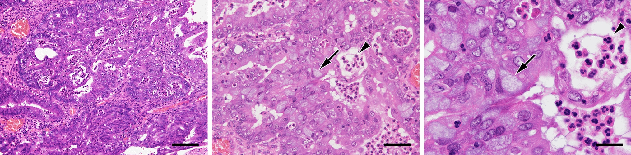
A 46-year-old Japanese woman, gravida 2, para 2, was referred to the hospital with complaints of dysmenorrhea. Systemic computed tomography (CT) and abdominal magnetic resonance imaging revealed a 6-cm-sized multilocular cystic tumor with solid lesions in the left ovary and a 5-cm-sized simple cystic tumor in the right ovary, along with a small amount of ascites. Serum cancer antigen 125 (CA125) and CA19-9 levels were 322 U/mL and 2,711 U/mL, respectively. These findings were suggestive of early-stage ovarian cancer with a background of endometriosis. A staging laparotomy including total abdominal hysterectomy, bilateral salpingo-oophorectomy, omentectomy and retroperitoneal lymphadenectomy was performed. Consequently, the patient was diagnosed with ovarian endometrioid carcinoma with a score of IC1, based on International Federation of Gynecology and Obstetrics (FIGO) staging. The patient was given six cycles of intravenous paclitaxel (175 mg/m2) and carboplatin (area under the concentration–time curve [AUC] of 6 mg·min/mL) on a 21-day cycle. Post-chemotherapy, systemic CT revealed complete response according to the Response Evaluation Criteria in Solid Tumors (RECIST version 1.1) [10]. The patient is currently living and has had no cancer recurrence for 40 months.
Preparation of recombinant lentiviruses
Lentiviral vectors harboring cDNAs encoding human CDK4R24C and cyclin D1 were constructed based on previous reports [8, 9]. Briefly, cDNA encoding CDK4 was amplified by polymerase chain reaction (PCR) and inserted into pcDNA3 (Thermo Fisher Scientific, Waltham, MA), and the R24C mutation was then introduced using inverse-PCR and DpnI digestion (New England Biolabs, Ipswich, MA). Resultant CDK4R24C cDNA was transferred into the pCDH-CMV-MCS-EF1-Hygro vector (System Biosciences, Mountain View, CA), which carries the hygromycin phosphotransferase (HPT) gene, resulting in pCDH-CMV-CDK4R24C-EF1-Hygro. Similarly, cDNA encoding cyclin D1 was PCR amplified and inserted into pCDH-CMV-MCS-EF1-Bsd, which was constructed by replacing the puromycin N-acetyltransferase gene in pCDH-CMV-MCS-EF1-Puro (System Biosciences) with the blasticidin S deaminase (BSD) gene, resulting in pCDH-CMV-cyclin D1-EF1-Bsd. pLOX-TERT-IRES-TK (Addgene plasmid #12,245), a lentiviral vector harboring cDNA encoding human TERT was a gift from Dr. Didier Trono. Human embryonic kidney (HEK) 293 T cells were cultured in three 10-cm dishes, and cells in each dish were transfected with one of the above three lentiviral vectors plus psPAX2 (Addgene plasmid #12,260, a gift from Dr. Didier Trono) and pCMV-VSV-G (Addgene plasmid #8454, a gift from Dr. Bob Weinberg) [11] at a ratio of 3:2:1 using the calcium phosphate–DNA co-precipitation method [12]. Forty-eight hours later, conditioned media containing infectious recombinant lentiviruses were recovered.
Isolation of carcinoma cells and gene transfer
Fragments of tumor tissue obtained at surgery were cut into small pieces, suspended in 10 mL of 1 mg/mL Collagenase/Dispase (Roche Diagnostics, Mannheim, Germany) and incubated at 37 °C for 30 min [13]. To remove undigested tissue fragments, the slurry was passed through a 40-μm pore cell strainer. After washing with phosphate-buffered saline (PBS), cells were cultured in RPMI 1640 medium supplemented with 10% fetal bovine serum (FBS), 100 U/mL penicillin, 100 μg/mL streptomycin and 0.25 μg/mL amphotericin B (Nacalai Tesque, Kyoto, Japan). Twenty-four hours later, cells were infected with the above three recombinant lentiviruses by replacing the regular medium with a mixture of the three conditioned media prepared above. After another 24 h, virus-containing medium was changed to regular medium supplemented with 400 μg/mL of hygromycin B and 10 μg/mL of blasticidin S (Thermo Fisher Scientific) to select transformed cells. Growth and morphology of cultured cells were observed with an inverted phase-contrast microscope IX71 (Olympus, Tokyo, Japan).
Confirmation of transgene incorporation
Incorporation of the three exogenous genes mentioned above into the host cell genome was confirmed by reverse transcription-PCR (RT-PCR), as described [14] using the following primer pairs: 5’-ATgTTCggggATTCCCAATACgAg-3’ and 5’-CTATCggCgAgTACTTCTACACAg-3’ for HPT in the plasmid harboring CDK4R24C; 5’-gCTACAATCAACAgCATCCCCATC-3’ and 5’-CACATAACCAgAgggCAgCAATTC-3’ for BSD in the plasmid harboring cyclin D1; and 5’-gTCTTCTTgACgAgCATTCCTAgg-3’ and 5’-ACATgTAAAgCATgTgCACCgAgg-3’ for the internal ribosome entry site (IRES) in the plasmid harboring TERT. As a control, endogenous glyceraldehyde-3-phosphate dehydrogenase (GAPDH) expression was detected using the primer pair 5’-ATggggAAggTgAAggTCggAgTC-3’ and 5’-CAgAgATgATgACCCTTTTggCTC-3’.
Transmission electron microscopy
Electron microscopy specimens were prepared as previously described [15] and observed with an H-7650 transmission electron microscope (Hitachi, Tokyo, Japan).
Growth curve analysis and doubling time
Cells were seeded into wells of six-well plates at a concentration of 1.0 × 10⁵ cells/well. Cells in wells were counted in triplicate at 48-h intervals over a 16-day period. Doubling times were calculated using a web-based doubling-time calculator (https://www.doubling-time.com/compute.php).
Cell cycle analysis
Trypsinized monodispersed cells were fixed in ice-cold 70% ethanol at 4 °C for 2 h. Cells were then re-suspended in 0.5 mL PBS, and 5 μL Cell Cycle Assay Solution Blue (Dojindo Laboratories, Mashiki, Japan) was added and incubated for 15 min at 37 °C under light-shielded conditions. Stained nuclei were analyzed using FACSCanto II (BD Biosciences, San Jose, CA) with FlowJo software (Tree Star, Ashland, OR).
Karyotyping
Chromosomal G-band analysis was performed as described previously [15]. Karyotypes were described according to the International System for Human Cytogenomic Nomenclature (ISCN) 2020 [16].
Sanger sequencing
Mutations in DNA polymerase ε (POLE) were detected by Sanger sequencing as described [15]. Briefly, total RNA was extracted from cells using ISOGEN reagent (Nippon Gene, Tokyo, Japan), and single-stranded cDNA was synthesized as described [17]. DNA fragments corresponding to amino acid residues 270–475 of POLE, the hotspot region of the exonuclease domain [18], were amplified by PCR and the products were purified using QIAquick® Gel Extraction Kit (QIAGEN, Venlo, The Netherlands). Sequencing reactions were carried out using a BigDye® Terminator v.1.1 Cycle Sequencing Kit (Thermo Fisher Scientific) and sequenced using an Applied Biosystems 3500 Genetic Analyzer (Thermo Fisher Scientific).
Short tandem repeat (STR) analysis
Genomic DNA was extracted from cells using a NucleoSpin® Tissue (Takara Bio), and 16 STR loci were detected by multiplex PCR using a PowerPlex® 16 HS System (Promega, Madison, WI). STR profiles were compared with those recorded in the Expasy Profile Database (https://www.cellosaurus.org/str-search/), as described [15].
Monoclonal antibodies
The following monoclonal antibodies (mouse IgG unless otherwise noted) served as primary antibodies: OV-TL 12/30 (Dako, Glostrup, Denmark), recognizing cytokeratin 7 (CK7); Ks20.8 (Dako), recognizing CK20; BC12 (Nichirei Biosciences, Tokyo, Japan), recognizing paired box 8 (PAX8); V9 (Dako), recognizing vimentin; WT49 (Leica Biosystems, Newcastle Upon Tyne, UK), recognizing Wilms tumor 1 (WT1); 1D5 (Dako), recognizing estrogen receptor (ER); 1A6 (Dako), recognizing progesterone receptor (PgR); DO-7 (Nichirei Biosciences), recognizing p53; SP218 (rabbit IgG; Abcam, Cambridge, UK), recognizing phosphatase and tensin homolog deleted on chromosome 10 (PTEN); ES05 (Leica Biosystems), recognizing mutL homolog 1 (MLH1); 79H11 (Leica Biosystems), recognizing mutS homolog 2 (MSH2); EP49 (rabbit IgG; Leica Biosystems), recognizing mutS homolog 6 (MSH6); and EP51 (rabbit IgG; Leica Biosystems), recognizing postmeiotic segregation increased 2 (PMS2).
Histological and immunohistochemical analyses
Formalin-fixed, paraffin-embedded tissue sections were stained with hematoxylin and eosin (H&E) or immunostained with the monoclonal antibodies noted above. Immunohistochemical staining was performed using the Histofine system (Nichirei Biosciences), according to the manufacturer’s protocol [19].
Immunofluorescence staining
Immunofluorescence staining of JFE-21 cells was performed as previously described [20, 21].
Anticancer drugs
Paclitaxel (catalog number Y0000698) and carboplatin (catalog number BP711) were purchased from Merck KGaA (Marmstadt, Germany). Paclitaxel was first dissolved in 100% ethanol to make a 10 mM solution, and then diluted 1:100 with sterile water to make a 100 μM stock solution. Carboplatin, on the other hand, was directly dissolved in sterile water to prepare a 50 mM stock solution. These stock solutions were further diluted with phenol red-free RPMI 1640 medium to the desired final concentration.
Cell viability assay
One hundred μL of phenol red-free RPMI 1640 medium containing 2 × 10⁴ cells was dispensed into each well of a 96-well culture plate and cultured overnight. Then, 100 μL of medium containing various concentrations of paclitaxel or carboplatin was added to the wells and further cultured. Seventy-two hours later, 20 μL of substrate solution containing 5 mM 2-(4-iodophenyl)−3-(4-nitrophenyl)−5-(2,4-disulfophenyl)−2H-tetrazolium (Dojindo Laboratories, Mashiki, Japan) and 0.2 mM 1-methoxy-5-methylphenazinium methylsulfate (Dojindo Laboratories) was added to the wells and incubated at 37 °C in a humidified atmosphere with 5% CO2. Four hours later, absorbance of each well was measured at 450 nm using a SpectraMax iD3 microplate reader (Molecular Device, Sunnyvale, CA). The percentage of viable cells was calculated by dividing the absorbance of treated wells by that of untreated wells. The half maximal inhibitory concentration (IC50) was calculated using GraphPad Prism 7 software (GraphPad Software, San Diego, CA).


留言 (0)