Antibodies
Anti-p70S6K (66,638-1-lg), anti-caspase-3 (19,677-1-AP), and anti-caspase-7 (27,155-1-AP) antibodies were purchased from ProteinTech (Rosemont, IL). An anti-phosphorylated p70S6K antibody (A7189) was obtained from Assay Biotechnology Company, Inc. (Fremont, CA). Anti-cleaved caspase-3 (9664) and anti-cleaved caspase-7 (8438) antibodies were supplied by Cell Signaling Technology (Danvers, MA). An anti-Ki-67/MKI67 (SP-6) antibody (NB600-1252) was purchased from Novus Biologicals (Centennial, CO).
Cell culture
The human PDAC cell line S2-013, a subline of SUIT-2, was donated by Dr. Michael Hollingsworth at the University of Nebraska (Omaha, NE). The human PDAC cell line PANC-1 was obtained from the American Type Culture Collection (Manassas, VA). S2-013 and PANC-1 cells were maintained in Dulbecco’s modified Eagle’s medium (Gibco-BRL, Carlsbad, CA) containing 10% fetal calf serum at 37 °C. Human endothelial cells derived from human umbilical vein endothelial cells (HUVECs) and human mesenchymal stem cells (MSCs) were cultured as previously reported [21].
In selected experiments, S2-013 and PANC-1 cells were cultured with rapamycin (20 and 100 nM; TCI, Tokyo, Japan) for 48 h, everolimus (0.1, 1.0, and 10 µM; FUJIFILM Wako Pure Chemical Corporation, Osaka, Japan) for 48 h [22], gemcitabine (20 and 100 nM; TAIHO PHARMA, Tokyo, Japan) for 48 h [23], paclitaxel (20 and 100 nM; FUJIFILM Wako Pure Chemical Corporation) for 48 h, and 5-FU (20 and 100 nM; Yakult, Tokyo, Japan) for 48 h [23].
Immunoblotting analysis of cell lysates
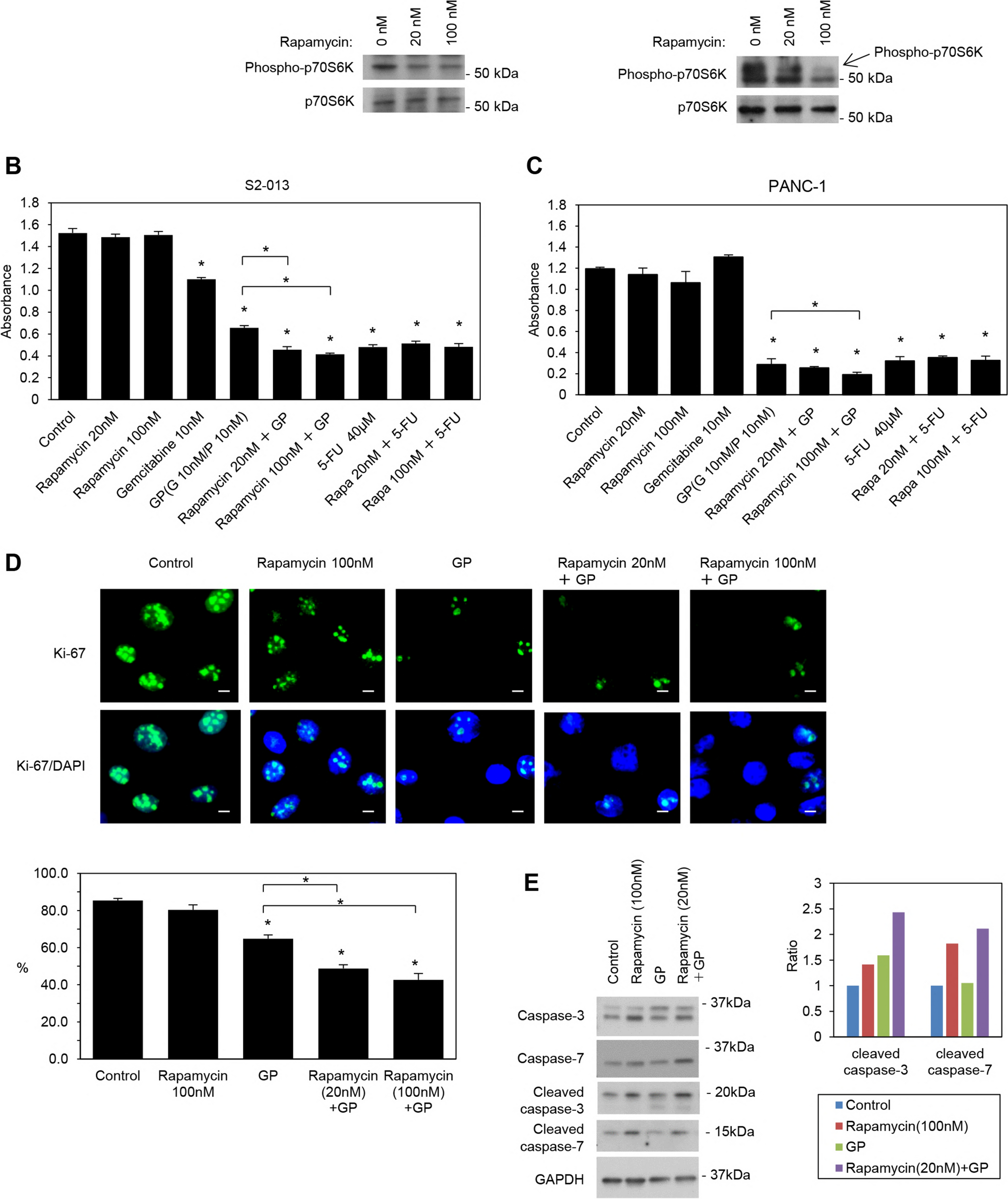
Immunoblotting was performed as previously reported [24]. Briefly, cell pellets were lysed using lysis buffer [Tris–HCl (pH 7.4), sodium dodecyl sulfate, mercaptoethanol, and glycerol], and protein concentrations were measured by the Bradford assay (#500–0006, Bio-Rad, Hercules, CA) using SpectraMax190 (Molecular Devices, San Jose, CA) or Bio Tek Cytation 5 (Agilent, Santa Clara, CA). Ten micrograms of protein was used along with primary antibodies at a dilution of 1:1,000 in 3% phosphoBLOCKER Blocking Reagent (AKR-103, Cell Biolabs, San Diego, CA) in TBST at 4 °C overnight. Following an incubation with appropriate secondary antibodies conjugated with horseradish peroxidase (sc-2371, sc-2385; Santa Cruz Biotechnology, Dallas, Texas) at a dilution of 1:8,000 at room temperature for 1 h, immunoreactive bands were visualized using the suitable ECL kit (GE Healthcare, Chicago, IL), ECL Plus kit (Thermo Fisher Scientific, Waltham, MA), or ECL Prime kit (GE Healthcare) according to the manufacturers’ instructions.
In vitro growth rate by the MTT assay
Cells were seeded at a concentration of 5 × 10⁴ cells per well using 6-well plates. After an incubation for 24 h, cells were separately treated with the following therapeutics: rapamycin, everolimus, GP, and 5-FU, as described above in the Cell culture section. Forty-eight hours after the first addition of therapeutics, cell counting kit-8 solution (Dojindo, Kumamoto, Japan) was added to each well at a concentration of 1/10 volume, and plates were incubated at 37 °C for an additional 4 h. Absorbance was then measured at 450 nm, with 630 nm as a reference, using a Molecular Devices SpectraMax190 (Molecular Devices, San Jose, CA) or BioTek Cytation 5 (Agilent). Results were displayed as mean absorbance relative to controls (untreated or the DMSO control as indicated). The assay was conducted 3 times independently.
Matrigel invasion assay
S2-013 and PANC-1 cells were seeded at densities of 3.5 × 10⁴ and 9.0 × 10⁴ cells per well, respectively, on 6-well plates. After an incubation for 24 h, cells were treated separately with the following therapeutics, including rapamycin, everolimus, GP, and 5-FU, as described above in the Cell culture section. A two-chamber invasion assay was used to assess cell invasion (24-well plates, membrane with a pore size of 8 µm coated with a layer of Matrigel extracellular matrix proteins; Becton Dickinson) as previously reported [25]. The assay was conducted 3 times independently.
Confocal immunofluorescence microscopy
Immunocytochemistry was performed as previously reported [26]. Briefly, cells were treated separately with the following therapeutics, including rapamycin, everolimus, GP, and 5-FU, as described above in the Cell culture section. Cells were fixed with 4% paraformaldehyde, permeabilized with 0.1% Triton X-100, covered with blocking solution (3% BSA/PBS), and then stained with Alexa 594-conjugated phalloidin (A12381, Thermo Fisher Scientific). In some experiments, cells were incubated with an anti-Ki-67 primary antibody followed by an Alexa 488-conjugated anti-rabbit IgG secondary antibody (A21206, Thermo Fisher Scientific). Coverslips were mounted using Vectashield Hard Set with DAPI (Vector Laboratories, Newark, CA). Each specimen was visualized under the All-in-One Fluorescence Microscope BZ-9000 (KEYENCE, Osaka, Japan). In some experiments, the percentage of positive immunolabeled cells was counted in ten selected areas in a masked manner.
Mice and xenografts
S2-013 cell line-derived organoids were established using cultured S2-013 cells, HUVECs, and MSCs as previously reported [21]. Animal experiments were approved by Kochi University (#R01-027), and mice were treated in accordance with the Institutional Animal Care and Use Committee guidelines of Kochi University. To generate subcutaneous tumors, a S2-013 cell line-derived organoid in 50 μL Matrigel matrix/DMEM mixture was subcutaneously implanted into the flanks of 7-week-old female athymic nude mice (BALB/cSlc-nu/nu) (Japan SLC, Inc., Shizuoka, Japan). After implantation, tumors were allowed to grow for 2 weeks before the initiation of treatment, as described below.
Treatment regimens
Tumors were allowed to grow for 2 weeks after the implantation of S2-013 cell line-derived organoids into mice, and treatment was then initiated with (a) 8 mg/kg/day rapamycin by an intraperitoneal injection (ip) 5 days per week for 6 weeks (n = 8); (b) 50 mg/kg gemcitabine plus 0.5 mg/kg paclitaxel on days 1, 8, and 15 every 28 days for 6 weeks (n = 8) [23]; (c) the ip combination of 8 mg/kg/day rapamycin 5 days per week with 50 mg/kg gemcitabine plus 0.5 mg/kg paclitaxel on days 1, 8, and 15 every 28 days for 6 weeks (n = 8); (d) 2 mg/kg everolimus administered orally 5 days per week for 6 weeks (n = 8) [22]; and (e) the combination of 2 mg/kg everolimus administered orally 5 days per week with 50 mg/kg gemcitabine plus 0.5 mg/kg paclitaxel ip on days 1, 8, and 15 every 28 days for 6 weeks (n = 8). In addition, tumors were allowed to grow for 2 weeks after the implantation of S2-013 cell line-derived organoids into mice, and treatment was then initiated with (a) S-1 administered orally 5 days per week for 28 days with a rest period of 2 weeks as one course for 8 weeks (n = 8) [23]; (b) the combination of 1.5 mg/kg/day rapamycin ip 5 days per week with S-1 administered orally 5 days per week for 28 days with a rest period of 2 weeks as one course for 6 weeks (n = 8); and (c) the combination of 2 mg/kg everolimus administered orally 5 days per week with S-1 administered orally 5 days per week for 28 days with a rest period of 2 weeks as one course for 6 weeks (n = 8). Tumor sizes were measured every week, and tumor volumes were calculated with the following formula: V (mm3) = 0.5 × a × b2, where a and b represent the long and perpendicular short diameters of tumors, respectively. Mice were sacrificed eight weeks after the implantation of S2-013 cell line-derived organoids, and solid tumors, the lungs, liver, and kidneys were dissected. Solid tumor, lung, and liver sections were stained with hematoxylin and eosin.
Immunohistochemical staining for Ki-67 and terminal deoxynucleotidyl transferase-mediated nick end labeling (TUNEL)
A proliferation analysis was performed on paraffin-embedded xenograft tumor tissues obtained from xenografts of S2-013 cell-line organoid-bearing mice by immunohistochemistry using the anti-Ki-67 antibody [15]. S2-013 cell line-derived organoids were implanted subcutaneously into the flanks of female athymic nude mice, as described in the Mice and xenografts section, followed by treatments described in the Treatment regimens section. Tissue sections from formalin-fixed paraffin-embedded xenograft tumors were stained with the anti-Ki-67 antibody, and the DAKO EnVision System (Glostrup, Denmark), containing a secondary horseradish peroxidase-conjugated anti-mouse antibody, was used with 3,3'-diaminobenzidine to detect Ki-67. TUNEL was performed on tissue sections from xenograft tumors of S2-013 cell-line organoid-bearing mice treated with the treatment regimens described above using the In Situ Cell Death Detection Kit (Roche Diagnostics, Basel, Switzerland), according to the manufacturer’s instructions. A positive reaction was defined as the intracellular distribution of brown coarse particles or the diffuse distribution of brownish yellow fine particles. Stained cells were counted in 10 random microscopic fields under a light microscope.
Statistical analysis
StatFlex software (Ver6; YUMIT, Osaka, Japan) and SAS software (Ver9.1.3; SAS Institute, Cary, NC) were used for statistical analyses. The significance of differences between groups was assessed using the two-tailed Student’s t-test or Fisher’s exact test, as appropriate. In all analyses, p < 0.05 was considered to be significant.


留言 (0)