
記住我
Oral cavity malignancies are the most prevalent among head and neck cancers and rank as the sixth most common cancer globally. Treatment typically involves surgery, radiotherapy, and chemotherapy, with cisplatin being a frequently utilized chemotherapeutic agent. Cisplatin functions by inducing DNA damage to inhibit cell proliferation and trigger apoptosis in cancer cells. However, its efficacy is often limited by the development of resistance, leading to relapse [6, 8, 30, 31].
Alternative or complementary therapies are being investigated to address cisplatin resistance, including the utilization of natural products to enhance cancer cell chemosensitivity. Herbal-based treatments, such as those derived from the Rumex genus, have demonstrated potential in this area. Rumex dentatus in particular has exhibited significant efficacy against various cancer cell lines [32].
The genus Rumex, belonging to the Polygonaceae family, exhibits a significant global distribution with approximately 200 known species within this genus, many of which demonstrate pharmacological effects. R. dentatus L. is particularly noteworthy for its efficacy. It has also been successfully evaluated against various cancer cell lines in research studies. While the roots of this plant have been extensively studied and screened, limited research has been conducted on the aerial part chemistry and biology, especially in the third stage of growth. Consequently, the aerial part of R. dentatus L. was selected in the third stage of growth to investigate its phenolic content and its in vitro potential synergistic effect when combined with cisplatin on tongue carcinoma cell line.
In this study, the phenolic aglycone profile of the aerial part of R. dentatus L. in the third growth stage was investigated for the first time using UPLC-ESI-MS/MS analysis. The extract comprises flavanol (quercetin, kaempferol), methylated flavanol (isorhamnetin, kaempferide), flavone (luteolin), flavanone (eriodictyol), and flavanol (taxifolin) compounds, which are phenolic structures of the flavonoid type. Additionally, the anthraquinone compounds detected were exclusively emodin and alo-emodin. These classes of compounds are well-established as anticancer agents.
The objective of the present study is to evaluate the in vitro potential positive impact of combining cisplatin with Rumex dentatus extract on the tongue carcinoma cell line. Our hypothesis posits that Rumex dentatus extract will enhance the anticancer efficacy of cisplatin by suppressing cell proliferation, inducing cell cycle arrest, facilitating programmed cell death, and inhibiting autophagy.
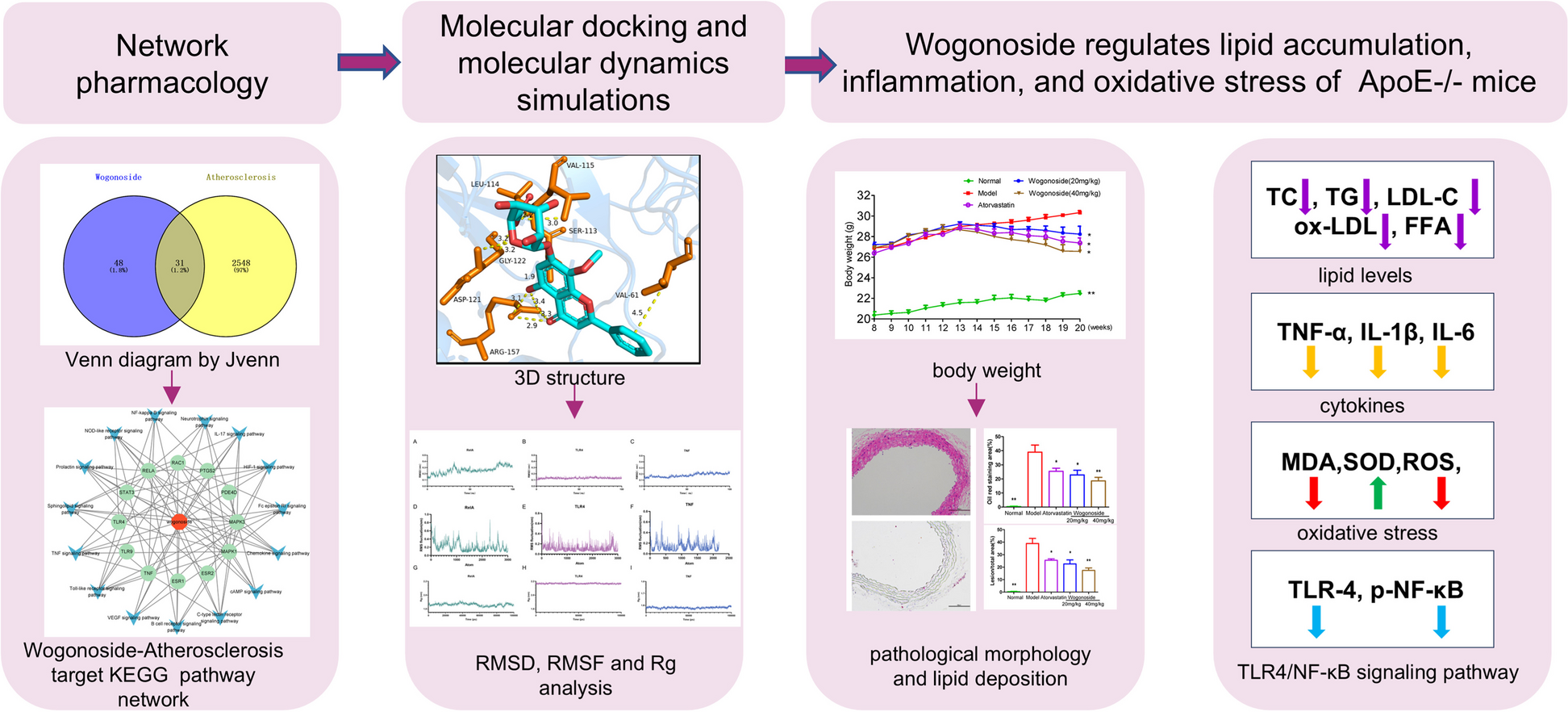
Network pharmacology was utilized to forecast the potential bioactive compounds of Rumex dentatus extract, their respective target genes, and the signaling pathways influenced by these bioactive compounds in the context of tongue carcinoma.
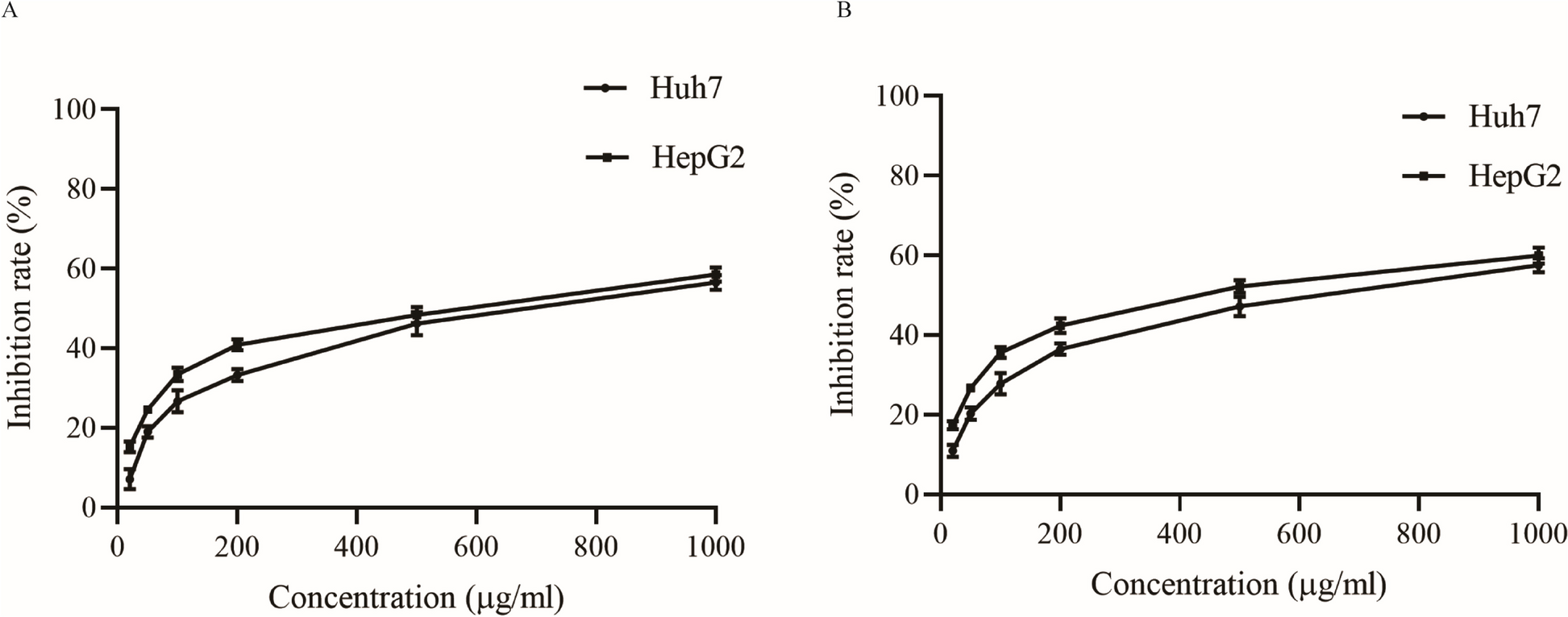
Our results revealed that a concentration-dependent inhibitory effect of R. dentatus extract and cisplatin on HNO-97 tongue carcinoma cells was observed when each was administered independently. The IC50 values for Rumex dentatus and cisplatin were 97.9 μg/mL and 0.328 μg/mL, respectively, indicating moderate cytotoxicity for Rumex dentatus. Rumex dentatus exhibited weak cytotoxicity against normal oral epithelial cells, suggesting its relative safety. Flow cytometry analysis revealed that Rumex dentatus significantly increased the sub-G1 population (indicative of apoptosis) in HNO-97 cells, with even higher levels observed when combined with cisplatin. The combination treatment significantly increased the expression of pro-apoptotic p53 and decreased anti-apoptotic BCL2 expression. Rumex dentatus also reduced the expression of autophagy protein ATG7, with greater reductions observed in the combination treatment.
Both Rumex dentatus and cisplatin exhibited dose-dependent inhibition of the HNO-97 cells. The IC50 value for Rumex dentatus was 97.9 µg/mL, which categorizes it as a moderately active agent, according to the classification by Atjanasuppat et al. This finding supports the hypothesis that Rumex dentatus, although less potent than cisplatin, demonstrates significant cytotoxic activity against tongue carcinoma cells. The moderate antiproliferative activity of Rumex dentatus suggests that while it is not as efficacious as cisplatin, it exerts a measurable impact on inhibiting cancer cell growth, rendering it a potential candidate for combination therapy to enhance cisplatin's effects. This combination approach holds the potential to decrease the required therapeutic concentration of cisplatin, thereby mitigating its associated side effects, while still preserving the intended therapeutic response.
In the current study Rumex dentatus was evaluated against the OEC normal oral epithelial cell line, demonstrating an IC50 > 100 µg/mL, which indicates minimal cytotoxicity against normal cells. This observation suggests a favorable safety profile for Rumex dentatus, as it exhibits selective toxicity against cancer cells while demonstrating limited effects on normal cells. This finding is significant, as a primary challenge in chemotherapy is achieving selective toxicity. The minimal effect of Rumex dentatus on normal cells, coupled with its moderate activity on cancer cells, underscores its potential as a safer adjunct to conventional chemotherapy, such as cisplatin, which is associated with severe side effects.
To our knowledge, it is the first study to investigate the synergistic effect of R. dentatus L. in association with cisplatin on tongue carcinoma.
Nevertheless, recent investigations have highlighted the anticancer properties of the leaves of R. dentatus L. alone and its ability to inhibit the proliferation of the MDA-MB-231 breast cancer cell line. Additionally, it has been documented to induce apoptosis and suppress cell survival in the MDA-MB-231 breast cancer cell line [33]. Flavonoids such as quercetin and taxifolin, as well as anthraquinones such as emodin, have been extensively documented for their anticancer properties, including the induction of apoptosis, cell cycle arrest, and inhibition of cancer cell proliferation in both prostate and breast cancer cells [34].
Flow cytometric analysis was employed to determine if Rumex enhances cisplatin's antiproliferative activity by disrupting cell division. The cell cycle phases include G1, S, G2, and mitosis (M). In G1, cell size increases, and RNA and proteins are produced for DNA synthesis. DNA replicates during the S phase, and new proteins are synthesized in G2. Nuclear and cytoplasmic divisions occur in the M phase. The reported antitumor efficacy of cisplatin has been closely associated with its capacity for DNA adduct formation, leading to cell-cycle arrest in numerous cancer cells [11].
The results of our study revealed that the untreated HNO-97 cells exhibited a normal cell cycle distribution, with low levels of cells in the sub-G1 phase, which is associated with apoptotic cell death. Normal cells maintained normal proliferation with low spontaneous apoptosis. Treatment with Rumex dentatus resulted in a significant increase in the sub-G1 cell population, indicating that the extract induces apoptotic cell death. This means that Rumex dentatus alone exhibits a pronounced pro-apoptotic effect on the HNO-97 cells, as evidenced by the accumulation of cells in the sub-G1 phase, this observation confirms its cytotoxic activity. The observed decrease in the G0/G1, S, and G2/M phases suggests that fewer cells are progressing through these stages of the cell cycle, as many are being directed toward apoptosis. This finding indicates that the mechanism of action of Rumex dentatus involves the induction of apoptosis, thereby inhibiting the proliferation of cancer cells.
The combination of Rumex dentatus and cisplatin resulted in a significant increase in the sub-G1 cell population compared to both the control and cisplatin alone, indicating enhanced apoptosis. This effect also led to further reductions in the number of cells in the G0/G1, S, and G2/M phases, as more cells were directed toward cell death rather than cell cycle progression.
Previous studies revealed that the methanol and chloroform extracts of R. dentatus may have anti-cancer compounds that are potentially useful in the treatment of human breast cancer [35]. Emodin was reported to induce cytotoxicity in SW480 and SW620 colorectal cancer cells, but not to a similar extent in normal human colon CCD 841 [36, 37].
The synergistic increase in the sub-G1 population when Rumex dentatus is combined with cisplatin is a crucial finding. It demonstrates that the combination treatment induces greater apoptosis than either agent alone, supporting the earlier conclusion that the combination exhibits a synergistic effect in cancer cell elimination. This observation is consistent with the Combination Index (CI) calculation, which suggested synergy between these two agents.
The B-cell lymphoma 2 (BCL-2) protein family consists of key regulators that control both pro-apoptotic and anti-apoptotic activities. Anti-apoptotic proteins, such as BCL-2 and BCL-XL, inhibit apoptosis and promote cell survival. Overexpression of BCL-2 enhances cell survival and proliferation, and recent advancements have led to the development of inhibitors targeting BCL2.
Additionally, p53, a nuclear transcription factor, plays a critical role in inducing cell cycle arrest and apoptosis. In many cancers, the p53 pathway is deactivated. Promising anticancer therapies aim to restore p53 function through gene therapy, RNA interference, and small molecules designed to target p53 [12, 14].
About 50% of known cancers exhibit p53 inactivation, and p53 induces apoptosis by activating the Bax gene, a critical member of the Bcl-2 family. Therefore, targeting Bcl-2 through the p53 pathway represents an effective strategy for combating cancer [38, 39]. Accordingly, our study tried to explore whether the apoptotic effect of Rumex dentatus could be achieved through the inhibition of Bcl-2.
Treatment with the combination of Rumex dentatus and cisplatin significantly upregulated the gene expression of the p53 protein. As a tumour suppressor, p53 plays a crucial role in inducing apoptosis by initiating the transcription of genes involved in cell cycle arrest and programmed cell death. The combination treatment also significantly downregulated BCL2 gene expression. BCL2, an anti-apoptotic protein that promotes cellular survival and proliferation through the inhibition of apoptotic signals, exhibits reduced expression, which is crucial for enhancing apoptosis in neoplastic cells.
These molecular alterations indicate that the combination of Rumex dentatus and cisplatin functions synergistically to induce apoptosis in tongue carcinoma cells. Through the concurrent upregulation of pro-apoptotic signals (via p53) and downregulation of anti-apoptotic defences (via BCL2), the combination enhances the efficacy of cisplatin, potentially overcoming resistance mechanisms and improving therapeutic outcomes. This suggests the potential of Rumex dentatus as a valuable adjuvant therapy to augment the apoptotic effect of cisplatin in the treatment of tongue cancer, which aligns with the research objective of identifying methods to enhance cisplatin's efficacy.
Studies showed that Rumex dentatus Inhibits Cell Proliferation, Arrests Cell Cycle, and Induces Apoptosis in MDA-MB-231 Cells through Suppression of the NF-κB Pathway and its subsequent transcripts, Bcl-xl, Bcl-2, Cyclin D1, survivin, and XIAP.
Studies have demonstrated that the beneficial impact of combining cisplatin with phytochemicals in the treatment of various cancer types including bladder cancer and gastric cancer [33, 40].
Autophagy is a cellular process that maintains homeostasis through the degradation of damaged components. In cancer, it exhibits a dual role: tumor suppression in normal cells and survival support in established tumors. ATG7, a key protein in autophagosome formation, plays a crucial role in cancer. Inhibition of ATG7 can prevent the development of precancerous lesions, and increased resistance to certain therapies. Thus, ATG7 is of significant importance in both cancer progression and treatment responses [41,42,43,44,45,46].
The findings of our study regarding the effects of Rumex dentatus extract on autophagy protein ATG7 expression in HNO-97 cells provide significant insights into the potential mechanisms underlying its therapeutic effects. A substantial reduction in ATG7 gene expression following treatment with Rumex dentatus extract suggests that the extract may inhibit autophagy in tongue carcinoma cells. This observation aligns with the broader understanding of autophagy's role in cancer, wherein autophagy can function as a survival mechanism in established tumors. By decreasing ATG7 expression, Rumex dentatus may disrupt this survival pathway, potentially increasing the vulnerability of cancer cells to apoptosis or other death signals. Previous investigations demonstrated that emodin inhibited cell metastasis in hepatocellular carcinoma (HCC) through the interplay between autophagy and epithelial-mesenchymal transition (EMT) [47]. On the other hand, quercetin-inhibition of autophagy contributes to apoptosis in A549 and H1299 lung cancer cells, which involves the SIRT1/AMPK signaling pathway. Quercetin has been shown to inhibit cancer through the modulation of apoptosis and autophagy through the targeting of the PI3K/Akt/mTOR, Wnt/-catenin, and MAPK pathways [18, 48].
Furthermore, when Rumex dentatus was combined with cisplatin, the decrease in ATG7 gene expression was more pronounced compared to either treatment alone. This observation suggests a synergistic interaction between Rumex dentatus and cisplatin in inhibiting autophagy. Given that cisplatin is known for inducing DNA damage and apoptosis, the combination may enhance the overall cytotoxic effects by simultaneously inhibiting a key survival mechanism (autophagy) and promoting cell death.
In this study, we employed network pharmacology to anticipate the mechanism responsible for the therapeutic benefits of R. dentatus L. in the treatment of tongue carcinoma in correlation to the results of the in vitro investigation. Through our analysis, we pinpointed and scrutinized 3 active antitumor compounds, taxifolin, quercetin, and emodin which constitute essential bioactive compounds contributing to the anti-tumor activities of R. dentatus L.
Taxifolin and emodin, phenolic compounds, have attracted significant interest due to their extensive pharmacological effects. They also exhibit anticancer properties. Taxifolin showed inhibition of angiogenesis, cytochrome P450 enzymes, P-glycoprotein, reactive oxidative species (ROS), and modulation of cell cycle regulators. Additionally, taxifolin is implicated in inducing apoptosis [45]. Studies have unveiled that quercetin effectively hinders the proliferation of a broad spectrum of cancer cell lines, demonstrating its capability to induce apoptosis and/or arrest the cell cycle [46].
Emodin, a natural anthraquinone derivative present in the roots and rhizomes of various plants, has been the subject to pharmacological studies that highlight its diverse biological functions [30]. Emodin showed anticancer activity, as evidenced by studies illustrating its ability to impede cell growth in multiple cancer cell types. Furthermore, it modulates genes associated with the regulation of cell apoptosis, oncogenesis, cell proliferation, as well as the invasion and metastasis of cancer cells [49].
The results of our network study showed that the three selected bioactive compounds targeted a total of 154 human genes. Venny shape revealed that there are 66 common target genes for Rumex, cisplatin, and tongue carcinoma. The PPI network comprising 66 intersection genes was established. Additionally, through the utilization of CytoHubba, it has been identified that PARP1, CDK2, MCL1, ESR1, MMP2, SRC, EGFR, PPARG, BCL2, and MMP9 could potentially serve as key target genes for bioactive compounds in the treatment of tongue carcinoma. Among those hub genes ESR1, EGFR, and BCL2 are deemed core targets within the PPI network as determined by network topology parameters. Research findings suggest a potential association between ESR1 and the risk of late-onset prostate cancer [50]. A thorough examination of the current literature on EGFR and cancer prognosis highlights a consistent correlation between elevated EGFR levels and unfavorable patient outcomes across various cancer types, including head and neck, ovarian, cervical, bladder, and esophageal cancer [51]. The elevated expression of the Bcl-2 protein in tumor cells, compared to normal cells, indicates that inhibitors targeting this protein have minimal impact on normal cells. Thus, a promising therapeutic approach for overcoming tumor cell resistance to apoptosis involves inhibiting the anti-apoptotic Bcl2 protein, aligning with novel strategies involved in tumor pathogenesis [52]
Gene ontology is a bioinformatics tool used to categorize genes based on their functions and roles in biological processes. GO analyses would have been employed to categorize the genes affected by bioactive compounds and identify the biological processes and pathways influenced by these compounds in the treatment of tongue carcinoma.
The findings from the GO analyses indicate that the bioactive compounds can modulate different biological processes and pathways. Our analysis highlighted that the principal enriched Biological Process (BP) categories included are GO:1,901,700: Response to oxygen-containing compound, GO:0043067: Regulation of programmed cell death, GO:0010035: Response to inorganic substance, GO:0008284: Positive regulation of cell population proliferation, GO:0006468: Protein phosphorylation, GO:0010647: Positive regulation of cell communication, GO:0023056: Positive regulation of signaling, GO:0016310: Phosphorylation. This modulation may involve influencing specific cellular activities, signaling pathways, or molecular interactions that play a crucial role in the development or progression of tongue carcinoma. The analysis of cellular components (CC) revealed that GO:0000307 cyclin-dependent protein kinase holoenzyme complex. GO:0043073 germ cell nucleus. GO:1,902,911 protein kinase complex, GO:0000781 chromosome telomeric region, GO:0043235 receptor complex, GO:0045121 membrane raft, GO:0098857 membrane microdomain, GO:0098687 chromosomal region, O:0005667 transcription regulator complex GO:0005739 mitochondrion.
KEGG pathway analysis was conducted to explore the interactions among target genes involved in tongue carcinoma. The results of the KEGG analysis revealed the specific pathways through which the target genes can exert their effects in the pathogenesis and treatment of tongue carcinoma. This analysis provides valuable insights into the molecular mechanisms and signaling cascades that contribute to the observed therapeutic actions of bioactive compounds against tongue carcinoma.
The KEGG analysis indicated the potential pathways through which bioactive compounds may exert therapeutic effects on tongue carcinoma are PI3K-Akt signaling pathway, MicroRNAs in cancer, EGFR tyrosine kinase inhibitor resistance and proteoglycans in cancer.
The PI3K-AKT signaling pathway is a key pathway for cancer therapy and is involved in various biological processes such as apoptosis, cell proliferation, and cell cycle [53]. The synergy between the PI3K-AKT pathway and various chemotherapeutic agents, including doxorubicin, etoposide, topotecan, cisplatin, vincristine, and taxol, is well-documented, leading to heightened tumor sensitivity to chemotherapy. Notably, inhibiting PI3K-AKT has been shown to trigger apoptosis and impede tumor growth in primary neuroblastoma cells derived from patients and in an in vivo neuroblastoma model. Furthermore, early clinical investigations have indicated that the combination of PI3K-AKT inhibition with chemotherapy is both safe and well-tolerated [54].
Data from several studies presented the impact of PI3K/AKT pathway dysregulation on the survival of SCC patients originating from different parts of the body, such as the cervix, oral cavity, head and neck, and skin. Furthermore, targeted therapies against this pathway have shown effectiveness in reducing tumor burden in both animal models and clinical settings. Lastly, several molecules that regulate the PI3K/AKT pathway can serve as diagnostic markers for different types of SCCs. The PI3K-Akt pathway also stimulates tumor angiogenesis by upregulating vascular endothelial growth factor (VEGF). The activation of the PI3K-Akt pathway has been shown to enhance the motility and invasiveness of oral squamous cell carcinoma (OSCC) cells. This effect is attributed to the regulation of proteins involved in cytoskeletal rearrangement, matrix degradation, and epithelial-mesenchymal transition (EMT), ultimately facilitating the invasion of surrounding tissues and metastasis by cancer cells [13, 22, 29].
In 2007, the correlation between MicroRNAs and the metastasis process was initially unveiled when Li Ma, Robert Weinberg, and their colleagues investigated MicroRNAs expression profiles in various breast cancer cells, distinguishing between metastatic and non-metastatic cells, alongside healthy human mammary epithelial cells. This investigation led to the identification of several MicroRNAs associated with metastasis. Among them, MicroRNAs −101 demonstrated its efficacy in hindering the progression of oral cancer by suppressing migration and invasion [47].
MicroRNAs (miRNAs) have become important prognostic biomarkers and potential therapeutic targets in oral cancer. Research indicates that specific miRNAs are associated with clinical stage, metastasis, and patient survival, suggesting their potential as indicators for disease progression and prognosis. miR-31-5p promotes OSCC cell migration and invasion, while high expression of miR-99a is linked to a better prognosis. Conversely, overexpression of miR-183 predicts poor outcomes [55].
Several miRNAs are implicated in oral cancer cell proliferation, apoptosis, and metastasis. For example, miR-155 promotes cell proliferation, whereas miR-34a-5p and miR-204-5p inhibit metastasis. Therapeutically, miRNAs like miR-375 and miR-494-3p enhance radio sensitivity, while others such as miR-23a-3p and miR-1254 inhibit cancer cell proliferation and aggressiveness [56].
Inhibitors of the epidermal growth factor receptor tyrosine kinase (EGFR-TKI), has gained widespread acceptance for the treatment of metastatic EGFR-mutant non-small cell lung cancer (NSCLC), leading to notable improvements in outcomes. In head and neck squamous cell carcinoma (HNSCC), where EGFR is overexpressed in over 90% of cases, the use of EGFR-TKI has shown promising results. The findings underscore a distinct positive response of tongue cancer to EGFR-TKI indicating its potential utility in the management of this specific type of cancer.
EGFR tyrosine kinase inhibitor (TKI) resistance in oral squamous cell carcinoma (OSCC) can arise from various mechanisms. These may involve the activation of alternative signaling pathways (such as MET, HER2, IGF-1R), epithelial-mesenchymal transition (EMT), mutations in downstream signaling pathways (such as PI3K/AKT), increased glycolytic activity (via PKM2, GLUT1), and drug efflux mechanisms. The tumor microenvironment also plays a role by providing support for alternative growth signals. To combat this resistance, strategies could involve combination therapies targeting alternative pathways, glycolysis inhibitors (such as quercetin), and combining TKIs with immune checkpoint inhibitors.
Proteoglycans, a class of high-molecular-weight glycoproteins, are prominently present in the extracellular matrix of connective tissue, providing structural support to the body. They constitute a significant portion of the extracellular matrix, filling intercellular spaces [57]. In the intricate process of tumor angiogenesis, various proteoglycans play a role in influencing cell growth by interacting with growth factors, thus regulating cancer cell proliferation. Unlike other body tissues, the extracellular matrix (ECM) stands out as a vital component of connective tissue [58]. Proteoglycans are intricately linked to every stage of the metastatic cascade. Notably, specific proteoglycans seem to actively participate in various aspects of cancer progression, underscoring their potential as key players on the cancer cell surface [59]. The most significant signaling pathway affected in primary OSCC was found to be "proteoglycans in cancer." These findings could potentially improve the prognosis for patients with early-stage OSCC and lead to more effective therapeutic strategies. Gene set enrichment analysis was conducted to illustrate the important pathways and GO annotations impacted in primary OSCC. This analysis utilized genes associated with prominent clusters in the PPI network related to the etiology of the disease. The key pathway identified through this analysis was "proteoglycans in cancer".
It is worth saying that the previously mentioned pathways are enriched with hub genes that serve as key target genes for bioactive compounds in the treatment of tongue carcinoma.
To verify the in vitro study that revealed that autophagy and induction of cancer cell apoptosis are major mechanisms involved in the inhibition of tongue carcinoma cell proliferation induced by the Rumex and cisplatin combination. We carried out KEGEG analysis for Bioactive − common target-drug-autophagy and Bioactive − common target-drug-apoptosis. Results showed that pathways involved in both processes are enriched with hub genes that act as major targets involved in the treatment of tongue carcinoma.
Previous studies indicated that plant-derived compounds could emerge as favorable candidates for drug discovery after undergoing evaluations using drug-likeness filters [60]. Drug likeness is a concept that indicates the similarity between bioactive compounds and known drugs.
Our results revealed that the three bioactive compounds taxifolin, quercetin, and emodin have drug-like properties. They also have high oral absorption and bioavailability.
In summary, Rumex dentatus and cisplatin inhibited HNO-97 cell growth dose-dependently, with Rumex dentatus showing moderate antiproliferative activity and low cytotoxicity against normal cells indicating a favorable safety profile. Their combination had synergistic effects. Rumex dentatus induced apoptotic cell death by increasing the sub-G1 phase in HNO-97 cells. The combination treatment notably enhanced apoptosis compared to either agent alone, as shown by flow cytometry. Rumex dentatus, alone or with cisplatin, significantly reduced autophagy protein ATG7 and anti-apoptotic BCL2 expression, while increasing pro-apoptotic p53 expression in HNO-97 cells. Three key bioactive compounds in Rumex dentatus (quercetin, taxifolin, and emodin) target 66 common genes linked with cisplatin and tongue cancer, including EGFR, BCL2, and ESR1 as the most important genes identified. Pathways related to OSCC were identified by using GO and KEGG pathway analyses The three bioactive compounds comply with Lipinski's rule of five, indicating good drug-like properties and high oral absorption potential. So it could be concluded that Rumex dentatus shows promise in enhancing cisplatin's anticancer effects, suggesting its potential as an adjunctive therapy for tongue cancer.
The importance of this study arises from its ability to prove that the combination of Rumex dentatus extract and cisplatin demonstrates significant therapeutic potential for the treatment of tongue cancer. The extract enhances cisplatin's anticancer effects through the induction of apoptosis, disruption of the cell cycle, and inhibition of autophagy. Network pharmacology and gene expression analyses elucidate that Rumex targets key OSCC-related pathways, emphasizing its role in the treatment of this type of cancer. These findings provide a foundation for further preclinical and clinical investigations to explore the utilization of Rumex dentatus as an adjunctive therapy to existing chemotherapeutic agents, particularly for tongue carcinoma.
While the investigation demonstrates promising outcomes, particularly regarding the synergistic effect between Rumex dentatus and cisplatin, several limitations necessitate further research. These include the requirement for in vivo validation as in vivo studies are crucial for confirming and extending the results obtained in vitro. future studies will build on this work by incorporating in vivo experiments to validate the in vitro results, more extensive mechanistic studies, and a more comprehensive evaluation of safety, drug delivery mechanisms, and clinical applicability is also important in future research studies.
留言 (0)