Study population
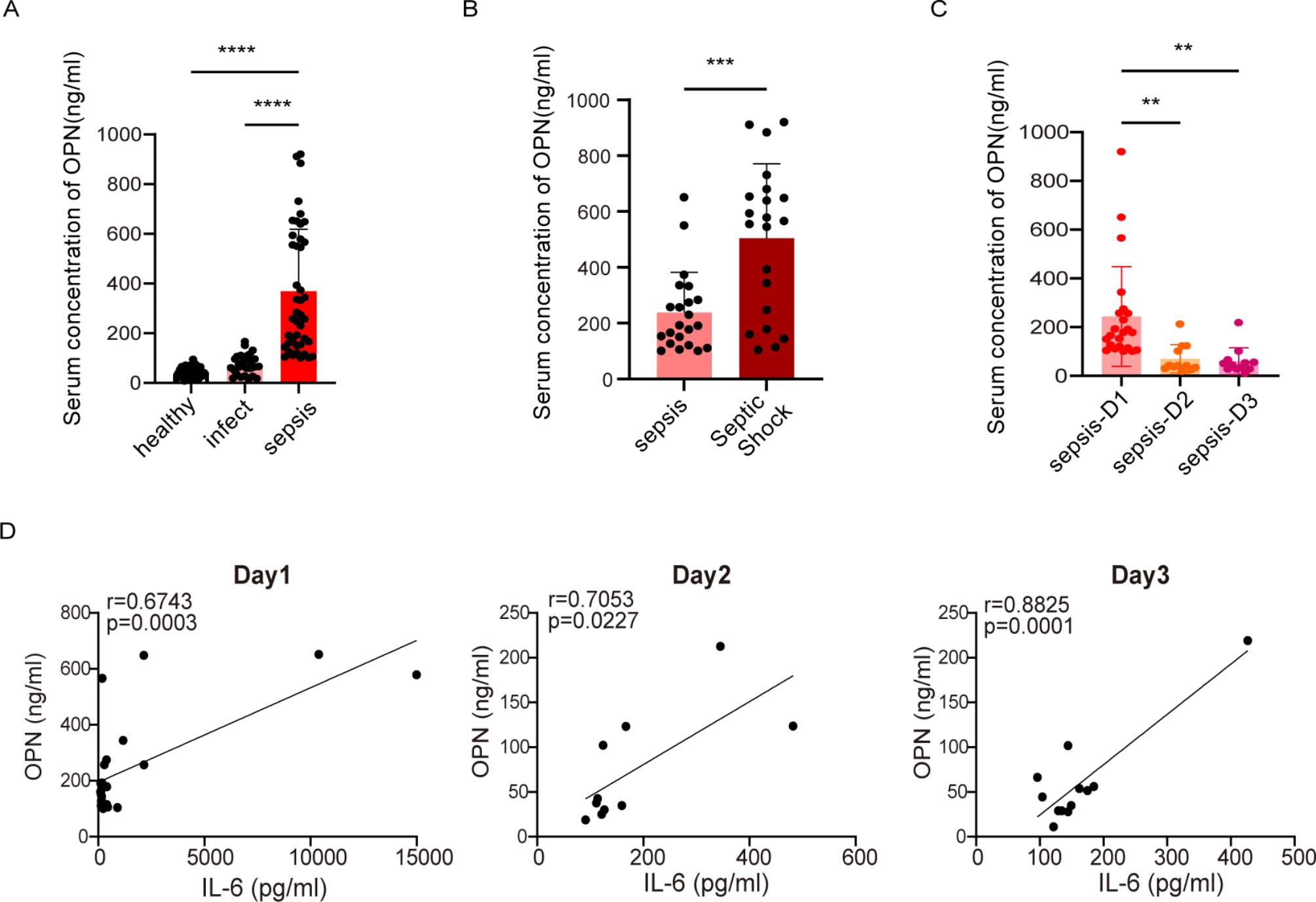
Serum samples were obtained from children fulfilling the inclusion criteria and hospitalized at the Children’s Hospital of Chongqing Medical University from January 2024 through December 2024. A total of 43 cases of sepsis were included, with the following criteria: a. Consistency with the 2024 International Consensus on Sepsis in Children [4]. During the same period, 28 cases of infected children were identified among children exhibiting signs of infection, yet these cases did not meet the diagnostic criteria for sepsis within the same age group at the same hospital. For the control group, consisting of 38 healthy children, the subjects were of the same age and were examined at the same hospital’s health examination center during a physical examination.
This study protocol has received approval from the Institutional Review Board of Children’s Hospital of Chongqing Medical University ( File No: (2021) Ethical Review Research No. 325-1). Informed consent has been obtained from all participants in accordance with the principles outlined in the Declaration of Helsinki.
Experimental animals
Male wild-type (WT) C57BL/6J mice, aged between 6 and 8 weeks, were procured from Chongqing Medical University. These mice were bred in a controlled environment characterized by a temperature range of 20–24 °C and a 12-hour light/dark cycle. They were provided with unrestricted access to standard food and water. All animal experiments were conducted in compliance with the regulations approved by the Chongqing Experimental Animal Center and the Animal Committee of Children’s Hospital (Approval No.: CHCMU-IACUC20231208004).
To create a model of polymicrobial sepsis, the CLP procedure was performed [13]. Mice were anesthetized using pentobarbital sodium at a dosage of 50 mg/kg. After sterilization, a 1-cm midline laparotomy was performed on the abdomen, the cecum was then ligated at 20% of its length and punctured with an 18-gauge needle, resulting in a slight extrusion of cecal contents. Subsequently, the cecum was repositioned into the abdominal cavity, and the incisions were sutured. Sham-operated animals underwent identical surgical procedures, except for the ligation and puncture of the cecum. After CLP surgery, the animals were resuscitated with an intraperitoneal injection of normal saline at a ratio of 1 ml per 20 g of body weight. Twenty-four hours post-surgery, the animals were humanely euthanized, after which serum and lung tissue samples were collected.
Cells experiments
MH-s cells (Procell, Wuhan, China) were cultured in RPMI1640 medium supplemented with 10% fetal bovine serum (FBS) and 1% penicillin-streptomycin, within a 37 °C, 5% CO2 incubator, and subcultured at intervals of 1 to 2 days. The cells were allocated into three distinct groups: negative control (NC) group, NC + LPS group, and OPN-siRNA + LPS (SiRNA + LPS) group. A concentration of 100nM of either NC or OPN-siRNA was complexed with an equal volume of transfection reagent for 10–15 min. Subsequently, these complexes were added to MH-s cells in RPMI1640 medium supplemented with 10% FBS and incubated for a period ranging from 24 to 72 h. Then, the medium was refreshed, and 100 ng/mL LPS was introduced to continue the culture for an additional 6 h, Ultimately, RNA or protein was extracted from the cells.
Inhibitor‑mediated blockade of OPN
To counteract the activity of OPN in CLP, mice in the CLP + OPN inhibitor (OI) group were administered 50 µg of an OPN inhibitor (MCE, OPN expression inhibitor 1, HY-146064, USA), which was dissolved in 100 µl of phosphate buffered saline (PBS). In contrast, a control group of mice received an equivalent volume of sterile PBS as a vehicle control.
In vitro administration of recombinant OPN
Prior to treatment, MH-s cells were subjected to OPN knockdown via OPN-siRNA transfection. The cells were then divided into two groups: the experimental group, which was treated with 200 ng/mL of recombinant mouse OPN (rmOPN) protein (MCE, Osteopontin, HY-P78358, USA) for 30 min, and the control group, which received no such treatment. Both groups were subsequently co-cultured with 100 ng/mL of LPS for 6 h. Ultimately, cell supernatant, RNA and protein were extracted from the cells.
Enzyme-linked immunosorbent assay (ELISA)
The concentrations of OPN in human samples were quantified by using commercial ELISA kits provided by FineTest (EH0248, China), and serum inflammatory factor IL-6 was quantified using Human IL-6 ELISA kit (Neobioscience, EHC007, China). Likewise, the levels of OPN (Jonin, JL10068, China) and various inflammatory cytokines in mice (with n = 5–8 per group) were evaluated. These cytokines encompassed tumor necrosis factor-alpha (TNF-α) (Neobioscience, EMC102a.96, China), interleukin (IL)-1β (Neobioscience, EMC001b.96, China), and IL-6 (Neobioscience, EMC004.96, China). Both serum and cell supernatant samples were analyzed with commercially available ELISA kits.
The dry-to-wet (D/W) ratio of lung tissue
The upper lobe of the right lung from the mice was excised. The blood on its surface was carefully absorbed using filter paper and then it was weighed. This weight was recorded as the wet weight (W). Subsequently, the tissue was placed in an oven set at 60 °C for a duration of 48 h. After that, it was weighed again, and the result was noted as the dry weight (D). Consequently, the dry-to-wet ratio of the lung tissue was calculated using the formula D/W.
Immunofluorescence detection
The paraffin sections of lung tissue were heated at 60 °C in an oven for 1 h. Subsequently, dewaxing and sodium citrate antigen retrieval were performed. Then, the sections were cooled and rinsed three times with 1× phosphate-buffered saline with tween-20 (PBST) for 5 min each. Incubation with goat serum was performed at room temperature for 30 min. After drying the slides, they were placed in a humid chamber with the primary antibody and incubated overnight at 4 °C. The primary antibodies employed were as follows: anti-Osteopontin (Abcam, ab283656, USA), NLRP3 Monoclonal antibody (Proteintech, 68102-1-Ig, China) and F4/80 Rat mAb (zenbio, 263101, China). On the next day, the slides were washed again with 1×PBST three times for 5 min each. Then, the secondary antibodies such as CoraLite 488-conjugated Goat Anti-Rabbit IgG (H+L), Fluorescein (FITC)-conjugated Goat Anti-Mouse IgG (H+L) (Proteintech, SA00003-1, China) and Rabbit anti-Rat IgG H&L (FITC) (zenbio, 550066, China) were then added and incubated at room temperature in the dark for 1 h. After washing three times with 1×PBST as before, a drop of 4′,6-diamidino-2-phenylindole(DAPI)-containing anti-fluorescence quenching mounting medium was applied to seal the slides, and images were captured using a fluorescence microscope.
Histopathology
Lung tissue was harvested, and hematoxylin and eosin (H&E) staining was utilized to evaluate pathological changes (n = 5). Fresh samples were rinsed with cold PBS and then fixed in 4% paraformaldehyde. Subsequently, the tissues were dehydrated, embedded in paraffin, sliced into 4 μm sections, and stained routinely. The pathology scores for the lung were determined based on the following aspects: alterations in lung histology, such as edema, congestion, interstitial inflammation, and inflammatory cell infiltration.
RNA extraction and quantitative real-time PCR
Total RNA was extracted from cells by employing the RNA Isolation Kit (Beyotime, R0027, China). Subsequently, 1 µg of RNA was reverse transcribed into cDNA using the ABScript III RT Master Mix for qPCR, which includes a gDNA remover kit (ABclonal, RK20429, China). Gene expression was analyzed through real-time quantitative PCR (qPCR) on a Bio-Rad CFX ConnectTM Real-Time System (Bio-Rad, USA), utilizing SYBR Green (ABclonal, Rk21203, China). The primer sequences are detailed in Table 1. The expression levels were quantified using the 2 − ΔΔCq method, with glyceraldehyde-3-phosphate dehydrogenase (GAPDH) serving as the internal control.
Table 1 q-PCR primer sequences. OPN osteopontin, TNF tumour necrosis factor, IL interleukin, NLRP3 NOD-, LRR- and pyrin domain-containing 3, GSDMD Gasdermin D, ASC apoptosis-associated speck-like protein containing a CARD, GAPDH glyceraldehyde-3-phosphate dehydrogenase, siRNA small interfering RNA, qPCR quantitative real-time reverse transcriptase-polymerase chain reaction, siRNA small interfering RNAWestern blotting analysis
Proteins were extracted from cells by using a radioimmunoprecipitation assay (RIPA) lysis buffer (MCE, HY-K1001, USA) supplemented with 1% phenylmethanesulfonylfluoride (PMSF) (MCE, HY-B0496, USA), 1% protease inhibitor (MCE, HY-K0010, USA), and 1% phosphatase inhibitors (MCE, HY-K0021, USA). The protein concentration was quantified using a NanoDrop spectrophotometer (Thermo Fisher). Subsequently, the proteins were separated by sodium dodecyl sulfate‒polyacrylamide (SDS‒PAGE) gel (EpiZyme Biotechnology, PG112, China) electrophoresis and then transferred onto a polyvinylidene fluoride (PVDF) membrane (Millipore, IPVH00010, USA). The membranes were blocked with NcmBlot blocking buffer (New Cell & Molecular Biotech, P30500, China) for 20 min and incubated with the appropriate primary antibodies overnight at 4 °C. The primary antibodies employed were as follows: anti-Osteopontin (Abcam, ab283656, USA), anti-IL-1β (Proteintech, 16806-1-AP, China), anti-IL-18 (Proteintech, 10663-1-AP, China), NLRP3 Rabbit mAb (Abclonal, A24294, China), GSDMD Polyclonal antibody (Proteintech, 20770-1-AP, China), CASP1 Rabbit pAb (Abclonal, A20470, China), ASC Rabbit mAb (Abclonal, A22046, China), anti-IL-6 (MCE, HY-P80723, USA), anti-TNF-α (Proteintech, 17590-1-AP, China) and beta Actin Rabbit mAb (zenbio, R23613, China). Thereafter, the membranes were incubated with goat anti-rabbit IgG, HRP-conjugated polyclonal antibody (CoWin Bio, CW0103S, China) and goat anti-mouse HRP-conjugated polyclonal antibody (Proteintech, 66009-1-Ig, China) as secondary antibodies at room temperature for 1 h, and the results were visualized using a Bio-Rad ChemiDoc™ Touch Imaging System (Bio-Rad, California, USA).
Statistical analysis
Statistical analyses were performed using GraphPad Prism 9 software. All data are presented as the mean ± standard deviation (SD) from at least three independents experiments. Group differences were evaluated by either a t-test (Mann-Whitney U test) or a one-way analysis of variance (Tukey’s multiple comparison test). A P value less than 0.05 was considered statistically significant, where “n” values indicate the number of cultures, tissue samples, or animals examined within each group.


留言 (0)