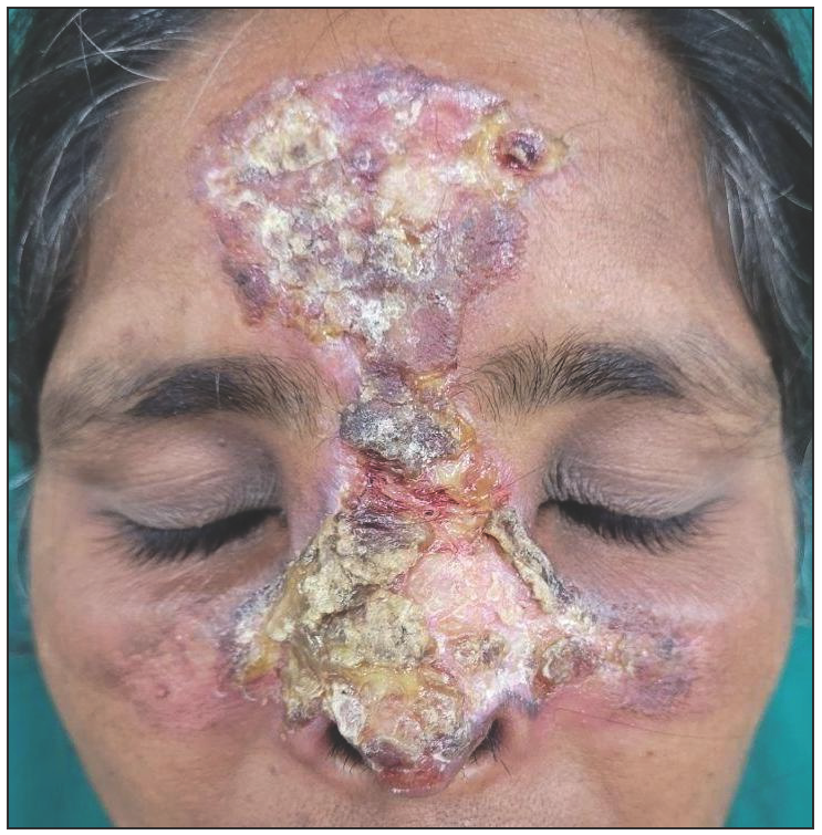
A woman in her forties presented with lesions over the face for the past 10 years. It initially presented as pea-sized lesions over the nose and gradually progressed in size. The delay in seeking medical advice was attributed to the superstitions. Examination revealed erythematous hyperkeratotic plaque over the forehead, nose, and cheeks with follicular plugging and adherent crusting [Figure 1]. Dermoscopy showed whitish scales, red globules, and white structureless areas. A clinical diagnosis of discoid lupus erythematosus was proven histopathologically that showed follicular plugging, dense lymphocytic infiltration, and vacuolar degeneration of basal keratinocytes. The patient improved following oral hydroxychloroquine, topical potent corticosteroids and photoprotection.

Export to PPT
留言 (0)