Animals
Male C57BL/6 (Medical Laboratory Animal Center, Guangzhou, China) mice (6–8 weeks old, 24–30 g) were housed in a temperature/humidity environment of 24 °C/50–60%, with normal ventilation and light/dark cycles (12 h), and fed and watered ad libitum. Animal experiments were ethically licensed by the Ethics Committee (Shengjing Hospital, China Medical University) and conducted in accordance with the guidelines.
Mouse CCI model establishment
Neuralgia pain model was simulated using CCI. CCI model was induced as described previously (Wang et al. 2020). In Brief, mice with anesthetized (0.8% pentobarbital sodium, ip), incisions were made to expose the left sciatic nerve, and loose ligations (1-mm spacing) were applied to the trunk nerves at the branches using ligature wires. The muscles of the ipsilateral hind limb were observed to show slight twitching. The sham-operated group was operated but not ligated.
AAV injection and experimental design
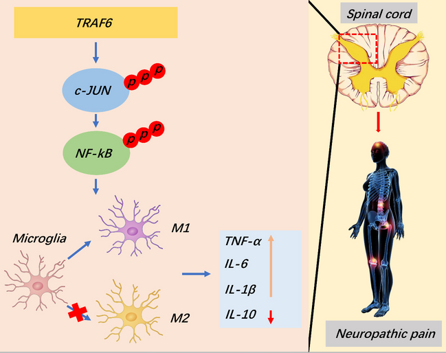
To verify the impact of TRAF6 on CCI, adeno-associated virus (AAV) was injected intrathecally using a glass micropipette (Hamilton). The dosages of AAV-shRNA or AAV-TRAF6 were chosen from previous reports (He et al. 2020). Specifically, AAV (10 μl) was injected 2 weeks before CCI. AAV-TRAF6 (5.0 × 10 12 vg/ml) vector and its control vector AAV-NC (5.0 × 10 12 vg/ml), AAV-shRNA-TRAF6 (1.5 × 10 12 vg/ml) and its control vector AAV-shRNA (1.6 × 10 12 vg/ml) were purchased from Viligo Biotechnology Co. Ltd (Hangzhou, China). c-JUN inhibitor SP600125 (15 mg/kg, i.p, MCE, USA) was injected on postoperative days 4–8. Thermal hyperalgesia and mechanical allodynia were performed using the CCI model (3, 5, 7, 14, and 21 days). To confirm the quality of transfection, we provided the results of AAV fluorescence identification (Figure S1).
Behavioral tests
The paw withdrawal threshold (PWT) was calculated using the thin filament stimulation method (Von Frey, 0.07 and 0.4 g, Stoelting Co.), as mentioned previously (He et al. 2020). For paw withdrawal latency (PWL), thermal testing was evaluated using IITC Inc./Life Science Instruments (Model 336, Science Instruments, Woodland Hills, CA). After behavioral tests, the spinal cord was saved for subsequent detection.
Cell culture and transfection
Ventilated BV2 microglia were cultured in DMEM medium (10% fetal bovine serum, 37 °C, 5% CO2). Lipopolysaccharide (LPS) (10, 100, 1000 ng/ml, Sigma-Aldrich) was then applied to BV2 cells for 24 h to imitate the activation of BV2 microglia.
In accordance with the instructions, H293T cells were transfected with AAV-shRNA, AAV-shNC, AAV-NC, or AAV-TRAF6 for 48 h. The TRAF6 or c-JUN recombinant AAV viral solution was incubated with BV-2 cells, and subsequent experiments were performed. BV-2 Cells (1 × 10⁵ cells/well) were transfected with 40 nM TRAF6, sh-TRAF6, sh-c-JUN, or negative shRNA. After 24 h, transfection efficiency was assessed using WB.
Flow cytometry
To study microglia polarization, spinal cord tissue was removed and cut up, prepared into single cell suspensions by digestion with 0.25% trypsin (Invitrogen), and incubated with CD86-FITC (555018, BD Biosciences, 1:200) and CD206-APC (550889, BD Biosciences, 1:200) for 30 min in the dark on ice. Analysis using flow cytometry (FACSVerse 8, BD) with visualization (FlowJo software version 7.6.1).
Quantitative real-time PCR
Total spinal cord RNA was extracted (Trizol, Thermofisher, USA), and then processed (SuperScript IV, Thermofisher, USA) for cDNA using a reverse transcription kit. Table S1 lists measurements, markers, and associated PCR primers employed in.this study.
Western blot
Total proteins(RIPA, Beyotime, China) were extracted for quantification, followed by electrophoresis on a gel (SDS-10% polyacrylamide). Subsequently transferred to PVDF membranes for containment (Tris, 3% skimmed milk, 1 h), primary antibodies were incubated overnight: anti-TRAF6 (A0973, ABclonal, 1:1000), anti-iNOS (ab178945, Abcam,1:1000), anti-Arg1 (ab96183, Abcam, 1:1000), anti-c-JUN (SC-74543, Santa Cruz, 1:10000), anti-NF-kB p65 (3034, Cell Signaling, 1:1000), anti-c-JUN (phosphor S70) (ab30620, Abcam, 1:1000), anti-NF-kB p65 (phosphor S536) (3033, Cell Signaling, 1:1000), anti-GAPDH (60008–1-Ig, Proteintech, 1:1000).
Washed in PBS, the secondary antibody (ab288151, Abcam, 1:3000) was incubated (37 °C, 1 h), followed by visualization and quantitative analysis.
Immunofluorescence assay
Fixed spinal cord tissues were paraffin-embedded, deparaffinized and repaired with antigens (boiled, 10 min), blocked in goat serum (room temperature, Solarbio,15 min), and incubated with primary antibodies (4 °C, 24 h): anti-TRAF6 (A0973, ABclonal, 1:1000), anti-iNOS (Abcam, ab178945, 1:500), anti-Arg1 (Abcam, ab96183, 1:500), anti-Iba-1 (Abcam, ab178846, 1:500), Neuronal Marker anti-NeuN (Abcam, ab177487, 1:1000), anti-GFAP (Proteintech, 16825–1-AP, 1:50). After PBS washing, the secondary antibody (Alexa Fluor 488/Alexa Fluor 546, Beyotime, China, 1:200) was reacted for 90 min, and observed under a fluorescence microscope.
ELISA assay
The IL-1β, TNF-α, IL-6, and IL-10 levels in the spinal cord were measured using Elisa kits (Beyotime). Quantification was performed using a microplate reader (Molecular Devices, USA, OD 450 nm). Samples concentrations were counted from the prepared standard curve.
Statistical analysis
Data were visualized and analyzed (means ± SD) using GraphPad Prism 7 (GraphPad Software, Inc.). Data were statically analyzed for differences using a t-test or one-way or two-way analysis of variance (ANOVA), with p < 0.05 or p < 0.01 considered significantly different.

留言 (0)