
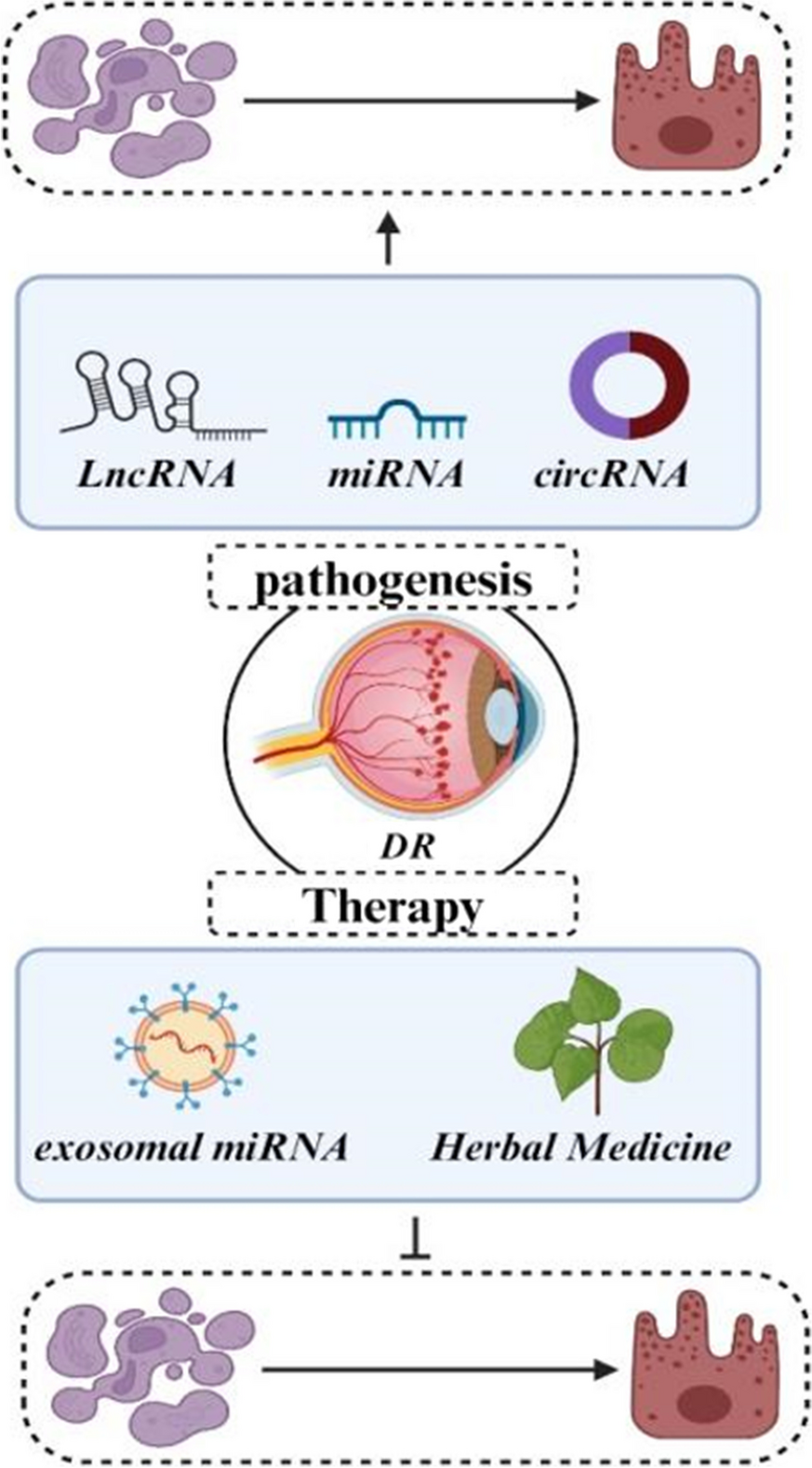
記住我
The PI3K/AKT/mTOR signaling cascade constitutes a pivotal pathway engaged in fundamental biological processes, including angiogenesis, apoptosis, proliferation, growth, and cellular metabolism (Karar and Maity 2011; Yu and Cui 2016). The PI3K/AKT signaling pathway entails a series of events, primarily involving the binding of extracellular factors to receptors, subsequent receptor activation leading to the phosphorylation of PI3K, PI3K-mediated phosphorylation of AKT, and the subsequent activation of downstream effector molecules. Following activation, PI3K facilitates PIP2 phosphorylation at the 3-position of the inositol ring, resulting in the production of PIP3 (Weernink et al. 2004). Subsequently, PIP3 functions as a docking site for two protein kinases, specifically AKT, also known as PDK1 (phosphoinositide-dependent protein kinase 1), and protein kinase B (PKB), which are recruited to the cellular membrane through their pleckstrin homology interaction domains (PH domains) (Cho and Park 2008; Powis et al. 2023). Upon being recruited to the cellular membrane, AKT is phosphorylated at Ser473 by mTORC2 (mTOR complex 2), which induces a structural alteration in AKT and facilitates its subsequent phosphorylation at Thr308 by PDK1 (Moore et al. 2011; Pullen et al. 1998). Upon being activated, AKT experiences a series of phosphorylation events on its target proteins. These initial phosphorylations take place at the cellular membrane, enabling the subsequent detachment of AKT. Subsequently, AKT relocates to the cytoplasm and nucleus, where it proceeds to phosphorylate further target proteins. Target proteins phosphorylation culminates in the activation of growth, cell survival, and proliferation (Chen et al. 2022). PI3K/AKT signaling pathway a critical influence on apoptosis within retinal cells, thereby potentially contributing to the pathogenesis of DR.. In this regard, Zeng et al. documented a concurrent decline in DJ-1 protein levels and a rise in the apoptotic activity of RRPs within the HG group. Their further investigation revealed that exposure to HG concentrations for a duration of two days led to noteworthy levels of apoptosis in RRPs. This cellular condition demonstrated increased levels of ROS, along with an increase in p-p53 and activation of caspase-3. Furthermore, there was evidence of mitochondrial impairment, coupled with reduced catalase (CAT) and manganese superoxide dismutase (MnSOD) activities. Furthermore, a decline in DJ-1 protein expression, along with diminished levels of its subsequent targets, phosphorylation state of AKT, and mTOR was evident. Conversely, elevation of DJ-1/PARK7 led to contrasting outcome. Additionally, it enhanced activities of MnSOD and CAT, thus improving mitochondrial functionality. Furthermore, the increased expression of DJ-1/PARK7 was linked to a decrease in the expression of genes involved in apoptosis, specifically p-p53 and activated caspase-3. This led to a decline in ROS production and a lower rate of apoptosis in renal proximal tubular cells exposed to HG levels. These findings posit a protective role for DJ-1 in shielding retinal pericytes from oxidative stress damage induced by HG exposure. Consequently, DJ-1 has the potential to augment mitochondrial function, mitigate ROS generation, and bolster antioxidant capacity, thereby mitigating apoptosis in retinal pericytes. This effect might be facilitated via the PI3K/AKT/mTOR signaling pathway, which possibility associated with the initial pathophysiological processes DR (Zeng et al. 2019).
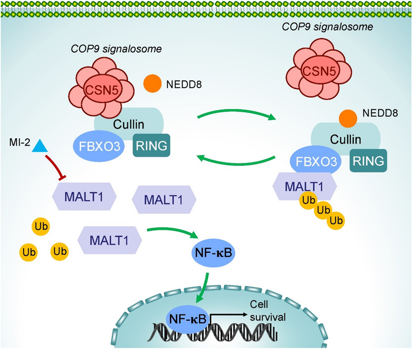
TLR4/NF-κB in Retinal cells apoptosisThe TLR4/NF-κB pathway is a pivotal inflammatory signaling cascade intricately associated with cellular apoptosis, proliferation, differentiation, and the activation of pro-inflammatory responses. Toll-like receptors (TLRs) constitute a category of receptors that are classified within transmembrane proteins known as pattern recognition receptors (PRRs) (Kawai and Akira 2010; Zhu et al. 2018a). Among mammals, PRRs are unique molecules capable of transmitting extracellular antigenic signals to cells, thereby initiating an inflammatory response. TLR4 holds the distinction of being the inaugural member identified within the Toll-like receptor family, and it functions in both immune regulation and immune defense (Zhang et al. 2022). When activated, TLR4 undergoes dimerization and initiates two primary signaling pathways: one dependent on myeloid differential factor 88 (MyD88) and the other on toll/interleukin 1 receptor domain-containing adaptor inducing interferon-beta (TRIF). These signaling cascades subsequently lead to the activation of downstream factors such as mitogen-activated protein kinases (MAPKs), and NF-κB, ultimately inducing a diverse array of pro-inflammatory genes, including cytokines and enzymes associated with inflammation (Yu et al. 2022; Fitzgerald et al. 2001; Piras and Selvarajoo 2014). Recent experimental findings suggests that the TLR4/NF-κB signaling pathway assumes a pivotal role in the regulation of apoptosis in retinal cells and contributes significantly to the progression of DR. In this context, according to recent experimental findings, HG elevated the expression levels of TLR4, and four downstream signaling molecules associated with TLR4 (NLRP3, TRAF6, NF-κB, and MyD88) and pro-inflammatory cytokines (IL-18 and IL-1β) in retinal ganglion cells (RGCs). Notably, HG trigger a conspicuous elevation in the apoptosis of RGCs. Furthermore, TAK-242 introduction, which functions as an antagonist of TLR4, effectively suppressed both inflammation and RGC apoptosis within the HG group. These findings unequivocally illustrated the pivotal involvement of TLR4 in the inflammatory response and apoptotic processes of RGCs triggered by HG (Hu et al. 2017). Furthermore, Zhai et al. revealed that Berberine (BBR) led to a reduction in the ganglion cell layer, mitigated cellular apoptosis, attenuated oxidative stress induced by diabetes, and suppressed the NF-κB signaling pathway in a DR rat model. They subsequently unveiled that HG heightened oxidative stress and prompted mitochondria-dependent apoptosis in Müller cells by activating the NF-κB signaling pathway. As well, BBR reversed the influences induced by HG by reducing IκB phosphorylation, restraining NF-κB nuclear translocation, and consequently suppressing the NF-κB signaling pathway. In this manner, BBR conferred protection against DR by suppressing oxidative stress and cellular apoptosis through NF-κB signaling pathway deactivation (Zhai et al. 2020).
SIRT1 in Retinal cells apoptosisSirtuins constitute a group of NAD+-dependent deacetylases that have maintained a high degree of conservation throughout the course of evolution. The sirtuin family, consisting of seven members, namely SIRT1-SIRT7, exhibits ubiquitous cellular distribution and is characterized by unique enzymatic properties, subcellular localization patterns, and physiological roles (Winnik et al. 2015). SIRT1 engages with protein substrates across diverse signaling pathways, including Wnt and Notch, and assumes a pivotal regulatory position in the control of numerous physiological functions within the body, prominently influencing processes such as metabolism, apoptosis, differentiation, and cell proliferation, thereby garnering significant interest from researchers across various fields of study. Recent experimental findings have revealed that agents promoting SIRT1 activation lead to reduced levels of apoptosis, inflammation, oxidative stress, and mitochondrial impairment, thereby conferring protection against DR (Karbasforooshan and Karimi 2018). In this regard, it was observed that lentiviral overexpression vector pLV5-Sirt increased cellular viability, and reduced apoptotic rate in RGCs, while pLV3-si-Sirt1 exert opposite effects. Also, pLV5-Sirt1 significantly reduce the expression levels of FOXO3a, p53, caspase-3, and NF-κB within RGCs, while pLV3-si-Sirt1 exert opposite effects. In this manner, Sirt1 has the capacity to suppress RGC apoptosis by modulating the expression of certain apoptotic cytokines, making it a potential candidate gene for therapeutic interventions in DR (Zhou et al. 2021) (Fig. 2).
Fig. 2
Integrative representation elucidating the pivotal signaling pathways orchestrating retinal cell apoptosis in the progression of DR. The diagram highlights key pathways, including SIRT1, TLR4/NF-κB, and PI3K/AKT/mTOR, and their intricate interplay in the regulation of apoptotic events within retinal cells
Apoptosis-related miRNA in DRmiRNAs constitute a class of short, ncRNA molecules, typically ranging from 19 to 25 nucleotides in length, that function as post-transcriptional regulators of gene expression via silencing mechanisms (Lu and Rothenberg 2018). Their biogenesis entails a two-step cleavage process wherein a progressively shorter hairpin structure is generated: the initial cleavage orchestrated by ribonuclease Drosha and Dgcr8, followed by a subsequent cleavage event led by Dicer alone, culminating in the formation of a miRNA duplex. The miRNA duplex is loaded onto an Argonaute protein, forming the basis of the miRNA-induced silencing complex (miRISC), designated as the passenger strand, undergoes extrusion. The miRISC associates with 3'-UTR of target mRNA sequences, subsequently initiating either mRNA degradation or translational inhibition (Cai et al. 2009; Liu et al. 2008). Recent experimental evidence has demonstrated that miRNAs significantly contribute to the development of DR by predominantly regulating apoptotic pathways. In this regard, the downregulation of miR-122 alleviated apoptosis triggered by HG in ARPE-19 cells. Additionally, HG resulted in downregulation of Bcl-2 and upregulation of cleaved caspase-3 in the cells. Interestingly, these effects were abrogated by a subsequent reduction in miR-122 expression. Furthermore, miR-122 predominantly functions by targeting Tissue inhibitor of metalloproteinases-3 (TIMP3). Also, simultaneous increase in TIMP3 expression counteracted the effect of elevated miR-122 levels on HG-induced apoptosis in ARPE-19 cells. In conclusion, miR-122 promoted apoptosis in ARPE-19 cells and accelerated the advancement of DR by suppressing TIMP3 (Wang et al. 2020a). In the subsequent section, we will discuss relevant literature concerning miRNAs function in apoptotic processes in DR.
MiR-200aThe miR-200 family comprises five members (miR-429, -141, and 200ca/b/c) which are arranged in two separate polycistronic primary miRNA transcripts, specifically miR-200c-141, and miR-200b-200a-429, located on human chromosomes 12 and 1, respectively (Fu et al. 2019). ARPE-19 cells subjected to HG demonstrated a notable decrease in the expression of miR-200a-3p, along with a simultaneous increase in transforming growth factor-β2 (TGF-β2) levels. This finding was similarly observed in the retinal tissues of rats with DR. ARPE-19 cells exposed to HG exhibited a significantly increased incidence of apoptosis relative to the control. Moreover, HG induced a marked upregulation of pro-apoptotic proteins, including Bax and Caspase-3. Conversely, a concomitant downregulation of the anti-apoptotic protein Bcl-2 was observed. Moreover, upregulation of miR-200a-3p resulted in a significant decrease in the apoptotic rate of ARPE-19 cells exposed to HG conditions. Mechanistically, increased miR-200a-3p levels were observed to attenuate apoptosis in ARPE-19 cells under HG conditions, mediated by the inhibition of the TGF-β2/Smad signaling pathway. In conclusion, increased expression of miR-200a-3p effectively suppress apoptosis in DR by blocking the TGF-β2/Smad pathway, indicating a promising therapeutic marker for the treatment of DR (Xue et al. 2020). Additionally, miR-200b display a substantial upregulation in the retinas of Akita mice. miR-200b directly regulates the expression of Oxidation resistance 1 (Oxr1). This regulatory interaction was demonstrated in a human Müller cell line (MIO-M1), where overexpression of miR-200b resulted in a decrease in Oxr1 expression levels. Also, upregulation of recombinant Oxr1 mitigated oxidative stress markers, specifically the nitration of cellular proteins, and also alleviated apoptosis triggered by 4-hydroxynonenal (4-HNE), which is an oxidative stress-inducing agent. Furthermore, miR-200b inhibitor reduced the number of apoptotic cells, while transfection with a miR-200b mimic increased the apoptotic cell count after exposure to 4-HNE treatment. Thereby, miR-200b, through the regulation of Oxr1 and its modulation of apoptosis, potentially plays a protective role in DR (Murray et al. 2013)..
MiR-423-5pMiR-423 is situated on chromosome 17 at the 17q11.2 locus (gene coding ID: 494335). Two mature miRNA sequences, specifically miR-423-5p and miR-423-3p, were discerned through an exploration of the miRBase sequence database (Ke et al. 2020). MiR-423-5p expression demonstrates a significant increase in RPE cells exposed to HG conditions, as well as in the plasma of DR patients. Furthermore, the upregulation of miR-423-5p worsens apoptosis induced by HG. Functionality, miR-423-5p directly targets TFF1, leading to the suppression of TFF1 expression, and that upregulating TFF1 mitigates apoptosis in RPE cells subjected to HG. Moreover, increased expression of TFF1 negates the pro-apoptotic effects induced by miR-423-5p in RPE cells exposed to HG. Hence, MiR-423-5p promotes apoptosis in RPE cells under HG conditions by inhibiting TFF1. Furthermore, reduction of TFF1 led to the activation of the NF-κB pathway and enhanced apoptosis induced by HG in RPE cells. Thereby, miR-423-5p modulate apoptosis induced by HG in RPE cells by suppressing the NF-κB signaling pathway mediated through TFF1. Importantly, NFE2, through miR-423-5p overexpression in RPE cells subjected to HG conditions, modulates the NF-κB signaling pathway mediated by TFF1. Therefore, a regulatory mechanism comprising NFE2, miR-423-5p, and NF-κB emerges as a significant determinant affecting apoptosis in RPE cells induced by HG (Xiao et al. 2019).
MiR-455-5pHuman miR-455 resides within the 9q32 locus on chromosome 9. Interestingly, the COL27A1 gene, encoding the collagen type XXVII alpha 1 chain, harbors the miR-455 sequence (Kumar and Reddy 2019). The involvement of miR-455 has been suggested in a range of human conditions, including DM. miR-455-5p markedly reduce in ARPE-19 cells subjected to HG. As well, enforced expression of miR-455-5p resulted in changes to the Bax/Bcl-2 ratio and cleaved caspase-3, leading to a notable increase in cellular viability and a reduction in apoptosis in HG conditions. Furthermore, elevation of miR-455-5p functioned as a negative regulator of intracellular ROS levels, decreased malondialdehyde (MDA) content, and decreased NADPH oxidase 4 expression, while enhanced the activities of GPX, catalase, and superoxide dismutase under HG conditions. So, miR-455-5p significantly attenuated oxidative stress injury induced by HG. Most importantly, miR-455-5p directly target suppressor of cytokine signaling 3 (SOCS3), and miR-455-5p exerts a negative regulatory influence on the expression of SOCS3. Moreover, SOCS3 restoration nullified the advantageous impacts of miR-455-5p on apoptosis and the buildup of ROS. Taken together, miR-455-5p mitigated HG-induced damage by inhibiting apoptosis and oxidative stress through the targeting of SOCS3, suggesting that miR-455-5p emerges as a promising candidate for future DR therapeutic development (Chen et al. 2019).
MiR-29a/bWithin the human genome, the miR-29 family comprises four members: miR-29b-1, miR-29b-2, miR-29a, and miR-29c. These miRNAs originate from two distinct primary transcripts: pri-miR-29a/b1 cluster located on chromosome 7q32.3 and pri-miR-29b2/c cluster situated on chromosome 1q32.2 (Horita et al. 2021). The miR-29 family is frequently associated with DM in various studies. In diabetic patients, miR-29a expression shows downregulation, while STAT3 and IL-6 levels exhibit upregulation in both the lens capsules and aqueous humor. As well, Overexpression of miR-29a combined with si-STAT3 transfection mitigated the increase in apoptosis and the disruption of MMP caused by HG. Notably, suppression of STAT3 resulted in an elevated Bcl-2/Bax ratio when compared to cells subjected to HG treatment alone. As well, it was observed that STAT3 upregulation countered the impacts of miR-29a transfection. Furthermore, by suppressing STAT3 signaling pathway, miR-29a alleviates the adverse impacts of HG on cell viability and mitochondrial dysfunction. Also, upregulation of miR-29a led to a partial decrease in ROS generation, elevated MDA levels, reduced SOD activity, and an upregulation of the Bcl-2/Bax ratio, which are linked to apoptotic pathways. These findings underscore miR-29a involvement in oxidative damage and apoptotic processes. In this manner, HG induces inflammatory responses and initiates mitochondrial dysfunction, thereby augmenting apoptosis via activation of the IL-6/STAT3 pathway (Li et al. 2021b). On the other hand, numerous studies have consistently shown that miR-29 functions as a proapoptotic factor in this particular condition. In this context, HG resulted in a concentration-dependent increase in apoptotic cell death within RPE cells, and miR-29 exhibited an upregulation in response to HG within RPE cells. Additionally, Downregulation of miR-29 expression mitigated HG-induced apoptosis in RPE cells, as evidenced by a reduction in caspase-7 protein generation. Also, miR-29 directly target PTEN, and downregulation of miR-29 increased PTEN expression. Thereby, miR-29 contributes to the apoptotic pathway triggered by HG in RPE cells. This effect is likely mediated by its inverse association with PTEN expression (Lin et al. 2016). Furthermore, Zeng et al. discovered that the miR-29b-3p expression exhibited a 3.2-fold increase, while the SIRT1 protein was reduced in patients with DR. In the HG-CoCl2 environment, they observed an elevation in miR-29b-3p and the Bax/Bcl-2 ratio, accompanied by a reduction in SIRT1 within HRMECs. They observed a decrease in the viability of HRMECs and a concomitant rise in apoptotic cell death when exposed to HG-CoCl2 treatment. Ultimately, they demonstrated that miR-29b directly target SIRT1, and that the Up-regulation of miR-29b-3p corresponded with a significant down-regulation of SIRT1 protein expression and a concomitant increase in the Bax/Bcl-2 ratio. Conversely, down-regulation of miR-29b-3p resulted in opposite effects. Furthermore, SRT1720, a SIRT1 agonist, mitigated the apoptosis of HRMECs induced by miR-29b-3p by enhancing the expression of SIRT1 protein. So, aberrant regulation of miR-29b-3p/SIRT1 axis is a crucial mechanism driving the apoptosis of HRMECs in DR (Zeng et al. 2020).
MiR-204The miR-204 resides within the sixth intron of the transient receptor potential melastatin 3 (TRPM3) gene.. MiR-204 assumes a pivotal role in both the developmental processes and functional aspects of the retina, with its dysregulation being implicated in the pathogenesis of various retinopathies (Bereimipour et al. 2021). Quantitative analysis of miR-204 expression in the retinal tissues of DR model rats revealed a significant downregulation in comparison to healthy controls. This observation suggests a dysregulation of miR-204 in DR pathogenesis, potentially contributing to the development of the disease. Moreover, Transfection with miR-204 mimics resulted in a marked upregulation of endogenous miR-204 expression. This, in turn, mediated a coordinated increase in the expression of Bcl-2 and SIRT1, while concurrently suppressing the production of pro-inflammatory mediators like TNF-α. Thus, miR-204 mitigates inflammation and cellular apoptosis in DR in rats via an increase in Bcl-2 and SIRT1 expression (Qi et al. 2020).
MiR-211MiR-211 is produced through the transcriptional process of Trpm1 gene. It is important to note that Trpm1 gene is located on chromosome 15q13-q14, a genomic region commonly associated with deletions found in various cancerous conditions. Furthermore, MiR-211 is situated specifically within the sixth intron of the Trpm1 gene (Yuan et al. 2022; Ye et al. 2022). MiR-211 may play a pivotal role in regulating the metabolic and catabolic processes of retinal cells, with its importance extending to the maintenance of adult visual functionality (Barbato et al. 2017). Diabetic cataract (DC) mice exhibited elevated levels of p53, Bax, and miR-211, alongside reduced levels of SIRT1 and Bcl-2. As well, miR-211 functions as a direct regulator of SIRT1 expression. Furthermore, upregulation of miR-211 trigger elevated expressions of p53, Bax, and miR-211 alongside reduced expressions of SIRT1 and Bcl-2. Notably, elevated expression of miR-211 led to decreased proliferation, and increased apoptosis of lens epithelial cells, while miR-211 depletion is associated with opposite effects. Therefore, miR-211 may function as a regulator of lens epithelial cell homeostasis in DC mice. miR-211 overexpression appears to promote apoptosis and inhibit proliferation, potentially through its interaction with SIRT1. These findings implies a potential therapeutic role for miR-211 in the treatment of DR (Fitzgerald et al. 2001).
MiR-365Zheng et al. explored miR-365 influence on retinal neuron apoptosis in rats with diabetes mellitus, primarily through its targeting of IGF-1. In their research, suppression of miR-365 through the administration of anti-miR-365 led to a reduction in apoptotic cells and Bax protein levels relative to control groups. Conversely, the groups treated with sh-IGF-1 and anti-miR-365 + sh-IGF-1 exhibited a contrasting pattern of results. Furthermore, the diabetes mellitus rat models displayed elevated levels of miR-365 and Bax expression, alongside reduced levels of Bcl-2 and IGF-1 expression. Additionally, in the diabetic rat models, a higher quantity of cells undergoing apoptosis was noted. Furthermore, they demonstrated that the sh-IGF-1 group exhibited decreased Bax expression, increased Bcl-2 and IGF-1 expressions, and a reduced number of apoptotic cells. Lastly, they observed that the anti-miR-365 + sh-IGF-1 groups displayed an upregulation of Bax expression, downregulation of IGF-1 and Bcl-2 expressions, and an increased count of apoptotic cells when compared to the anti-miR-365 group. Their findings indicate that the inhibition of miR-365 results in heightened expression of IGF-1, which subsequently exerts anti-apoptotic effects on retinal neurons in diabetic rats. These results underscore the potential of miR-365 as a viable therapeutic target in the context of DR (Zheng et al. 2018).
MiR-126MiR-126, predominantly known as miR-126-3p,is situated within intron 7 of the EGFL7 gene on chromosome 9 of the human genome (Nikolic et al. 2010). Dysregulation of miR-126 constitutes a key element in the etiology and development of DM and its associated complications. IL-17A increased, while miR-126 displayed upregulation in HRECs exposed to HG. Functionality, miR-126 upregulation stimulated cell proliferation and hindered apoptosis process in HRECs subjected to HG conditions. Interestingly, introduction of IL-17A abrogated the effects mediated by miR-126. Additionally, miR-126 exerted suppressive effects on the expression of caspase-3, Bax, and IL-17A, concurrently promoting survivin expression and inducing phosphorylation of both AKT and PI3K. Importantly, the reintroduction of IL-17A mitigated these effects. Notably, IL-17A function as a direct target of miR-126, and overexpression of miR-126 decreased IL-17A. In this manner, miR-126 enhances cell growth and prevents cellular apoptosis in HRECs exposed to HG by stimulating the PI3K-AKT pathway, elevating survivin levels, and reducing the levels of caspase-3 and Bax through the regulation of IL-17A (Chen et al. 2020a).
MiR-21The mature form of miR-21 displays complete conservation among mammals, similar to numerous other microRNAs, and is generated from a single gene. The human miR-21 gene has been thoroughly characterized and is situated on chromosome 17q23.2 (Olivieri et al. 2021). Recent investigations have supplied supporting evidence that the expression levels of plasma miR-21 can serve as a reliable marker to gauge the severity of T2D accompanied by DR (Jiang et al. 2017). Chen et al. via experimental investigation revealed a decreased level of retinal cell apoptosis in dKO mice when contrasted with control db/db mice. This observation implies that the deletion of miR-21 led to a reduction in diabetes-induced retinal cell apoptosis. They also revealed an upregulation of PPARα in miR-21 knockout (miR-21−/−) OIR retinas when compared to wild-type (WT) OIR retinas, thereby confirming the involvement of miR-21 in the downregulation of PPARα in retinal tissue subjected to ischemic conditions. Furthermore, a notable reduction in retinal cell apoptosis, as evidenced by reduced DNA fragmentation, was observed in miR-21−/− OIR retinas in comparison to WT OIR retinas. These results indicate that the miR-21 silencing mitigated the reduction of PPARα induced by ischemia and improved the condition of retinal apoptosis. In summary, downregulation of PPARα and the induction of apoptosis in DR are, to some extent, mediated by the heightened expression of miR-21 within the diabetic retina (Chen et al. 2017a).
MiR-146aThe miR-146a gene is situated on human chromosomes 19, 10, and 5, whereas in mice, it is located on chromosome 11. A substantial body of medical research has consistently identified dysregulation of miR-146a in individuals with T2D, underscoring the significant role of miR-146a in the development of T2D and its associated complications (Ghaffari et al. 2023). In human RECs exposed to HG conditions, upregulation of miR-146a expression led to diminished levels of IL-6, reduced STAT3 phosphorylation, and decreased VEGF. Additionally, it resulted in attenuated cellular apoptosis. More importantly, in RECs, miR-146a modulates STAT3/VEGF signaling and apoptosis via the IL-6 receptor signaling pathway. Importantly, miR-146a effectively inhibited IL-6 signaling, consequently resulting in diminished levels of VEGF and STAT3 in RECs under HG conditions, ultimately leading to a reduction in apoptosis. Thereby, miR-146a emerges as a potential therapeutic target in DR due to its capacity to suppress the IL-6-mediated STAT3/VEGF pathway, thereby mitigating both apoptosis and inflammation (Ye and Steinle 2017).
MiR-219-5pHG treatment induced a significant upregulation of miR-219-5p expression. Also, miR-219-5p was found to directly target the liver receptor homolog-1 (LRH-1), resulting in a significant decrease in LRH-1 levels in ARPE-19 cells under HG conditions. As well, inhibition of miR-219-5p significantly attenuated HG-induced apoptosis in ARPE-19 cells, accompanied by a concomitant increase in cell viability. Furthermore, miR-219-5p silencing activate LRH-1/Wnt/β-Catenin signaling pathway. In conclusion, involvement of miR-219-5p in DR pathogenesis is attributed to its impact on the apoptosis of human RPE cells via LRH-1/Wnt/β-Catenin signaling pathway (Zhao et al. 2018).
MiR-495MiR-495 represents a small non-coding RNA entity originating from a genetic locus situated on chromosome 14, specifically at the chromosomal region 14q32.31 (Chen et al. 2017b). Existing finding has suggested that miR-495-3p plays a crucial function in cancer progression (Liang et al. 2015). miR-495 substantially increased in RGCs subjected to HG treatment. MiR-495 reduction provided protection to RGCs against HG-induced apoptosis, while conversely, the miR-495 upregulation yielded the opposite effect. Notably, miR-495 serves as a direct regulator of Notch1, as miR-495 exhibited a negative regulatory effect on Notch1 expression and the associated Notch signaling pathway. Moreover, miR-495 reduction resulted in the suppression of PTEN expression and the activation of Akt. Importantly, the inhibition of miR-495 provides a protective effect against HG-induced apoptosis; however, this effect is effectively nullified upon Notch1 silencing. Overall, miR-495 reduction mitigates apoptosis in RGCs induced by HG through the regulation of the Notch1-mediated PTEN/Akt signaling pathway (Zhang et al. 2018).
MiR-383miR-383 consists of two unique mature variants, namely miR-383-3p and miR-383-5p. The miR-383 gene is situated on chromosome 8p22, specifically positioned within the third intron of the sarcoglycan zeta (SGCZ) gene (Yi et al. 2022). miR-383 levels increase significantly in ARPE-19 human RPE cell lines in response to HG exposure. Additionally, overexpression of miR-383 by altering the expression of Bcl-2 and Bax led to diminished cell viability, enhanced apoptosis, and increased ROS production in ARPE-19 cells. Importantly, miR-383 directly target peroxiredoxin 3 (PRDX3), and downregulated its expression in ARPE-19 cells. Furthermore, reintroduction of PRDX3 counteracted the miR-383-induced increase in ROS production and apoptosis. Conversely, the suppression of PRDX3 mimicked the miR-383 deleterious effects on ARPE-19 cells. In summary, HG-induced upregulation of miR-383 mechanistically contributes to apoptosis in RPE cells, likely by repressing PRDX3 expression (Jiang et al. 2017).
MiR-218MiR-218 is a type of intronic miRNA that is uniquely present in vertebrates. This gene exhibits co-expression with its host genes, the tumor suppressor genes SLIT2 and SLIT3. miR-218 arises from two discrete genomic loci: miR-218–1 and miR-218–2. These genetic loci have been assigned to chromosomal regions 4p15.31 and 5q35.1 (Lu et al. 2015). Elevated glucose levels caused a rise in miR-218 expression, which subsequently inhibited cell proliferation and triggered apoptosis in ARPE-19 cells. Furthermore, miR-218 overexpression inhibited ARPE-19 cell proliferation and promoted apoptosis, while miR-218 depletion led to contrary outcomes. Mechanistically, Runx2 acted as a target of miR-218 to assist in restraining cell proliferation and promoting apoptosis in ARPE-19 cells. Therefore, miR-218 promotes apoptosis in RPEs through direct targeting of RUNX2, suggesting the miR-218/RUNX2 regulatory axis as a promising target for DR (Yao et al. 2019).
MiR-542-5pMiR-542-5p, situated in a genomic region on chromosome Xq, possesses the capability to suppress hyperlipidemia and hyperglycemia (Tian et al. 2020). MiR-542-5p display a downregulation in individuals with DR and in retinal pigment epithelial (RPE) cells treated with HG. Upregulation of miR-542-5p effectively curbed apoptosis in RPE cells subjected to HG conditions. Furthermore, miR-542-5p was found to directly interact with co-activator-associated arginine methyltransferase 1 (CARM1). Overexpression of miR-542-5p caused a decrease in CARM1 expression levels, whereas suppression of miR-542-5p led to an elevation in CARM1 levels. Importantly, upregulation of CARM1 promoted apoptosis mediated by miR-542-5p in RPE cells exposed to HG conditions. Thus, miR-542-5p's direct targeting of CARM1 suggests a protective role for miR-542-5p in RPE cells, opening new avenues for therapeutic interventions in DR (Guo et al. 2020).
MiR-133bThe miR-206/miR-133b cluster is remarkably preserved in the muscle tissues of various organisms, including flies, mice, and humans, and it is situated on chromosome 6p12.2 (Nohata et al. 2012). Yao et al. noted that texposure to HG or miR-133b silencing result in a decrease in apoptosis in hRECs. Furthermore, HG resulted in an increase in the mRNA and protein levels of LIMK, ROCK1, RhoA, and the p-MLC protein in hRECs. On contrary, elevated expression of miR-133b suppressed cellular proliferation, enhanced apoptosis, and led to a downregulation of both mRNA and protein quantities of RhoA, ROCK1, LIMK, and p-MLC in hRECs subjected to HG conditions. Thereby, heightened expression of miR-133b hindered cell proliferation and stimulated apoptosis in a DR cell model through the reduction of RhoA expression (Yao et al. 2018).
MiR-499-3pLiu et al. demonstrated a decrease in IFNA2 levels and an increase in miR-499-3p expression in retinal tissues and cells of diabetic rats. This molecular alteration resulted in the activation of the TLR4 signaling pathway. They ascertained that miR-499-3p function as a direct regulator of IFNA2, and exerting an inhibitory effect on its expression. Also, miR-499-3p may function as a potential regulator of the TLR4 signaling pathway by targeting IFNA2. Their further experiments revealed that the reduction in miR-499-3p levels facilitated the proliferation of retinal cells and concurrently inhibited apoptosis, thereby mitigating the effects of DR. In conclusion, miR-499-3p facilitated the progression of retinopathy by activating TLR4 signaling pathway and promoting apoptosis, underscoring its potential for exploration as a therapeutic target for further research in DR (Liu et al. 2020).
MiR-34aResiding on chromosome 1p36, miR-34a) has been implicated in the pathophysiology of DM (Mone et al. 2022). MiR-34a upregulated, while SIRT1 downregulated in retinal tissues of DR-afflicted rats and in HG-induced RVECs. Suppression of miR-34a had a beneficial impact on DR, exerting its influence by modulating apoptosis and the expression of VEGF in DR rats. As well, miR-34a directly target SIRT1, and overexpression of miR-34a suppressed proliferation and induced apoptosis in RVECs, and these effects successfully reversed by increasing SIRT1 expression. Therefore, miR-34a drives apoptosis in RVECs by modulating SIRT1 in DR rats, underscoring the potential significance of the miR-34a/SIRT1 axis as a potential avenue for DR treatment (Ji et al.
留言 (0)