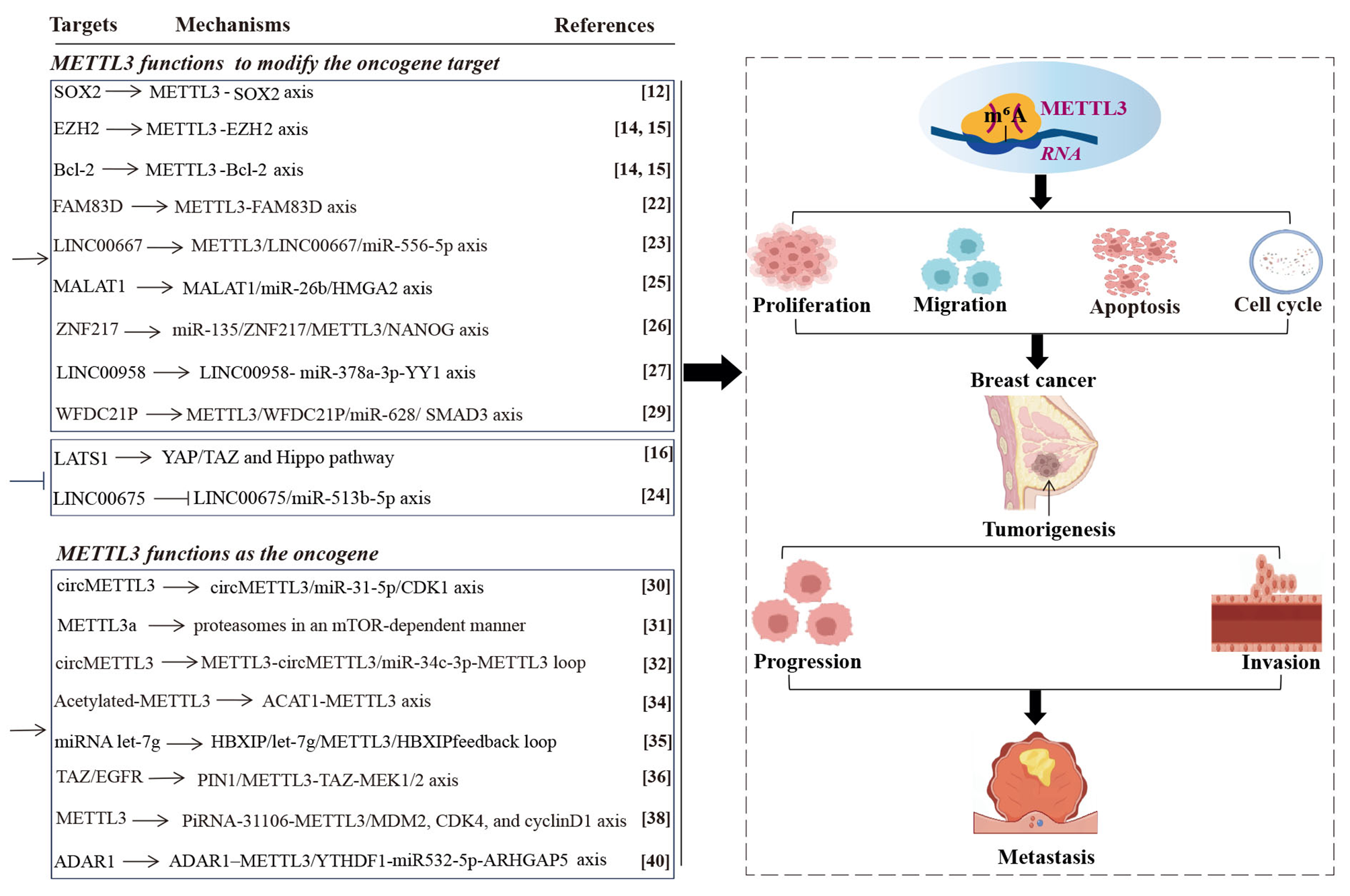
Materials
Pyrotinib was ordered from Jiangsu Hengrui Pharmaceutical Co., Ltd (Jiangsu, China) with > 99% purity. Herceptin was purchased from Roche Pharmaceuticals Co., Ltd. (Shanghai, China). N-hydroxysuccinimide (NHS), 1-ethyl-3-[3-dimethylaminopropyl] carbodiimide hydrochloride (EDC), dimethyl sulfoxide (DMSO), sodium dodecyl sulfate (SDS), 98% soy phosphatidylcholine, 99% cholesterol, chloroform, ethanol, methanol, and paraformaldehyde were purchased from Aladdin Chemical Reagent Co., Ltd. (Shanghai, China). Thiazolyl blue tetrazolium bromide (MTT), 7-AAD, Annexin V-FITC apoptosis detection kit, trypsin, bovine serum albumin, and Triton X-100 were supplied by Nanjing Jiancheng Bioengineering Institute (Nanjing, China). SK-BR-3 cell lines were obtained from Keygen Biotech (Jiangsu, China). McCoy's 5A medium and fetal bovine serum (FBS) were purchased from Beijing Dingguo Changsheng Biotech Co., Ltd. (Beijing, China). Phenylmethanesulfonyl fluoride (PMSF), a mitochondrial membrane potential assay kit (JC-1), and calcein-AM/PI were provided by Beyotime Biotechnology Co., Ltd. (Shanghai, China). Anti-Bcl-2, anti-Bax, and anti-Caspase-3 antibodies were obtained from Abcam (Cambridge, UK). N-cadherin, galectin-3, EpCAM, ErbB-2, and EGFR were purchased from Wanleibio (Liaoning, China).
Preparation of Ptb-M-Lip-HerSK-BR-3 cell membrane derivation
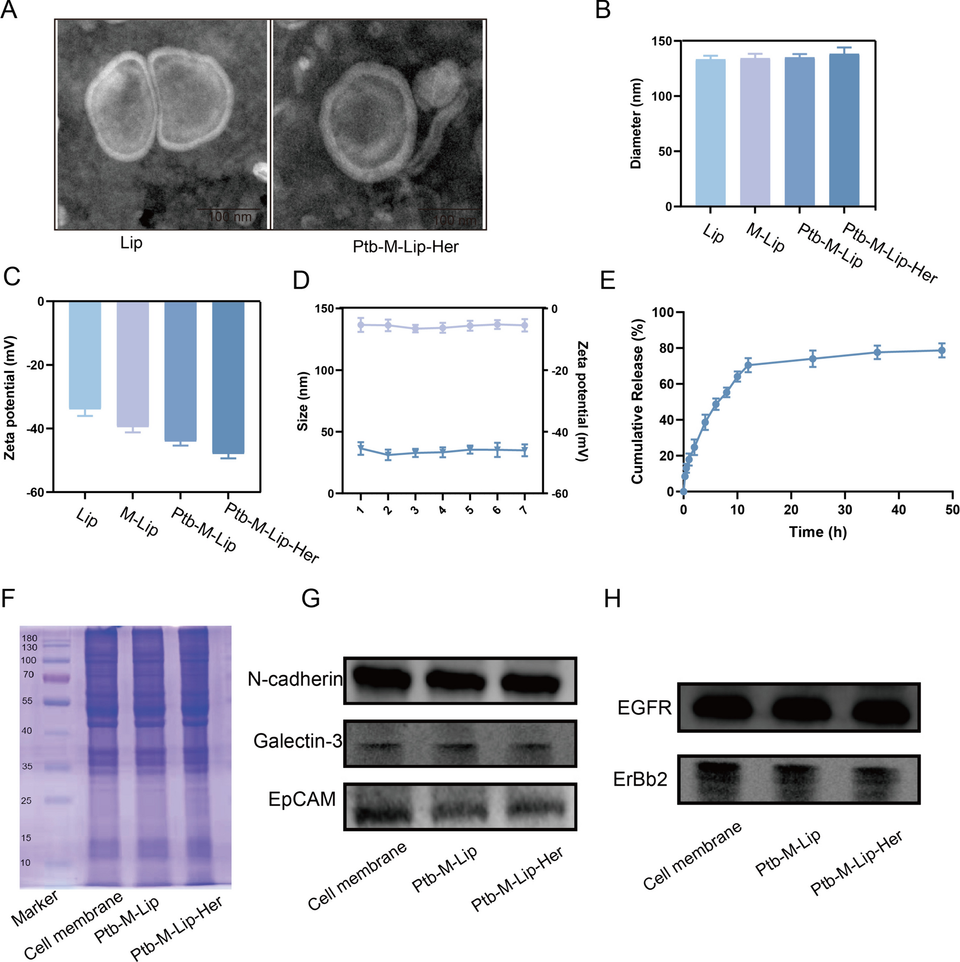
SK-BR-3 cells were cultured in McCoy's 5A medium containing 10% FBS (37 °C, 5% carbon dioxide). When SK-BR-3 cells were overgrown, they were digested with trypsin and collected by centrifugation at 1000 rpm/min. The cells were washed three times with precooled PBS and then treated with 0.2% PBS for 24 h. The cell suspension was centrifuged at 1250 rpm for 15 min, and the supernatant was discarded. NaHCO3 (1 mM), EDTA (0.2 mM) and PMSF (100 mM) were added to the precipitate, and the cell suspension was transferred to a homogenizer and ground 30 times. The supernatant was collected after the suspension was centrifuged at 1250 rpm for 30 min. The supernatant from both collections was centrifuged at 12,000 rpm for 20 min, and the supernatant was discarded. The remaining precipitate was the SK-BR-3 cell membrane, which was stored at − 20 °C for further use.
Preparation of Ptb-M-Lip-Her and related particles
NHS and EDC (5 mg/mL) were added to the cell membrane to activate carboxyl groups on the cell membrane. Then, 1 mg of Herceptin was dissolved in 1 mL of PBS and mixed with the cell membrane after activation of the carboxyl group. The mixture was incubated at room temperature for 6 h to obtain M-Her. Then, the sample was centrifuged at 12,000×g for 30 min, and the supernatant was collected. The content of Herceptin in the supernatant was determined with a BCA kit. The grafting amount of Herceptin was calculated using the following formula.
Pac is the amount of connected HCT (mg), Pi is the initial amount of HCT (mg), and Pnc is the amount of nonconnected HCT (mg) in the supernatant.
Soy phosphatidylcholine and cholesterol were added to the round-bottomed flask at a mass ratio of 3:1, and chloroform was added to dissolve it fully [36]. Then, in a 37 °C constant temperature water bath, the chloroform was evaporated by rotary evaporation, and finally, the lipid membrane was formed. The lipid membrane was hydrated for 30 min with a saline solution containing Herceptin-modified cell membrane and pyrotinib for 30 min to preliminarily mix with lipids. The system was then ultrasonicated in an ice bath for 5 min at 10-s intervals and incubated at 37 °C for 30 min to achieve complete fusion of the cell membrane and lipid. Finally, the whole nanoparticle was successively extruded by the liposome extruder at 0.4 μm, 0.2 μm and 0.1 μm. Then, Ptb-M-Lip-Her was isolated by a dextran gel column. To prepare the drug-loaded liposomes without biomimetics modification (Ptb-Lip), the normal saline solution did not contain the SK-BR-3 cell membrane but only contained Ptb. To prepare FITC-labeled liposomes, the normal saline solution contained Ptb, SK-BR-3 cell membrane and FITC. The next steps in the preparation process were the same as those described above.
Determination of drug loading
The prepared Ptb-M-Lip-Her was ultrasonicated with an ultrasonic probe at 200 W for 5 min at intervals of 10 s and then centrifuged at a rate of 80,000 r/min for 20 min. The supernatant was filtered through a 0.22 μm filter membrane, and the absorbance of Ptb was determined by ultraviolet spectrophotometry (UV-2000, languages, Franksville, WI).
$$}\,}\% = (}\,}/}\,}\,}) \times 100\%$$
CharacterizationMorphology and characterization of nanomaterials
The morphologies of Lip and Ptb-M-Lip-Her were observed by transmission electron microscopy (TEM, Jem-1400 Flash, Japan). The particle size, zeta potential and stability of Lip, Ptb-M-Lip and Ptb-M-Lip-Her were measured using a laser diffraction particle size analyzer (Nano-ZS90, Malvern, Malvern, UK).
Membrane protein characterization
The membrane protein of the SK-BR-3 cell membrane was characterized by sodium dodecyl sulfate-polyacrylamide gel electrophoresis (SDS-PAGE), and the successful encapsulation of the SK-BR-3 cell membrane was verified. To put it simply, SK-BR-3 cell membrane, Ptb-M-Lip and Ptb-M-Lip were lysed by radioimmunoprecipitation assay (RIPA) lysis buffer (Dingguo, China). A bicinchoninic acid protein assay (BCA) kit was then used to assay the protein concentration. The sample was mixed with SDS-PAGE sample loading buffer (Dingguo, China) and heated at 100 °C for 5 min. Samples with the same amount of protein (10 μL/well) were then loaded on an 8% SDS-PAGE gel and electrophoresed for 2 h under constant pressure. The obtained gels were stained with Komas blue for 2 h and analyzed with Quantity One 1-D analysis software (Bio-Rad, Hercules, USA). Western blot analysis of SK-BR-3 cell membrane, Ptb-M-Lip and Ptb-M-Lip-Her surface proteins (ErbB-2, EGFR, EpCAM, N-cadherin, and Galectin-3) further verified the successful encapsulation of SK-BR-3 cell membrane.
In vitro drug release study
The release properties of Ptb-M-Lip-Her in vitro were investigated using phosphate buffer solution as the release medium. Ptb-M-Lip-Her was placed in a dialysis bag (MWCO 3500). At the predetermined time, 3 mL of solution was taken and then injected into the same volume of PBS. The absorbance of Ptb was measured at 260 nm by an ultraviolet spectrophotometer (UV-2000, languages, Franksville, WI), and the cumulative release was calculated. The experiment was repeated three times, and the results were averaged and expressed as the standard deviation (± SD).
In vitro cell assayCell culture
SK-BR-3 cells were cultured in McCoy's 5A medium containing 10% FBS and 1% penicillin-streptomycin in a 37 °C incubator with 5% CO2 under saturated humidity. SK-BR-3 cells were digested with 2.5% trypsin at passage. In addition, FBS supplemented with 10% DMSO was used to preserve cells at − 80 °C.
Cell uptake
SK-BR-3 cells were inoculated in a confocal dish and cultured for 12 h in an incubator containing 5% CO2 and saturated humidity at 37 °C. Then, the medium was removed, and FITC-labeled Ptb-Lip, Ptb-M-Lip and Ptb-M-lip-Her were added. The cells were incubated at 37 °C for 2 h and washed three times with PBS. The cells were then fixed with 4% paraformaldehyde for 15 min, permeabilized with 0.1% Triton for 10 min, and sealed with 1% BSA at 37 °C for 30 min. Finally, the cytoskeleton and nucleus were stained with phalloidin and Hoechst 33,342 at 37 °C, respectively, and cell uptake was observed by confocal laser scanning microscope (CLSM, BioTek Instruments, Winooski, VT).
Flow cytometry was used to assess cell uptake. SK-BR-3 cells were digested and inoculated in 6-well plates for 24 h. Then, the medium was removed, and FITC-labeled Ptb-Lip, Ptb-M-Lip and Ptb-M-Lip-Her were added for incubation for 2 h. Cell uptake was measured by flow cytometry (Agilent Biosciences Inc., Santa Clara, CA).
Homotypic targeting
To verify the homologous targeting of Ptb-M-Lip, we evaluated the uptake of Ptb-M-Lip in different cancer cells. SK-BR-3, A549, and C6 cells were seeded in confocal dishes at a density of 5 × 10³ and then incubated with Ptb-M-Lip for 2 h for cellular uptake as described above. Finally, the uptake of Ptb-M-Lip by different cancer cells was observed by CLSM.
Cytotoxicity analysis
To evaluate the toxicity of Ptb, Ptb-M-Lip and Ptb-M-Lip-Her on SK-BR-3 cells, an MTT assay was used to detect the inhibitory effect of Ptb on SK-BR-3 cell proliferation. After the cells were digested, cell counting plates were used to count the cells, and according to the counting results, the cell suspensions were diluted into cell suspensions with a concentration of 5 × 10⁴ cells/mL by medium. Cell suspensions (100 μL) were inoculated into 96-well plates and cultured for 24 h. After cell adhesion, Ptb, Ptb-M-Lip and Ptb-M-Lip-Her were diluted to different concentrations (equivalent concentrations of Ptb were 0.2, 0.4, 0.8, 1, 4, and 8 μg/mL), added to 96-well plates and incubated for 48 h. MTT (5 mg/mL) solution was added away from the light and incubated in the dark for 4 h. Then, the supernatant in the 96-well plate was discarded, and 100 μL DMSO was added and shaken in the dark at a low speed for 15 min. The optical density (OD) was measured and recorded at 492 nm using a microplate reader (VERSA max, Molecular Devices, Sunnyvale, CA) after the formazan was completely dissolved. The following formula was used to calculate cell viability:
$$}\,} = }_}} /}_}} \times 100\%$$
Live and dead cell staining
SK-BR-3 cells were inoculated into 24-well plates and cultured for 24 h. Ptb, Ptb-M-Lip and Ptb-M-Lip-Her (equivalent to 0.8 μg/mL Ptb) were added for 24 h. Calcein-AM/PI detection working solution (500 μL) was added to each well. Then, the cells were incubated in 37 °C darkness for 40 min, rinsed with PBS and observed under a fluorescence microscope (Leica, Wetzlar, Germany).
Flow cytometric detection of apoptosis
SK-BR-3 cells were digested and inoculated into 6-well plates at 1 × 10⁵ cells/well for 24 h. Then, Ptb, Ptb-M-Lip and Ptb-M-Lip-Her (equivalent to 0.8 µg/mL Ptb) were added to the 6-well plate for 48 h. The cells were digested with trypsin and then washed with PBS 3 times. The cells were resuspended in 500 μL binding buffer, added to 5 μL Annexin V-FITC and 7-AAD, and incubated for 15 min away from light. Finally, apoptosis was detected by flow cytometry (Agilent Biosciences Inc., Santa Clara, CA).
Detection of mitochondrial membrane potential
SK-BR-3 cells were inoculated into 12-well plates and cultured for 24 h. Then, Ptb, Ptb-M-Lip and Ptb-M-Lip-Her (equivalent to 0.8 µg/mL Ptb) were added for 24 h. JC-1 staining solution was added and incubated at 37 °C for 30 min in the dark. Finally, SK-BR-3 cells were observed with a fluorescence microscope (Leica, Wetzlar, Germany).
Immunofluorescence to detect apoptosis
SK-BR-3 cells were inoculated into 24-well plates and cultured for 24 h. Then, Ptb, Ptb-M-Lip and Ptb-M-Lip-Her (equivalent to 0.8 µg/mL Ptb) were added for 24 h. The cells were washed with PBS 3 times, fixed with 4% paraformaldehyde for 30 min, and drilled with 0.5% Triton for 15 min. The cells were then sealed with goat serum for 2 h and incubated at 4 °C overnight with anti-Caspase-3. The next day, the cells were incubated with the secondary antibody at room temperature for 2 h, and Hoechst 33,342 and phalloidin were added for 15 min. Finally, SK-BR-3 cells were observed under a fluorescence microscope (Leica, Wetzlar, Germany).
Western blot
Western blotting was used to detect the expression level of apoptotic proteins in SK-BR-3 cells. SK-BR-3 cells were first cultured in a culture flask, and then Ptb, Ptb-M-Lip and Ptb-M-Lip-Her (equivalent to 0.8 µg/mL Ptb) were added for 48 h. The cells were gently scraped from the flask over ice. Cells were collected and washed with PBS 3 times. RIPA lysis buffer was added to each sample and incubated on ice for 30 min. After centrifugation at 4 °C and 12,000 rpm for 30 min, the protein concentration in each sample was determined using a BCA protein assay kit. For western blot analysis, the protein (10 μL) in each sample was electrophoretically separated on a polyacrylamide gel and then transferred to a polyvinylidene fluoride (PVDF) film. The PVDF membrane was incubated in 1% BSA for 2 h and then incubated with primary antibodies (anti-Bcl-2, anti-Bax, and anti-Caspase-3) overnight at 4 °C. The next day, the secondary antibody was added and incubated at room temperature for 2 h. The membrane was treated with ECL chromogenic agent. Expression analysis was performed by Quantity One 1-D analysis software (Bio-Rad, Hercules, USA).
In vivo experimentEstablishment of the mouse tumor model
BALB/c nude mice (females, 18–20 g) were purchased from Beijing Weitonglihua Experimental Animal Technology Co., Ltd. (Beijing, China). All mice were housed in the SPF Laboratory Animal Center of Jinzhou Medical University. The experiment was conducted in accordance with the Animal Management Regulations of Jinzhou Medical College. SK-BR-3 cells (5 × 10⁶) were injected into the adipose pad of the right mammary gland of nude mice. One week later, there was a mass in the fat pad of the right breast, indicating the successful establishment of the tumor model.
In vivo antitumor effect and safety
When the tumor volume reached 100 mm3, the nude mice were randomly divided into four groups: the control group, Ptb group, Ptb-M-Lip group and Ptb-M-Lip-Her group. Different preparations (equivalent to Ptb 10 mg/kg) were injected into the caudal vein, and the control group was given normal saline. The drug was administered every three days for a total of 7 doses. We measured the longest and shortest tumor diameters and mouse body weight before each administration. The tumor volume was calculated as follows:
$$}\,}\,} = \left( }\,}} \right) \times \left( }\,}} \right)^ /2.$$
The tumor inhibition rate was obtained according to the following formula:
$$}\,}\,} = (1 - }_}} /}_}} ) \times 100\%$$
where Wt is the mean weight of the tumor for each drug treatment group and Wc is the mean weight of the tumor for the control group.
The mice were killed after the last administration, and the tumor tissue and major organs (heart, liver, spleen, lung, and kidney) were removed. The tissues were fixed with 4% paraformaldehyde for 24 h, dehydrated and embedded in paraffin. Sections were stained with hematoxylin and eosin (H&E) and Ki-67 immunohistochemical staining. The sections were observed with a fluorescence microscope (Leica DMI 4000B, Wetzlar, Germany).
In vivo imaging
To further verify the targeting ability of Ptb-M-Lip-Her, FITC-labeled Ptb, Ptb-M-Lip, and Ptb-M-Lip-Her were injected into the tail vein of tumor-bearing nude mice. The mice were killed 3 h later, and the heart, liver, spleen, lung, kidney and tumor were removed. Fluorescence signal intensity in tumors and major organs was observed at 518 nm and 494 nm using an in vivo imaging system (IVIS Spectrum, PerkinElmer, Waltham, MA).
Statistical analysis
The statistical analysis was performed using GraphPad Prism (version 8.0), and the results of all experiments were reported as the mean ± SD. When the P value was less than 0.05, it was considered statistically significant.

留言 (0)