Keys findings
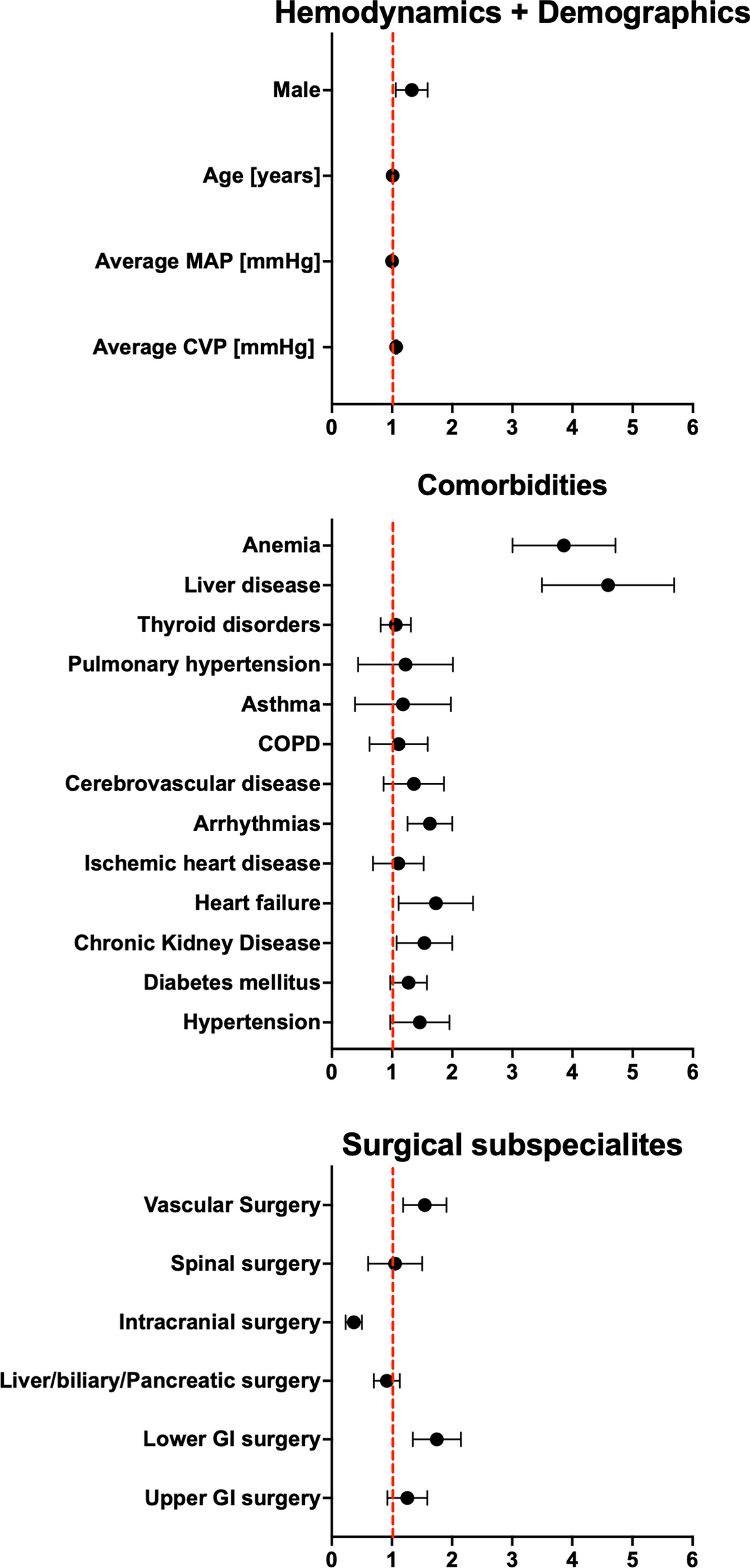
In our study, using a fully resuscitated large animal model, we demonstrated that inhibition of DPP3 with PCZ was associated with 1/ Decreased catecholamine and fluid requirements with comparable organ perfusion, as assessed by an isotopic method, 2/ Lower vascular and myocardial inflammation, 3/ Lower myocardial injury and 4/ Better pulmonary function, as reflected by higher PaO2/FiO2 ratio.
In the PCZ group, the reduction in catecholamine requirement was associated with an increase in Ang II concentration and prevention of the increase in Ang I/Ang II ratio seen in the septic control animals, as assessed by a mass spectrometry method. There was also improved Ang II signaling at the tissue level, with higher AT1 protein expression in the left ventricle. Interleukin-6 expression levels were reduced in the vascular and myocardial samples.
Interpretation of the data and implications of the study findings
The reduction in catecholamine requirements was associated with an increase in the RAS activity, as shown by increased concentration of Ang II and a preserved Ang I/Ang II ratio. Additionally, there was an increase in Ang III, a metabolite of Ang II that can activate AT1 and hence increase blood pressure [24]. These findings suggest that PCZ may prevent the reduction in Ang II signaling, thus increasing blood Ang II concentrations. The increase in the Ang I/Ang II ratio was associated with worse outcomes in the ATHOS-3 trial [7].
Although an expected reduction in vasopressor requirement was observed with exogenous Ang II administration in the ATHOS-3 trial [28], some patients did not have this vasoconstrictive response, leading to the hypothesis that peptidases, such as DPP3, could be involved in the lack of response to exogenous Ang II [12, 29]. This provides a rationale for PCZ administration in this setting.
Increased Ang II levels and AT1 expression had a beneficial impact on cardiovascular function, with a possible inotropic effect [26] and/or reducing catecholamines exposure on the septic heart [30]. Adrenergic stimulation can increase myocardial oxygen consumption, lead to the downregulation of β-adrenergic receptors, and reverse adrenergic G protein coupling, resulting in impaired myocardial contractility and septic cardiomyopathy, particularly during prolonged administration [31, 32].
In the current study, cardiac output and heart rate were lower in the PCZ group, which may appear contradictory to the increased contractility observed with PCZ in the study by Deniau et al. [21]. In this study, the increased contractility was hypothesized to be related to an increase in blood Ang II concentration in the treated animals. However, norepinephrine was not administered in the animal model used by Deniau et al. [21] whereas, in our study, the strong beta-adrenergic stimulation from the norepinephrine infusion—in both groups—masked any potential moderate inotropic modulation exerted by changes in Ang II signaling. The higher mRNA β1-adrenergic receptor expression in the PCZ group, along with a reduced myocardial IL-6 mRNA expression and myocardial injury compared to the septic control group is consistent with a cardioprotective effect, which aligns with previous observations of PCZ reducing myocardial oxidative stress in mice [20]. Moreover, in a clinical pilot study, the use of Ang II as a primary vasopressor was associated with reduced troponin levels, in line with a possible cardioprotective effect of Ang II signaling during septic shock [33].
Modulation of inflammation is evidenced by a significant reduction in myocardial and radial artery IL-6 mRNA expression in the PCZ-treated animals. Inflammation is one of the mechanisms implicated in the downregulation of angiotensin receptors and the increase in inducible nitric oxide synthase (iNOS) and NO release, both involved in the development of vasoplegia [10, 34, 35]. Increased Ang II concentrations could potentially lead to decreased renin release via a bio-feedback mechanism. Alternatively, Ang II may be converted to anti-inflammatory metabolites, such as Ang 1–7, thereby improving vascular function [36, 37].
Finally, reduction in catecholamine exposure might also contribute to a local reduction in inflammatory response, considering the numerous interactions between immune cells and adrenergic stimulation during sepsis [38], and may improve host defense, as norepinephrine has been shown to dysregulate the immune response and increase bacterial dissemination in experimental models [39].
Other peptides of the RAS were increased in the PCZ group, including Ang III, Ang IV and Ang-(1–5), in line with a study in mice reporting that DPP3 and PCZ administration resulted in regulation of RAS peptides [40]. Increased signaling of the classical and the alternative RAS may contribute to the effects observed in our study [6, 36, 41].
Fluid management was titrated based on the PPV when MAP decreased [42]. The animals in the PCZ group needed less fluid during the experiment. PCZ could potentially prevent fluid overload, which is associated with a worse prognosis in septic shock [43]. The decreased fluid requirement might be due to an increase in vascular tone itself, and/or in AT1 signaling [44].
PCZ administration was associated with an improvement in gas exchange, reflected by a higher PaO2/FiO2 ratio. This was also observed by Wieruszewski et al. in a small retrospective clinical study on exogenous Ang II administration [45] and in a post hoc analysis of the ATHOS 3 trial [46]. This could be related to improved ventilation-perfusion matching, or related to local RAS modulation.
There was no difference in regional blood flow to the kidney and intestine as evaluated using 99mTc albumin micro-aggregates. These findings are consistent with the normalization of tissue perfusion indexes after proper resuscitation in both groups. Arterial lactate was lower in the PCZ group, a result that may be related to higher muscular beta-adrenergic stimulation in the septic control group than to tissue hypoperfusion, given the normal SvO2 and P-(v-a) CO2 values [47].
Sepsis-associated acute kidney injury, as assessed by creatinine levels, was observed in all animals, with cortical perfusion being higher than medullar perfusion, similar to what has been described in other experimental studies [48]. Impaired Ang II signaling due to decreased AT1 expression in the kidney has been documented in sepsis [49]. This was described in a cecal ligature and puncture model, in which sepsis was associated with increased renal blood flow and reduced renal AT1 expression [9]. In a large experimental sepsis model performed in sheep, Ang II compared to placebo restored renal blood flow and enhanced creatinine clearance [50]. This effect was not observed in our study. Nonetheless, similar renal function was achieved with a lower fluid balance, which could reduce the risk of venous congestion over an extended observation period [51].
Limitations
Our study does have several limitations. The open-label study design is an obvious one, even though randomization was performed prior to the start of the experiment, all interventions conducted according to a standardized protocol, with specific targets achieved in the two groups. Additionally, no differences were observed in the PPV or left ventricle end diastolic pressure during the resuscitation phase. The initiation of full resuscitation occurred later in the protocol and was relatively brief, but this timing was chosen to intensify the severity of organ hypoperfusion. However, beneficial effects were observed early in the resuscitation phase, which may be interesting in clinical practice, as the early phase of sepsis is of paramount importance in determining the outcome. Although no quantification of the bacterial load was performed, the progression and severity of sepsis were similar between the two groups. Finally, only mRNA expression was assessed in the tissue samples, given the relatively short duration of the experiments and no time-dependent measurements were made for the sham-operated control group, as this group was used as a reference for the septic tissue samples.


留言 (0)