
記住我


Male Sprague–Dawley rats (6–8 weeks old; weight, 220 ± 20 g) were obtained from the Experimental Animal Center of the Zhejiang Academy of Medical Sciences. Rats were housed in groups (3–4 per cage) at a temperature of 24 ± 2 °C under a 12-h light/dark cycle, with food and water provided ad libitum. The rats were randomly allocated to each group and allowed a week to adapt to the new environment before initiating the experiment. All animal experiments complied with the ARRIVE guidelines, internationally accredited guidelines, and ethical regulations on animal research [45]. The study protocol was approved by the Research Ethics Committee of the First Affiliated Hospital of Zhejiang University. All efforts were made to minimize the number of animals used and their suffering. The experimental timeline used in this study is shown in Fig. 8.
Fig. 8
Illustration of the experimental timelines
Induction of NPAfter baseline had been obtained, the rats were randomly assigned to the sham and chronic constriction injury (CCI) groups. Under isoflurane anesthesia, as reported by Bennett and Xie and in our previous studies [46,47,48], the left sciatic nerve of the rat was exposed through blunt dissection of the middle thigh. In the CCI group, the sciatic nerve was isolated and ligated using three strands of 4 − 0 chromic gut sutures (Pudong Jinhuan Co. Ltd., Shanghai, China) placed 1 mm apart. The muscles and skin were closed layer-by-layer with 4 − 0 sutures. In the sham group, the left sciatic nerve was visualized but not ligated. After surgery, the rats were subcutaneously injected with 80,000 U of penicillin to prevent infection.
Behavioral testsThe rats were allowed to acclimate for 3 consecutive days (30 min per day) in a plastic box (12 cm × 15 cm × 22 cm) on an elevated wire mesh before behavioral tests. The experimenter was blinded to the treatment received by the rats.
Mechanical withdrawal thresholdThe mechanical withdrawal threshold (MWT) was determined using a set of von Frey filaments. Briefly, the left plantar surface was stimulated with filaments of increasing stiffness (0.4–26 g) until a quick withdrawal or licking of the paw was noted, and the magnitude of the filaments was recorded as the MWT. The testing was repeated three times with an interval of 5 min, and the average value was considered the final MWT.
Acetone test scoreCold allodynia was tested using the acetone test score (ATS), as described by Farsi et al. [49]. on the same apparatus as the MWT test. Briefly, 100 µL of acetone was sprayed onto the left plantar surface, and the responses were observed for 20 s after application. The results were scored on a 4-point scale as follows: 0, no response; 1, startle response without paw withdrawal; 2, brief withdrawal of the paw; 3, prolonged withdrawal (5–30 s); and 4, prolonged and repetitive withdrawal along with flinching and/or licking. The testing was repeated three times at 5-min intervals, and the average value was considered as the final ATS.
ACC catheterization and drug administrationFor intra-ACC drug administration, ACC catheterization was performed under anesthesia with pentobarbital sodium (60 mg/kg) according to our previous report [27]. Briefly, the head was fixed on a stereotactic apparatus in the prone position, and a 1-cm longitudinal incision was made in the middle of the head to expose the skull. Two holes were drilled on each side (Bregma forward, 1.7 mm; lateral, 0.6 mm) and a trocar (Shenzhen Ruiwode Life Science and Technology Co., Ltd, Guangdong, China) was inserted. Two small screws were installed superficially in the occipital bone, dental methyl methacrylate was used to fix the trocar with screws, and the rats were administered a subcutaneous injection of 80,000 U penicillin to prevent infection. CCI was performed 7 days after catheterization. The BIP and IRE-1 inhibitors 4-PBA and Kira-6 were purchased from Selleck (Houston, TX, USA) and dissolved in 10% dimethyl sulfoxide (DMSO). The rats were randomly assigned to the following four groups: CCI, CCI + DMSO, CCI + 4-PBA, and CCI + Kira-6. Both 4-PBA and Kira-6 were dissolved in 10% DMSO at a concentration of 20 µg/µL. Drugs or vehicle were bilaterally injected into the ACC (1 µL per side) from days 0 to 6 after CCI surgery.
Systemic administrationFor systemic administration, intraperitoneal injections of 4-PBA (dissolved in 10% DMSO) were administered on the day of CCI and continued once a day for the following 7 days at a dosage of 20 mg/kg.
For ERS activation, a single intraperitoneal injection of tunicamycin, a recognized activator of ERS, was administered to naive rats. Tunicamycin was obtained from Aladdin Reagent (Shanghai, China) and dissolved in 10% DMSO. The rats were randomly assigned to the naive, DMSO (10% DMSO, 1 mL), and tunicamycin (2 µg/kg, in a 1 mL volume) groups. All animals underwent behavioral testing before and 2 and 24 h after administration.
Locomotive abilityWe used the YLS-13 A grasp tester (Jinan Yiyan Science Co. Ltd., Shandong, China) to measure the grip force of the anterior claw and evaluate locomotive ability. The experimental protocol involved horizontally situating the grip tester on the ground, placing the rat onto a flat plate, and firmly fastening the front paw onto the steel wire. The incremental force was gradually applied against gravity until the front paw released its grip on the wire. Subsequently, the grip force was automatically recorded. During each session, three measurements were collected at 5-min intervals, and the average was computed to determine the final grip force.
ElectroacupunctureThe EA procedure was performed according to the methods outlined in a previous study [50]. After baseline was obtained, the rats were randomly allocated to the following three groups: CCI, CCI + sham EA (CCI + SEA), and CCI + EA (CCI + EA). Briefly, acupuncture needles (diameter, 0.25 mm; depth, 4 mm) were inserted into the left Zusanli (ST36, 5 mm lateral to the anterior tubercle of the tibia) and left Kunlun (BL60; the sunken area between the lateral malleolus and the Achilles tendon) acupoints. The needles were connected to a HuaTuo acupuncture nerve stimulator (HuaTuo-SDZ-II; Suzhou Medical Appliance Co., Ltd., Suzhou, Jiangsu). The EA parameters were set as follows: 2 Hz, consecutive wave output, which lasted for 30 min, with intensities ranging from 0.5 to 1.5 mA (increased by 0.5 mA every 10 min). For sham EA treatment, needles were inserted as in the EA group, but without electrical stimulation. EA treatment was performed once daily for 7 consecutive days, from days 0 to 6 after CCI.
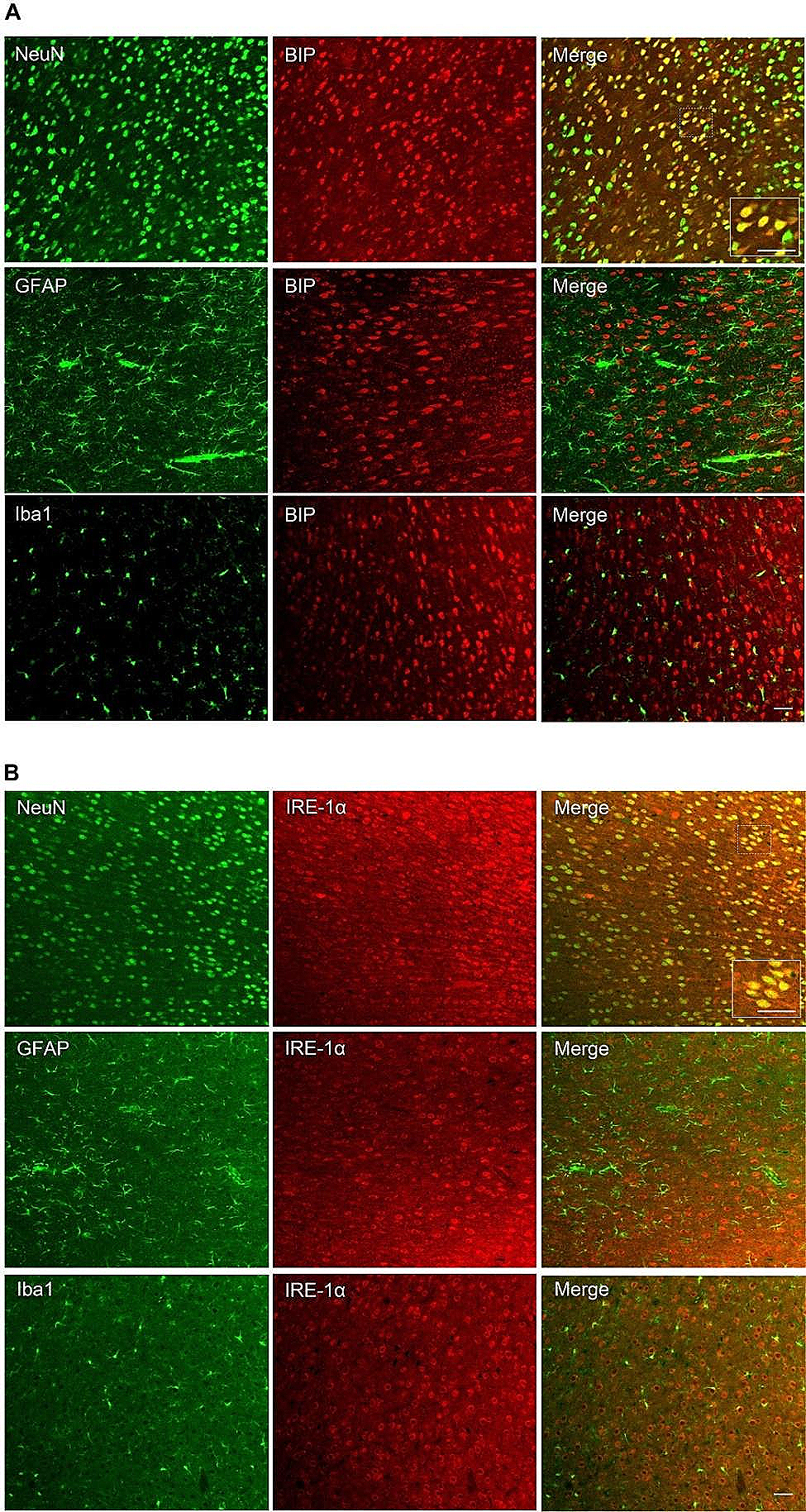
Immunofluorescence assayAfter deep anesthesia with pentobarbital sodium, the rats were transcardially perfused with 150 mL of 1 × phosphate buffered saline (PBS) (4 °C), followed by 150 mL of 4% paraformaldehyde (4 °C). The ACC was harvested, fixed with 4% paraformaldehyde for 48 h, and then dehydrated with 30% sucrose for 3 days at 4 °C. Subsequently, the ACC was transversely cut into slices (30-µm thick). The sections were blocked with 10% sheep or donkey serum for 2 h at room temperature and incubated with the following primary antibodies for 48 h at 4 °C: goat-anti-Iba1 (1:200, Abcam, Cambridge, UK), mouse anti-GFAP (1:500, Cell Signaling Technology), mouse anti-NeuN (1:2000, Abcam), rabbit anti-BIP (1:500, Cell Signaling Technology), and rabbit-anti-IRE-1 (1:1000, Proteintech). The sections were washed with 1 × PBS and incubated with fluorescent secondary antibodies in the dark for 2 h at room temperature. Finally, the sections were examined under a fluorescence microscope (FV3000; Olympus, Tokyo, Japan).
Western blot analysisAfter deep anesthesia with pentobarbital sodium, the rats were decapitated, the brain was harvested, and the ACC was divided into the left (Ipsi.) and right (Contra.) sections as described in our previous study [27]. The ipsilateral protein samples were separated by SDS-PAGE and transferred to a PVDF membrane. Subsequently, the membranes were blocked in 5% skim milk at room temperature for 1 h before incubation with the following primary antibodies for 24 h at 4 °C: rabbit-anti-BIP (1:500, Cell Signaling Technology, Danvers, MA, USA), rabbit-anti-IRE-1 (1:1000, Proteintech, Rosemont, IL, USA), rabbit-anti-pIRE-1 (1:1000, Proteintech), and mouse-anti-GAPDH (1:10,000, Proteintech). After washing with TBST, the membrane was incubated with an HRP-conjugated secondary antibody for 2 h at room temperature. The ChemiDoc MP System (Bio-Rad, Hercules, CA, USA) was used to detect complex immune bands.
Statistical analysisAll data are expressed as the mean ± SEM and were analyzed using GraphPad Prism 8.0 (GraphPad, San Diego, CA, USA). Behavioral data were compared between the two groups using independent t-tests. The results of behavioral tests across different time points were analyzed using repeated measures two-way analysis of variance (ANOVA), followed by Bonferroni’s post hoc test. Western blot data were analyzed using one-way ANOVA, followed by Tukey’s multiple comparisons test. Statistical significance was set at p < 0.05.
留言 (0)