
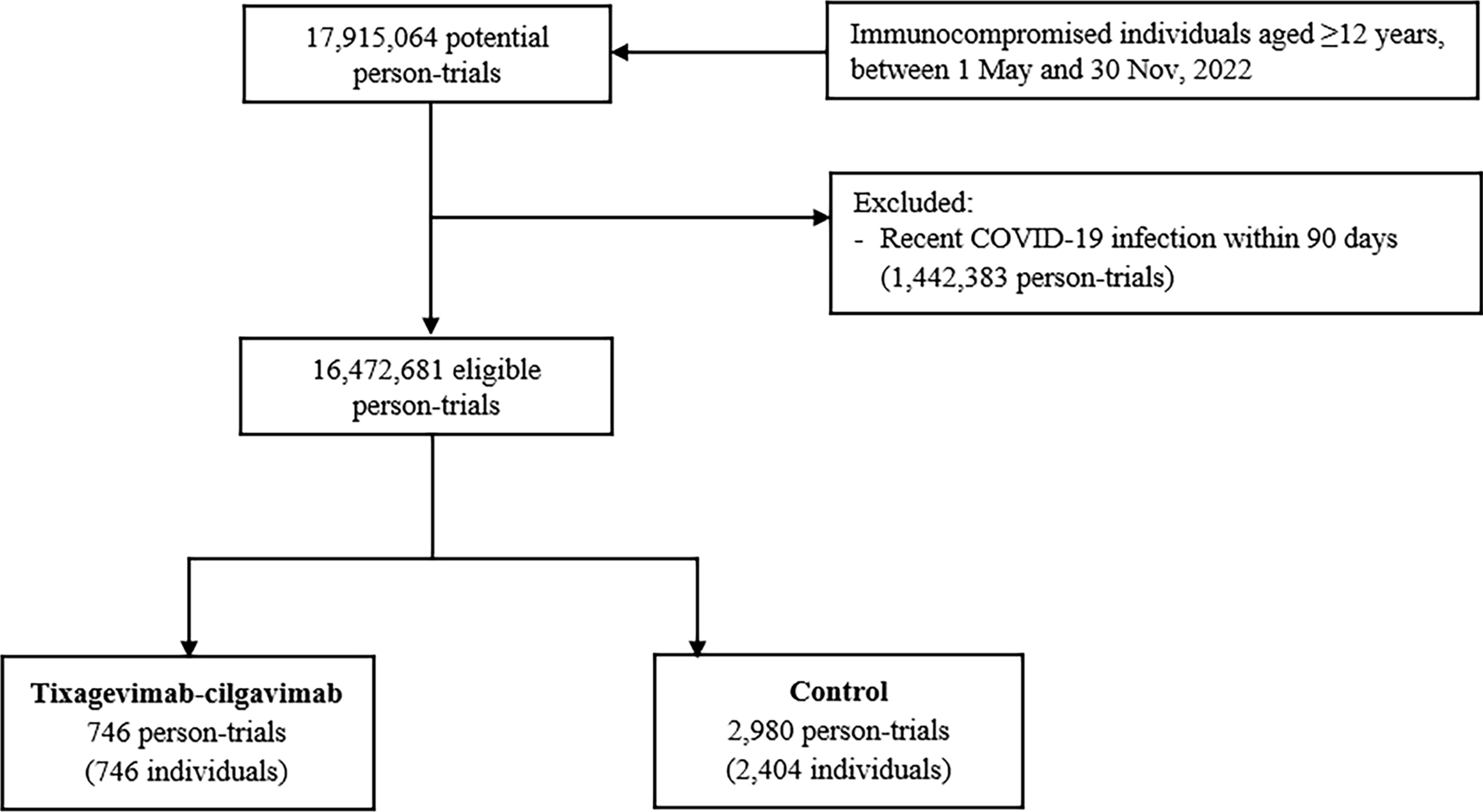
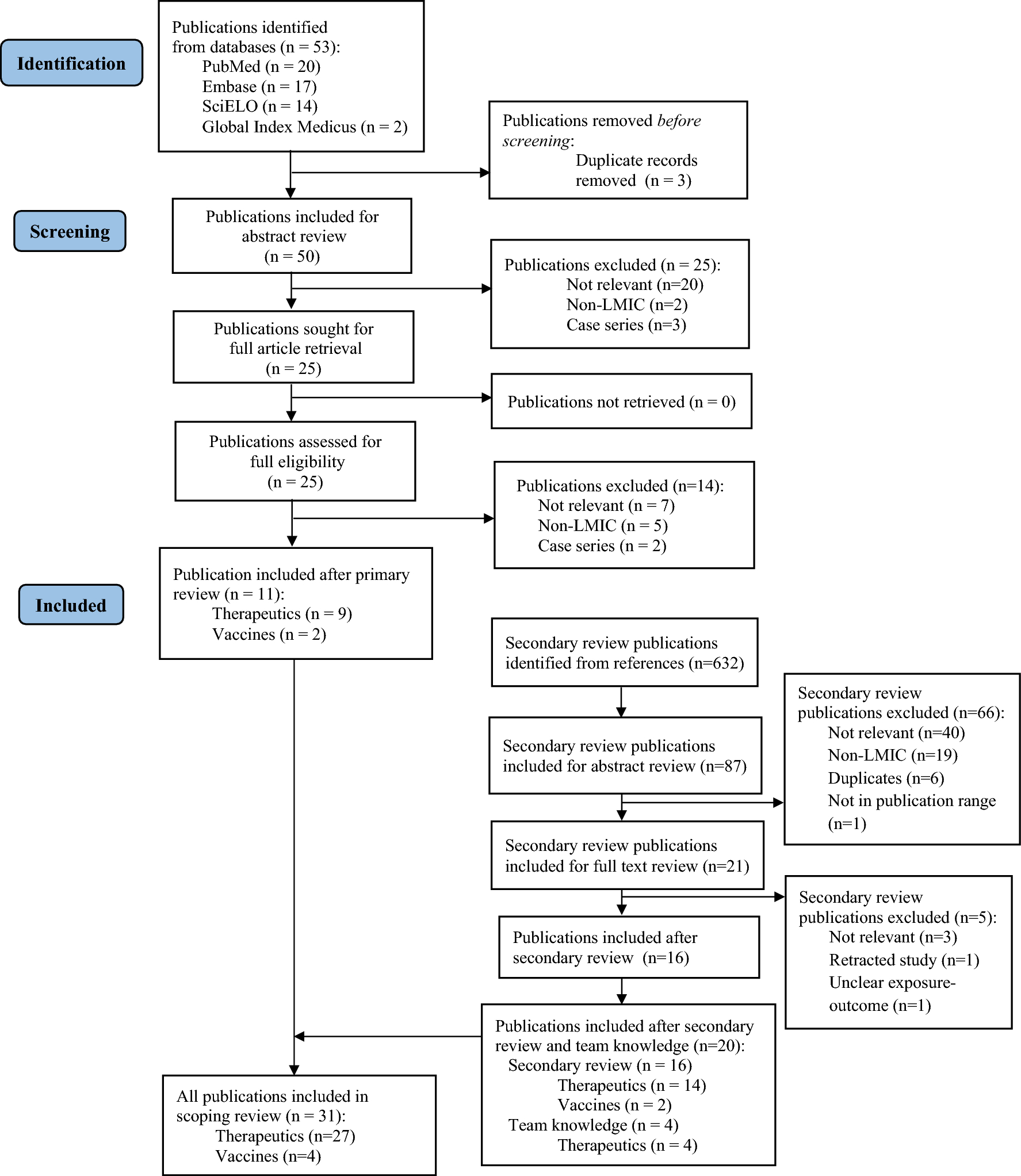
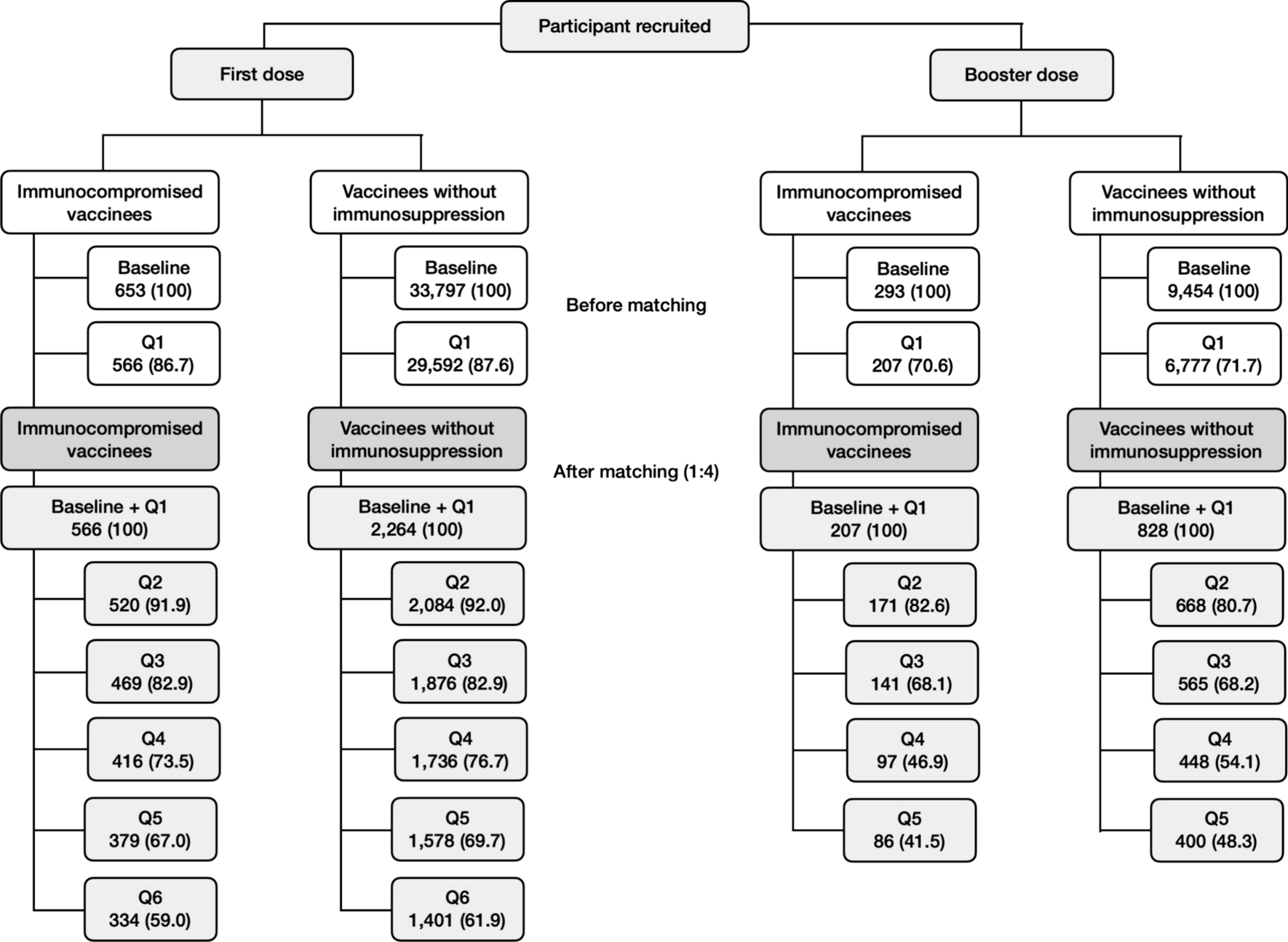
記住我


The eDISH plot is a log-log scatter plot where the x-axis is the peak on-treatment alanine aminotransferase (ALT) and the y-axis is the peak on-treatment total bilirubin (BILI), both depicted in multiples of the ULN (Fig. 1) [8,9,10]. A vertical line at 3×ULN and a horizontal line at 2×ULN indicate eDISH cutoff points and divide the plot area into four quadrants. The upper right quadrant is labeled as Hy’s Law quadrant and includes subjects with ALT elevations greater than 3×ULN and BILI elevations greater than 2×ULN. Subjects in this quadrant have laboratory abnormalities that indicate a drug’s potential for causing serious liver injury. The lower right quadrant is labeled as Temple’s Corollary and includes subjects with ALT elevation greater than 3×ULN and BILI not more than 2×ULN. Subjects in this quadrant are at an increased risk for hepatocellular injury. The upper left quadrant is labeled as Cholestasis quadrant and includes subjects with ALT not more than 3×ULN and BILI elevation greater than 2×ULN. Subjects in this quadrant are at an increased risk for cholestatic liver injury. The lower left quadrant includes subjects with neither ALT greater than 3×ULN nor BILI greater than 2×ULN. Since this quadrant contains subjects with normal and near normal elevations in both ALT and BILI, we labeled it as Normal & NN quadrant. The eDISH plot therefore categorizes a trial population at risk for DILI into these 4 groups: Normal & NN, Cholestasis, Temple’s Corollary and Hy’s Law.
Fig. 1
eDISH plot for a placebo-controlled phase III trial submitted to the US FDA in support of a new drug for CHC. ALT alanine aminotransferase, BILI total bilirubin, CHC chronic hepatitis C, eDISH electronic drug-induced serious hepatotoxicity, NN near normal, ULN upper limit of normal
2.2 The Composite PlotThe composite plot was generated through three steps (Table 1). To explain the steps and introduce the composite plot, we used a hypothetical dataset of four subjects. In Step 1, to understand the extent of liver injury on study entry, pretreatment liver tests of subjects were analyzed to determine their level of elevation using the eDISH plot thresholds for ALT and BILI (Fig. 2a) [9]. In this and all subsequent steps, subjects were grouped using colored symbols assigned according to their pseudo eDISH quadrants based on their pretreatment tests values, i.e., normal or near normal (Normal & NN) group (green square), Temple’s Corollary group (blue plus), Cholestasis group (yellow circle), and Hy’s Law group (red triangle).
Table 1 Sequential analytic steps in developing the composite plot, their function and use in reviewFig. 2
eDISH plot of pretreatment (baseline) liver tests (a) and peak on-treatment liver tests (b) where each subject is identified by a colored symbol corresponding to its pretreatment eDISH quadrant. ALT alanine aminotransferase, BILI total bilirubin, eDISH electronic drug-induced serious hepatotoxicity, NN near normal, ULN upper limit of normal
In Step 2, to characterize response to intervention, a classical eDISH plot was generated to identify subjects’ peak on-treatment liver test quadrants (Fig. 2b). This step captures individual subjects’ migration from baseline to on-treatment peak values by identifying subjects by their quadrant of origin through the colored symbols assigned to them in Step 1. For instance, in Fig. 2b, subject 3 was in the Hy’s Law quadrant where both ALT and BILI were elevated at baseline, and then moved to Cholestasis quadrant on treatment where only BILI is elevated based on the eDISH thresholds as indicated by the arrows in the figure. Identifying such migration is critical to understanding potential DILI and improvement in liver tests from baseline values.
In Step 3, subjects within each quadrant of the eDISH plot in Step 2 are plotted into a scatterplot where each axis represents peak on-treatment liver test in ×BLN. Fig. 3 shows such a scatter plot for the subject in the Hy’s Law quadrant of the eDISH plot in Fig. 2b. This two-dimensional shift plot captures both the magnitude and direction of change in peak on-treatment liver test values of a subject compared with its corresponding baseline values by referencing the horizontal (BILI) and vertical (ALT) lines at 1×BLN. Compared with their baseline, subjects located to the left of the 1×BLN vertical line had reductions in ALT during treatment while subjects to the right had an elevation. Similarly, subjects located below the 1×BLN line had reductions in BILI on-treatment, while those above it had elevations. The additional horizontal and vertical cut-off lines help visualize the extent of the liver test changes. For instance, in Fig. 3, subject 1 experienced an increase in both ALT and BILI by more than three times its respective baseline values.
Fig. 3
Shift plot from pretreatment to peak on-treatment liver test values for the subject in Hy’s Law quadrant of the eDISH plot depicted in Fig. 2b. ALT alanine aminotransferase, BILI total bilirubin, BLN baseline liver test value, eDISH electronic drug-induced serious hepatotoxicity, NN near normal
Putting these three elements together—the colored symbol that represents a subject’s pretreatment elevation, the panel where the subject belongs, and the location of the subject within the panel—the composite plot enables visualization of migration of subjects from pretreatment to peak on-treatment in terms of eDISH quadrant changes, as well as the magnitude of these changes in terms of their respective baseline test values. For instance, in Fig. 4, subject 1 migrated from Normal & NN quadrant at baseline to Hy’s Law quadrant on treatment and experienced an increase in both ALT and BILI by more than three times its respective baseline values, whereas subject 3 migrated from Hy’s Law quadrant at baseline to Cholestasis quadrant on-treatment and experienced reduction in ALT, but BILI remained the same compared with its respective baseline values.
Fig. 4
The composite plot for the hypothetical dataset. ALT alanine aminotransferase, BILI total bilirubin, BLN baseline liver test value, NN near normal
Quadrant tallies from the composite plots provide quantitative estimates of the relative proportion of potentially beneficial or concerning migrations during the trial. The number of subjects that move from the pretreatment quadrants to the on-treatment eDISH quadrants can be summarized in an eDISH migration table such as Table 2. Migrations are color coded for level of concern for potential DILI. The subjects that are retained in their pretreatment quadrants are shown in gray. The differences in migration into a quadrant of concern for DILI risk (red) or a quadrant of benefit or less concern (green) between treatment arms can be assessed by comparing tables derived from the separate composite plots. Interpretation of migration into the yellow boxes can be assessed on a case-by-case basis in the context of the trial, the drug in question, and the underlying disease. For example, a patient enrolled in a trial for primary biliary cholangitis (PBC) might move from the Cholestasis to Temple’s Corollary by resolving their jaundice but then experience DILI with elevation of ALT. Conversely, a subject in a viral hepatitis trial may improve their ALT (migrate out of Temple’s Corollary) but experience a cholestatic DILI with jaundice. Case level assessment of subjects in the yellow boxes may be needed within the context of the disease and trial.
Table 2 eDISH quadrants migration from pretreatment to peak on-treatmentIn the final step of the composite plot, the magnitude of change relative to the individual’s pretreatment test values is provided. Summary tallies of the shifts in on-treatment ALT and BILI in reference to the individual subject’s pretreatment levels (×BLN) can be compared by study arm. Differences in the proportion of subjects with an increase or decrease ×BLN between treatment arms for ALT (to the right or left of the vertical ×BLN line) and BILI (above or below the horizontal ×BLN line) may help discern benefit or harm attributable to the intervention. A visual inspection of the distribution of subjects in the shift ×BLN plots may help define multiples of BLN that result in greater separation of effect between study arms.
留言 (0)