Ethics statement
The research involving animals was carried out in alignment with established ethical standards for animal experimentation. Approval for the animal study protocol was granted by the Institutional Animal Care and Use Committee at the General Hospital of the Northern Theatre.
Sample preparation for sequencing
The isolation of total RNA from brain and kidney tissues of both normal mice (n = 3) and uremic mice (n = 3) was accomplished using Trizol reagent (Thermo). Quantification of RNA, along with assessments of its purity and structural integrity, was conducted through the utilization of a Qubit® 2.0 Fluorometer® in conjunction with a Qubit® RNA Assay Kit for concentration measurements, an IMPLEN Nanodrop spectrophotometer for evaluating RNA purity, and an RNA Nano 6000 Assay Kit employed on an Agilent Bioanalyzer 2100 system for determining the structural integrity of the RNA samples. The measurements yielded results indicating that RNA concentration was equal to or greater than 20 ng/μL, purity exceeded an OD260/280 ratio of 2.0, and integrity showed a RIN value equal to or greater than 7.0 with a 28S/18S ratio equal to or greater than 1.0 (Arunachalam et al. 2022).
High-throughput transcriptome sample sequencing
For the preparation of RNA samples, 3 μg of total RNA from each sample served as the input for cDNA library preparation using the NEBNext® Ultra™ RNA Library Prep Kit for Illumina® (NEB, E7435L, Beijing, China). The library quality was assessed based on indexing and clustering using the TruSeq PE Cluster Kit v3 cBot HS (Illumina) on the cBot system. Sequencing was conducted on the Illumina HiSeq 550. Moreover, the quality control was performed using FastQC v0.11.8, with data preprocessing by Cutadapt v1.18 to remove adapter and poly(A) sequences. Reads with more than 5% N bases were discarded via a Perl script, and those with base quality over 20 were selected with FASTX Toolkit v0.0.13. BBMap software was used for read pairing, and HISAT2 v0.7.12 aligned the high-quality reads to the mouse genome, ensuring data integrity and preparation for analysis (Deng et al. 2020; Peng et al. 2019).
miRNA high-throughput sequencing and quality control
For the small RNA library construction, 3 μg of total RNA from each sample served as the starting material for small RNA library construction using the NEBNext® Multiplex Small RNA Library Prep Set for Illumina® (E7300S, NEB), with unique index codes assigned for sample identification. The method involved direct ligation of NEB 3’ SR Adaptors to the 3’ ends of miRNAs, siRNAs, and piRNAs, conversion of single-stranded adaptors to double-stranded DNA, and first-strand cDNA synthesis using M-MuLV Reverse Transcriptase. PCR amplification used LongAmp Taq 2X Master Mix with the SR Primer serving as the Illumina and index primer. PCR products were purified on an 8% polyacrylamide gel, isolating DNA fragments of 140–160 base pairs, and resuspended in 8 μL elution buffer. The library’s quality was assessed with the Agilent Bioanalyzer 2100, followed by clustering with the TruSeq SR Cluster Kit v3-cBot-HS (GD-401–3001, Illumina) and sequencing on the Illumina HiSeq 2500/2000 platform to produce 50 bp reads.
The raw data presented in Fastq format underwent processing through the deployment of Perl and Python scripts. This crucial step entailed the exclusion of reads characterized by the presence of poly-N sequences, contamination with a 5’ adaptor, absence of a 3’ adaptor or insert tag, presence of homopolymeric runs of A, T, G, or C, and those of inferior quality, thereby resulting in the acquisition of purified data. Furthermore, critical metrics, including Q20 and Q30 scores, and the GC content pertaining to the raw data were meticulously computed. Following the initial purification, reads falling within a predetermined length range were isolated from the cleansed reads for further analytical procedures. The construction of a reference genome index was achieved via Bowtie2 version 2.2.8, allowing for the alignment of paired-end purified reads against the reference genome. The identification of established miRNAs was facilitated through the use of corresponding miRNA tags. Additionally, the potential miRNAs were discerned by referencing miRBase 20.0, utilizing the capabilities of mirdeep2 and srna-tools-cli software for this purpose (Ma et al. 2020).
CircRNA high-throughput sample sequencing
For circRNA sequencing, 5 μg of total RNA was prepared, with ribosomal RNA removal via the Epicentre Ribo-zero™ kit and ethanol precipitation to clear residuals. The RNA was treated with RNase R to degrade linear RNAs before constructing sequencing libraries using the NEBNext® Ultra™ Directional RNA Library Prep Kit for Illumina®, following the manufacturer’s instructions. This procedure included RNA fragmentation, first-strand cDNA synthesis using random hexamers and M-MuLV Reverse Transcriptase, followed by second-strand synthesis with DNA Polymerase I and RNase H, substituting dUTP for dTTP. After DNA end adenylation and adaptor ligation, cDNA fragments (150–200 bp) were purified with the AMPure XP system and PCR-amplified, with the final products assessed on the Agilent Bioanalyzer 2100. The indexed samples were then clustered and sequenced on the Illumina HiSeq 4000, generating 150 bp paired-end reads.
Preprocessing of raw Fastq data involved removing adaptor sequences, poly-N, and low-quality reads via Perl scripts, leading to purified data. Quality metrics (Q20, Q30, GC content) of this data were calculated. The cleaned data underpinned further analysis, with reference genome and gene annotation files sourced directly from genome databases. A reference genome index was created with Bowtie2 v2.2.8 for read alignment. Circular RNAs were identified using find_circ and CIRI2 software, and raw counts were normalized to TPM, with normalized expression calculated as (readCount*1,000,000)/libsize, where libsize is the sum of circRNA read counts (Ma et al. 2019).
Sequencing data analysis
The R software package "Limma" was employed for the analysis of miRNAs and circRNAs that exhibited differential expression between control and uremic groups, adopting thresholds of absolute log fold change (|logFc|) greater than 1 and a P-value less than 0.05. For the selection of differentially expressed genes (DEGs), the criteria set were an absolute log fold change (|logFC|) exceeding 1 and a P-value below 0.001. Visualization of DEGs was achieved through the "heatmap" package for heatmaps and "ggplot2" for constructing volcano plots to illustrate the variations in gene expression. The construction of Venn diagrams to display gene overlap utilized the "vennDiagram" package. Functional enrichment analyses, including GO and KEGG pathways, were conducted using the "clusterProfiler" package. Protein–protein interaction (PPI) networks were explored using the STRING database online, while circRNA-miRNA interaction predictions were made via the ERCOI web resource. Predictions of miRNA target genes were facilitated through both the miRmap and Tarbase databases (Qi et al. 2022).
In vitro cell culture
Human brain microvascular endothelial cells (HBMECs) (CRL-3245) were maintained in a culture medium consisting of DMEM/F12, enriched with 40 μg/ml endothelial cell growth supplement (ECGS) and 10% FBS. Meanwhile, HEK-293 T cells (CRL-3216), utilized in this research, were sourced from ATCC, United States. These cells were maintained at optimal growth conditions in a Thermo Fisher incubator set at 37 °C with an atmosphere of 5% CO2. The culture media components, FBS and DMEM/F-12, were acquired from Gibco (Chen et al. 2012).
Cell transfection
Lentiviral particles were packaged in HEK-293 T cells via transfection with the target plasmid along with the auxiliary plasmids pMD2.G (12,259, Addgene) and psPAX2 (12,260, Addgene) using the Lentiviral Packaging Kit (V48820, Invitrogen). Then, 48 h post-transfection, supernatants were collected, concentrated using Lentivirus Concentration Solution (Takara), and stored at -80 °C. Cells in the logarithmic growth phase were digested with trypsin and seeded at a density of 1 × 10⁵ cells per well in 6-well plates. Then, 48 h following infection, 10 μg/mL puromycin (540,222, Sigma-Aldrich) was used for selection, maintained for at least 1 week to establish stably transfected cell lines. The construction of target plasmids was undertaken by Shanghai Hanheng Biotechnology Co., Ltd., with sh-RNA and si-RNA sequences detailed in Table S1.
Transfection of mimics-NC/miR-301a-3p-mimics and inhibitors-NC/miR-301a-3p-inhibitors was performed using lipofection, with both mimics and inhibitors procured from Sangon Biotech (Shanghai, China). The sequences were as follows: mimics-NC: UUGUACUACACAAAAGUACUG; miR-301a-3p-mimics: CAGUGCAAUAGUAUUGUCAAAGC; miR-301a-3p-inhibitors: GCUUUGACAAUACUAUUGCAC. For lipofection-mediated transfection, 5 × 10⁵ cells were seeded in 6-well plates, and at 70–90% confluency, transfection was carried out using Lipofectamine™ 3000 (Thermo Fisher, L3000150). Then, 2 to 4 days post-transfection, the cells were used for identification and further analyses (Zeng et al. 2023; Nishiyama et al. 2022; Ouyang et al. 2020).
Cell groups for the experiment were organized as follows: for circRNA-PTPN4: si-NC, si-circ-1, si-circ-2, oe-NC, oe-circ; for miR-301a-3p: Mock, inhibitor NC, miR-301a-3p inhibitor, mimics NC, miR-301a-3p mimics; for FOXO3: sh-NC, sh-FOXO3-1, sh-FOXO3-2, oe-NC, oe-FOXO3, oe-circ + sh-FOXO3, si-circ + oe-FOXO3.
Gene and protein expression profiling
Total RNA was isolated from both tissues and cellular samples utilizing the Trizol reagent provided by Thermo Fisher Scientific, and subsequently reverse-transcribed into cDNA with the First Strand cDNA Synthesis Kit (D7168L, Beyotime, Shanghai, China). In the case of miRNAs, cDNA synthesis was facilitated through a PolyA Tailing Kit (Sangon Biotech, Shanghai, China), enabling the generation of miRNAs appended with PolyA tails. RT-qPCR was performed using an RT-qPCR Kit (Q511-02, Vazyme Biotech, Nanjing, China) following the manufacturer’s instructions. Primer sequences were designed and supplied by Sangon Biotech (Shanghai, China), with details in (Table S2). GAPDH served as the internal reference for mRNA, and U6 for miRNA. Gene expression quantification was achieved using the 2-ΔΔCt method (Ayuk et al. 2016).
For protein extraction from tissues and cells, RIPA lysis buffer containing 1% PMSF (Beyotime) was used. SDS-PAGE gels of 8%-12% were prepared according to the size of the target protein bands, and proteins were separated by electrophoresis. Proteins immobilized on the gel were subsequently transferred onto a PVDF membrane. The membrane was incubated with a 5% solution of non-fat milk at ambient temperature for a duration of 1 h. Primary antibodies (Table S3) were added and incubated overnight at 4 °C. HRP-conjugated goat anti-rabbit IgG secondary antibody (ab6721, 1:2000, Abcam, UK, and Cell Signaling Technology) was applied and incubated for 1 h at room temperature. The bands were visualized using ECL solution (1,705,062, Bio-Rad) on an Image Quant LAS 4000C gel documentation system (GE). As normalized to GAPDH, band intensities were quantified using ImageJ software to determine protein levels (Ban et al. 2019).
Assessment of cell viability, proliferation, migration, and apoptosis
The evaluation of cell viability was conducted utilizing the Cell Counting Kit-8 (CCK-8, Beyotime). Cells, resuspended and adjusted to a density of 1 × 10³ per well, were seeded into 96-well plates and cultured overnight. Subsequent to culture intervals of 24, 48, and 72 h, each well received 10 μL of the CCK-8 solution, which was then incubated for 1 h prior to the assessment of optical density at 450 nm using a microplate spectrophotometer (E8051, Promega) (Yang et al. 2016).
For proliferation rates, the EdU labeling assay was utilized. Following seeding in 24-well plates, cells were treated with 10 µmol/L EdU (Beyotime) and allowed to incubate for 2 h. Subsequently, the cells were immobilized using 4% paraformaldehyde, rendered permeable with 0.5% Triton X-100, and subjected to staining via the EdU click reaction protocol. DAPI staining was applied to visualize nuclei. Fluorescence microscopy (FV-1000/ES, Olympus, Japan) quantified the percentage of EdU-positive cells in random fields (Yang et al. 2022).
Wound healing assays were conducted by seeding cells in 6-well plates until reaching 90–100% confluence, followed by creating a scratch with a pipette tip. Images were taken at 24 h to measure migration distances using Image J software, calculating relative migration rates (Chen et al. 2022).
TUNEL assays was used to detect apoptosis with the Beyotime TUNEL Apoptosis Detection Kit. Fixed and permeabilized cells were incubated with a TUNEL reaction mixture and counterstained with DAPI. Fluorescent microscopy identified TUNEL-positive cells, and the apoptosis rate was calculated by counting positive cells in five random fields per sample (Cheyuo et al. 2012). Each experiment was replicated three times to ensure the reliability and reproducibility of the findings.
Luciferase activity assay
The cDNA fragments of circRNA-PTPN4 and FOXO3 containing miR-301a-3p binding sites, along with a DNA fragment of ZO-1 harboring a FOXO3 binding site, were cloned into the pmirGLO vector. Mutated versions of these fragments, synthesized through site-directed mutagenesis, were also inserted into the pmirGLO vector. The sequences for each construct were as follows: circRNA-PTPN4 Wild Type (Wt): AAAACUUCAGCACUGUUGCACUU; circRNA-PTPN4 Mutant (Mut): AAAACUUGUGCUGAGAACGUGAU; FOXO3 Wt: GCCGAGAUCAUGCCAGUGCACUC; FOXO3 Mut: GCCGAGAUCAUGCCAGUGCACUC; ZO-1 Wt: TGTAAACA; ZO-1 Mut: ACATTTGT. Based on lipofection, HEK293T cells were co-transfected with either circRNA-PTPN4-Wt/Mut or FOXO3-Wt/Mut recombinant vectors and either mimics NC or mimics miR-301a-3p; ZO-1 Wt/Mut recombinant vectors with either oe-NC or oe-FOXO3. After a 48-h incubation, the Dual-Luciferase® Reporter Assay System (E1910, Promega) facilitated the measurement of reporter gene activity, using Renilla luciferase as an internal reference (Jin et al. 2020).
RNA/DNA pull-down assay
Biotinylated constructs of circRNA-PTPN4 WT/Mut, FOXO3 WT/Mut, and ZO-1 WT/Mut (KeyGEN BioTECH, Wuhan, China) were transfected into HBMEC cells for 48 h. Cells were harvested, washed with PBS, and lysed. Lysates were incubated with RNase-free BSA and yeast tRNA-precoated streptavidin magnetic beads (Merck, LSKMAGT) at 4 °C overnight. Following washes with lysis, low-salt, and high-salt buffers, bound RNAs were purified using Trizol and analyzed for miR-301a-3p and FOXO3 enrichment via RT-qPCR (Luan et al. 2018).
ChIP-qPCR
Cells fixed with 1% formaldehyde were sonicated, generating appropriately sized DNA fragments, and centrifuged. Supernatants were incubated with anti-rabbit IgG (negative control) or anti-FOXO3 antibody (Abcam, ab70315, UK) overnight at 4 °C. DNA–protein complexes were precipitated, decrosslinked at 65 °C overnight, and purified. ChIP-qPCR products were analyzed on a 3% agarose gel, with primer sequences in Table S4 (Yang et al. 2016).
Fluorescence in situ hybridization (FISH)
Probes for circRNA-PTPN4 and miR-301a-3p were obtained from Sangon Biotech (Shanghai, China). After denaturing and fixation, cells were treated with sodium bisulfite and proteinase K, followed by dehydration through an ethanol series. Post-hybridization overnight at 37 °C in a humidified chamber, slides were washed and counterstained with DAPI. Fluorescence microscopy observed the hybridization signals (Ye et al. 2018).
Transendothelial electrical resistance (TEER) measurement
TEER was assessed prior to FITC-dextran permeability evaluation, following previously described protocols. Briefly, the culture medium in both dishes and Transwells was replaced with 0.1 M KCl. The EndOhm chamber’s cap was inserted at the top of the chamber, connecting the Transwell with a connector cable, and resistance was measured using an EVOM resistance meter (World Precision Instruments, Sarasota, FL). A new Transwell containing 0.1 M KCl, devoid of cells, served as a blank control (Zhao et al. 2019).
FITC-dextran endothelial permeability assay
For FITC-dextran permeability assessment, HBMECs in the logarithmic growth phase were seeded at a density of 1 × 10⁵ cells onto the upper chamber of 24-well Transwell plates (3524, Corning). Each chamber was supplemented with 100 µL and 600 µL of medium, respectively, and incubated at 37 °C in a 5% CO2 incubator. Upon reaching confluence, 1 mg/mL FITC-dextran (Sc-263323, SANTA CRUZ) was introduced and incubated at 37 °C for 5 min in a 5% CO2 incubator. A 200 µL sample of the baseline medium was taken to determine the baseline value. After an additional 24-h incubation with the supplemented medium, another 200 µL of the baseline medium was collected to measure FITC fluorescence intensity using a microplate reader (Wang et al. 2021).
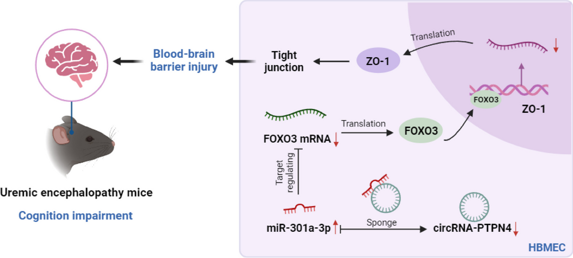
UE mouse model
A total of 126 male C57BL/6 mice, aged 5–6 weeks (sourced from Vital River Laboratory Animal Technology Co. Ltd., Beijing, China), were maintained under pathogen-free conditions at 26–28 °C and 50–65% humidity. An adenine-rich diet was utilized to establish a mouse model of UE. Mice were initially fed a diet containing 20% casein (C7906-5G, Sigma) for 7 days, followed by a diet supplemented with 0.2% adenine (Adenine code A0230000, Sigma) mixed with casein for another 7 days to induce tubular injury. This process was continued for 7 weeks with a diet containing 0.15% adenine. Control mice were fed a standard diet. After 9 weeks, blood samples from the tail vein were tested using a creatinine assay kit (DICT-500, BioAssay Systems) and a urea nitrogen assay kit (BC1535, Solarbio). Elevated creatinine and urea nitrogen levels in the uremic mice compared to the normal mice indicated successful model establishment.
From week 8 onwards, mice underwent daily Y-maze and Morris water maze tests to assess their cognitive functions. Upon exhibiting cognitive impairments, the mice were deeply anesthetized with isoflurane and perfused with ice-cold PBS. Brain and kidney tissues were collected for sequencing and histological analysis. Stereotaxic microinjections (Dorsal -0.26 cm, Lateral -0.15 cm, Anterior -0.02 cm) delivered 4 μL of lentivirus at a titer of 2 × 10⁸ units/mL into the mouse ventricles at a rate of 1 μL/min for 7 consecutive days.
Animal grouping for sequencing involved random assignment into a normal group (Normal) and a UE group (Uremia), with 3 mice in each group. For in vivo experiments, mice were divided into the normal group (Normal), UE group (Uremia), UE plus circRNA-PTPN4 overexpression and silence control group (oe-circ + sh-NC), and UE plus circRNA-PTPN4 overexpression and FOXO3 silencing group (oe-circ + sh-FOXO3), with 30 mice in each group. Fifteen mice from each group underwent Evans blue staining (Kim et al. 2021; Fang et al. 2023; Zhang and Liu 2023).
Y-maze test
The apparatus consisted of a maze with a central starting zone and three branching arms, each measuring 45 cm in length, 10 cm in width, and 15 cm in height, with reward food placed at the end of each arm. Mice were initially allowed to adapt to the experimental setup freely. They were then placed in the starting zone, with their initial positions standardized, and their behavior, including exploration, dwell time, and arm entry frequency, was recorded using a camera. After familiarization with the maze layout and food locations, the testing phase evaluated spatial learning and memory by altering the food’s placement to observe if mice could locate the correct position. Data analysis included exploration time, the number of entries into different arms, and the ability to find the reward location (Sun et al. 2021).
Morris water maze test
The experiment was conducted in a 120 cm diameter, 50 cm high pool filled with clear, body-temperature water. The test included a 15 cm diameter circular escape platform submerged 2 cm below the water’s surface and camouflaged with opaque color. Mice were acclimatized to reduce stress before the trial, involving familiarization with the lab and pool. Experiments commenced from varying starting points around the pool, with mouse behavior meticulously recorded. Training sessions enabled mice to locate the submerged platform, initially marked with cues later removed to assess if mice could independently find the platform. Metrics such as escape latency, occupancy in the target quadrant, platform crossings, and total swimming distance were statistically evaluated (Wei et al. 2021).
Histological and immunological analysis
For hematoxylin and eosin (H&E) staining, coronal sections of brain tissues, 20 µm thick, were stained using H&E, with incubation of 2 min for hematoxylin and 1 min for eosin. Sections underwent dehydration and permeabilization before microscopic observation (BX63, Olympus, Japan) (Li et al. 2020b).
For immunofluorescence assay, fixed cells or tissue sections were treated to allow penetration of primary antibodies targeting ZO-1 (ab307799, Abcam, UK), Occludin (ab216327, Abcam, UK), Claudin-5 (ab131259, Abcam, UK), and CD31 (Sc-376764, SANTA CRUZ, US), followed by incubation with Alexa Fluor-conjugated secondary antibodies and DAPI staining. Confocal microscopy facilitated the visualization of these markers (Li et al. 2019).
The immunohistochemistry protocol entailed the use of antibodies specific to NeuN (ab177487, Abcam), TNF-α (ab307164, Abcam), and IL-1β (ab283818, Abcam). Following secondary antibody application and SABC amplification, the DAB chromogen revealed the localization of target proteins, which was counterstained with hematoxylin. Observations were made under an upright microscope (BX63, Olympus, Japan) (Li et al. 2020b).
Evans blue assay
To assess vascular leakage, mice were intravenously injected with 100 µL of 2% Evans Blue (E2129, Sigma Aldrich, MO). Then, 2 h post-injection, mice were euthanized and perfused with PBS. Brain tissues were then dissected and examined under a stereo microscope for horizontal and coronal sections. After removing the cerebellum and olfactory bulbs, half-brains were weighed, homogenized in 1 mL formamide (F274368, Aladdin, Shanghai, China), and incubated at 60 °C overnight. Brain homogenates were centrifuged at 14,000 rpm for 30 min, and the supernatant containing Evans Blue was transferred to a 96-well plate, with 200 µL per well, including replicates. Optical density at 620 nm was measured using a pre-warmed microplate reader (Li et al. 2022).


留言 (0)