Patients and samples
The GC and corresponding adjacent tissue samples that were collected in this study were surgically removed from patients in Xijing Hospital during 2015–2020. Diagnosis of GC patients was in line with tissue biopsy examination. All patients did not receive chemotherapy or radiation therapy prior to surgical resection. After surgical resection, the tissue specimens were instantly frozen in liquid nitrogen. The present study gained approval from the Ethics Review Committee of Xijing Hospital.
Animal studies
The 4–6-week-old female nude mice were applied in this study. The study was conducted according to a protocol reviewed by Animal Experiment Management Committee. After establishing a stable YTHDC1-knockdown cell line, 2 × 10⁶ cells were subject to subcutaneous injection in right side of each mouse, and the tumors were imaged and weighed after the mice were euthanized 2 weeks later. Each mouse was given injection of 2 × 10⁶ cells via tail veins in lung metastasis model, and at 4 weeks later, euthanasia was completed. Then, the lungs were removed, with metastatic nodules being counted.
Cell culture, RNA extraction, qRT–PCR
Cell lines (GES-1, MGC803, AGS, BGC823, MKN28, MKN45, HGC27 and SGC7901 cells) were used in this study. All the cells were placed in humid incubator with 5% CO2 at 37 °C. The extraction of RNA was carried out using a Total RNA Kit (Omega, USA). SYBR Green Pro Taq HS kit (Agbio, China) and ABI7500 instrument were adopted for real-time fluorescence quantitative PCR. Supplementary Table 1 presents the gene-specific primer sequences.
WB analysis
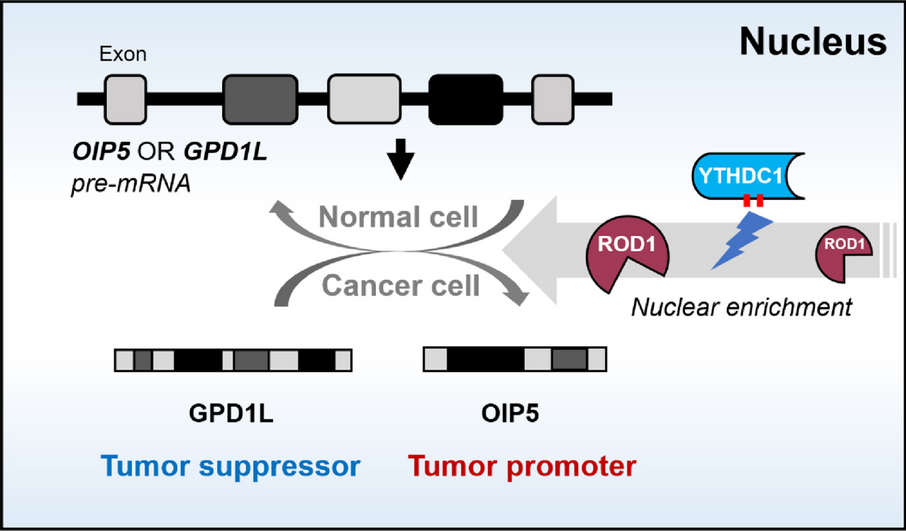
The proteins were transferred to NC membranes (Beyotime, China), and the membranes were incubated with primary antibodies against ROD1 (1:1000, # ab56918, Abcam, UK), OIP5 (1:1000, # TD12217S, Abmart, China), YTHDC1 (1:1000, # ab259990, Abcam, UK) and GPD1L (1:1000, # PS18323S, Abmart, China) at 4 °C overnight, followed by incubation with goat anti-rabbit secondary antibodies (1:2500, # EK020, ZZBIO, China) for 1 h. Then, antibody binding was identified with an ECL Ultrasensitive Chemiluminescent Solution (AccuRef Scientific, China).
Vectors and cell transfection
A YTHDC1 knockdown lentivirus was designed, constructed and generated with hU6-MCS-Ubiquitin-EGFP-IRES-puromycin (GeneChem, China), and stably infected cell lines were selected by incubation in 2 μg/mL puromycin (MCE, China) for 5 days. The YTHDC1 shRNA sequences used were 5′-GCUGGGAGGUGUCUUUAAATT-3′ (sense) and 5′-UUUAAAGACACCUCCCAGCTT-3′ (antisense). The following oligonucleotides were synthesized (GenePharma, China): hsa-ROD1 siRNA (sense: 5′-CCAAUCACAGAGAACUUAATT-3′, antisense: 5′-UUAAGUUCUCUGUGAUUGGTT-3′); hsa-YTHDC1–1 siRNA (sense: 5′-GCUGGGAGGUGUCUUUAAATT-3′, antisense: 5′-UUUAAAGACACCUCCCAGCTT-3′); hsa-YTHDC1–2 siRNA (sense: 5′-GGAGGAAGAAGAAGAAUAUTT-3′, antisense: 5′-AUAUUCUUCUUCUUCCUCCTT-3′); hsa-OIP5–1 siRNA (sense: 5′-GGUCUUCUCCAGAGUUACA-3′, antisense: 5′-UGUAACUCUGGAGAAGACC-3′); hsa-OIP5–2 siRNA (sense: 5′-UAUCAGAGAUGGAUAUUCA-3′, antisense: 5′-UGAAUAUCCAUCUCUGAUG-3′); and hsa-OIP5–3 siRNA (sense: 5′-GCUAACGCACAAUCGCUUA-3′, antisense: 5′-UAAGCGAUUGUGCGUUAGC-3′). The GPD1L overexpression plasmid was synthesized by GenePharma (China).
Transwell assay
In terms of the cell migration assay, the cells were adjusted to a concentration of 1 × 10⁵ cells/ml, 200 μl was supplemented to Transwell chambers (Corning, USA), and 500 μl of 1640 medium supplemented with 20 % fetal bovine serum was supplemented to the 24-well plates. After 24 h, remove the chamber from the 24-well plates, and gently remove any cells that have not passed through the chamber membrane using a cotton swab. Subsequently, the cells on the bottom of the membranes were fixed with 4% paraformaldehyde for 20 minutes and stained with crystal violet for 15 minutes. After drying, the number of cells that passed through the bottom of the chamber was counted after images were captured. Regarding the cell invasion assay, 5 × 10⁵ cells/ml were added to chambers that were precoated with Matrigel (Corning, USA), and the remaining steps remained the same as those in the migration experiment.
Colony formation assay
After reaching 80–90% cell density, we collected cells and resuspended them in culture medium, with cell concentration being adjusted to 3 × 10³ cells/ml. Around 3,00 cells (100 μl) were added to each well of a 24-well plate. Following approximately 10-day culture, the culture medium was removed, and PBS slow-release solution supplemented with 0.5% crystal violet was added to each well of the 24-well plate. In addition, the staining time was approximately 30 min. After completion, cells were washed by PBS three times and observed using an inverted microscope.
Wound-healing assay
After inoculation within the 6-well plate, cells were incubated until they reached around 90% confluence. Then, a scratch was gently made by a 200 μl pipette tip along the bottom of the 6-well plate. Images were obtained at the selected time points (0, 24 and 48 h). Finally, the degree of wound healing was analyzed with ImageJ software.
CCK8 assay
Cells (100 μl of the 1 × 10⁴ cells/ml suspension) were supplemented into 96-well plates. After the cells attached to the wall, 10 μl of CCK8 solution (APExBIO, USA) was added to the well. 0, 24, 48, 72 and 96 h after adhesion were measured for absorbance by a microplate reader (Thermo, USA).
EdU assay
A BeyoClick™ EdU Cell Proliferation Kit (Beyotime, China) was adopted for this experiment. The cells were incubated in 6-well plates with 1× EdU working solution at a final concentration for 2 hours. Then removed the culture medium, 1 ml of fixative was added, and the cells were fixed at room temperature for 15 minutes. The fixative was removed, with the cells being washed 3 times with washing solution. Then the cells were incubated with 1 ml of permeabilization solution per well for 10–15 minutes. Then permeabilization solution was removed, and the cells were rinsed with 1 ml of washing solution for 1–2 times. Then, the EdU assay was performed, and 0.5 ml of Click reaction solution was added to each well and incubated for 30 min in the dark. Then, the cell nuclei were stained with Hoechst. Finally, a fluorescence microscope was used for detecting the fluorescence intensity.
Immunohistochemistry
After paraffin embedding, samples were prepared in 4-μm slices, followed by dehydration and dewaxing overnight in a drying oven at 65 °C and then completely dewaxed in xylene and gradient alcohol solutions. After high-pressure antigen repair with EDTA alkaline or sodium citrate repair solution, the sections were subjected to 12-min incubation with 30% hydrogen peroxide, and another 30-min incubation using goat serum. Then, antibodies against ROD1 (1:500, # ab56918, Abcam, UK), YTHDC1 (1:500, # ab259990, Abcam, UK), OIP5 (1:300, # TD12217S, Abmart, China), GPD1L (1:200, # PS18323S, Abmart, China), N-cadherin (1:400, # bs1172R, Bioss, China), E-cadherin (1:500, # ab40772, Abcam, UK), and vimentin (1:400, # bs0756R, Bioss, China) were supplemented for overnight incubation under 4 °C. The next day, we introduced the secondary antibody for 1-h incubation, DAB color developing solution was supplemented, and hematoxylin was used to restain the nucleus.
Nucleoplasmic protein separation
The experiment was conducted using the Nuclear and Cytoplasmic Protein Extraction Kit (Beyotime, China). First, 200 μl of cytosolic protein extraction reagent A spiked with PMSF was supplemented to the precipitate, followed by 5 s of vigorous vortexing at maximum speed and 10–15 minutes. Then, 10 μl of cytosolic protein extraction reagent B was added and incubated for 1 minute, followed by 5 s of vigorous vortexing at maximum speed and 5 min of centrifugation at 12,000–16,000 g. The supernatant was immediately aspirated and transferred to a precooled plastic tube containing the extracted cytosolic proteins. The supernatant was immediately aspirated into a precooled plastic tube, and this supernatant was considered the extracted cytoplasmic proteins. For precipitation, the residual supernatant was fully aspirated, and 50 μl of cytosolic protein extraction reagent supplemented with the PMSF was added. Then vortexed vigorously at maximum speed for 15–30 s and later returned to the ice bath for 1 min; this process was repeated approximately 10 times. The sample was subject to centrifugation for 10 min at 4 °C. Then, the supernatant was immediately aspirated into a precooled plastic tube, and this sample was considered the extracted nucleoplasmic protein. Finally, the extracted nucleoplasmic proteins were subjected to WB.
Statistical analysis
SPSS 27.0.1 and GraphPad Prism 8.0.1 were used for the statistical analysis. Survival analysis was caried out by the Kaplan–Meier method, and P values were determined by the logarithmic rank test. Differences between two independent groups were evaluated based on Student’s t test (unpaired, two-tailed). The data are indicated to be the mean ± SEM or mean ± SD, and P < 0.05 was though to represent a statistically significant difference. In addition, the experiments were repeated at least three times.

留言 (0)