Study population. This study recruited 95 surgical patients aged 2-39 years old from 2 different institutions in total (Suppl. Fig. 1 and Table 1 for gender and age distribution). Patients below 18 years old (children and teenagers, N=76) were enrolled from the Clinical Hospital ‘José de San Martín’. Adult patients (N=19) were enlisted from the Institute of Otolaryngology Arauz. We excluded samples from patients with any kind of immunodeficiency, neither primary nor secondary. We also excluded any patient taking medication a month prior the surgery (antibiotics, corticoids, etc).
Due to a number of reasons, we were not able to perform the whole set of determinations described in the previous sections to all the samples (Suppl. Fig. 1). We detailed in the corresponding figure legend the precise number of samples used in the experiments shown in each of those figures. Adults were recruited later in the study than children and teenagers.
Isolation of cells. Primary human mononuclear cells were isolated from tonsils obtained from patients undergoing tonsillectomy. The particular number of samples per experiment were detailed in the corresponding Figure legends. TMC were prepared as follows. Briefly, tonsils were collected in phosphate buffered saline (PBS) buffer containing 50 µg ml− 1 amphotericin B (Richet, BA, Arg). Tissues were chopped with a scalpel and passed through a 70 μm-pore-size cell strainer (Falcon, Thermo Fisher, BA, Arg). TMC were purified by density gradient centrifugation with Ficoll-Hypaque (GE Healthcare, Uppsala, Sweden). The viability of primary cells, as determined by trypan blue exclusion was greater than 99% in all preparations. Informed consent was obtained from subjects before the study. The institutional ethics committee (Clinical Hospital, School of Medicine, Buenos Aires and Institute of Otolaryngology Arauz, Buenos Aires) approved the collection and use of clinical material, conformed to the provisions of the Declaration of Helsinki (as revised in Edinburgh 2000). Informed consent was obtained from all participants and/or their legal guardian/s. FACS experiments were performed with freshly isolated cells and cultured cells.
Cell culture. TMC were cultured in IMDM medium (Life Technologies, CA, USA) containing 10% heat-inactivated fetal calf serum, 2mM L-glutamine, 100 U/ml penicillin, 100 µg/ml streptomycin, 20 mM 4-(2-hydroxyethyl)-1-piperazineethanesulfonic acid buffer (HEPES), 1 mM sodium pyruvate and 50 µM 2-mercaptoethanol (all from Invitrogen, CA, USA). Human IL2 (20 ng/ml; R&D Systems, MN, USA) and human IL4 (20 ng/ml; R&D Systems, MN, USA) were added immediately before experiments also as supplements. When indicated, human recombinant CD40L (250 ng/ml; R&D Systems, MN, USA) and 25 µM CpG-ODN 2006 (InvivoGen, CA, USA) were used. Cells were cultured at 1 × 10⁶ cells/ml either in 24-well culture plates (1 ml) or 48-well culture plates (0.5 ml).
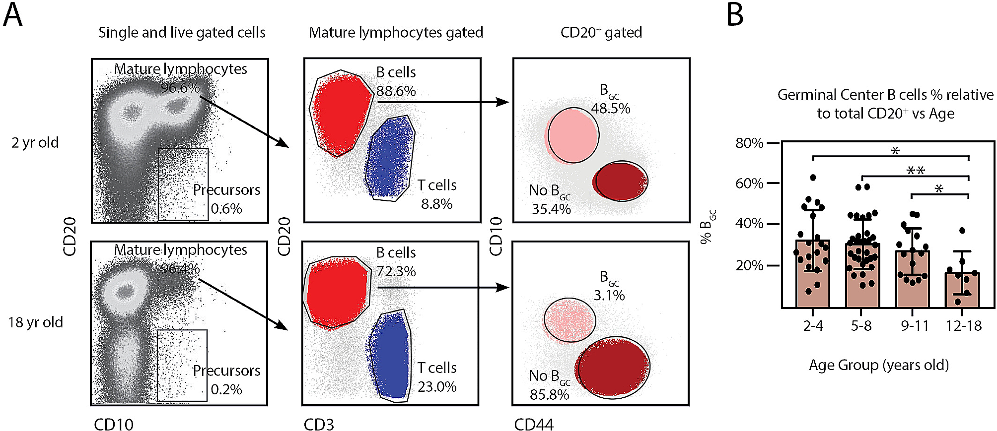
Antibodies and fluorescence-activated cell sorting (FACS). Fluorochrome conjugated mAbs specific for human CD3 (Pacific Blue, clone SK7, BioLegend), human CD20 (FITC, clone L27 and APC H7 clone 2H7), human CD4 (PerCP, clone SK3, BioLegend), CD8 (APC Cy7 clone SK1, BioLegend), CD39 (APC, clone TU66, BD Pharmingen), CD73 (PE, clone AD2, BD Pharmingen), CD27 (FITC, clone M-T271, BD Pharmingen), CD38 (APC, clone HIT2, BD Pharmingen), CD44 (Bv510, clone IM7, BioLegend), CXCR5 (AF488 clone RF8B2, BD Pharmingen), PD1 (Bv711, clone EH12.2H7, BioLegend), CD10 (PE, clone ALB1, Beckman Coulter), Ki67 (FITC, clone B56 RUO, BD Pharmingen) and respective isotype control mAbs were purchased from BD Biosciences (CA, USA) and Biolegend (CA, USA).
To detect Ki-67 transcription factor in the cells, the latter were incubated with Fixation/Permeabilization (eBioscience FOXP3/Transcription, Invitrogen) for 45 min and washed with Permeabilization Buffer (eBioscience FOXP3/Transcription, Invitrogen). Then, the cells were stained with anti-Ki-67 mAb.
Cells were acquired using FACSAria II (BD Biosciences, CA, USA) and analyzed with FlowJo software (Treestar, OR, USA). Single stained controls were used to set compensation parameters. Fluorescence minus one and isotype-matched Ab controls were used to set analysis gates.
Immunohistochemistry. The 5 μm tissue sections mounted on silanized glass slides were deparaffinizated by two consecutive 5 min incubations xylene each, hydrated in decreasing concentrations of alcohol (100%, 96% and 70%) for 5 min each, followed by antigenic unmasking with Sodium Citrate Buffer (0,05 M, pH 6.0) in a thermostatic water bath at 95 °C for 45 min.
The endogenous peroxidase was blocked by incubating tissue sections with 35% H2O2, 98.8% methanol PA (Synth) diluted in PBS pH 7.6 for 30 min, then washed with PBS-1X pH 7.6. The nonspecific binding sites were blocked with Bovine Serum Albumin (BSA) 5%. Ki-67 + cells were detected by incubating with the primary antibody. The antibody dilution was 1/100 Ki-67 (Rabbit monoclonal clone SP6, TecnoLab, 275R-15). Slides were incubated one hour at 4 °C and washed three times with PBS 1X, pH 7.6.
For the immunohistochemical staining system and counterstaining with hematoxylin, a fully automatic staining device, the Ventana BenchMark XT (Ventana Medical Systems, Roche Diagnostics Division) was used. The device uses a biotin-free, HRP multimer-based hydrogen peroxide substrate and 3, 3’-diaminobenzidine tetrahydrochloride (DAB) chromogen (UltraVIEW Universal DAB Detection, Catalog number 760 − 500, Ventana Medical Systems, Tucson, USA).
The slides were examined using the Leica DM500 optical microscope (Leica Camera, Wetzlar, Germany) at magnification of 20x and 40x.
Statistics. The results were analyzed using GraphPad Prism 7.0 and GraphPad Prism 8.0 software. The normality of variable distribution was assessed by the Shapiro-Wilk test. The statistical analysis of the results was performed using the unpaired t test, and a p value of < 0.05 was considered significant unless indicated otherwise.

留言 (0)