
記住我
In a visionary novel about the desperate quest for human immortality in 1930s Los Angeles, British novelist Aldous Huxley imagines a group of scientists running a secret experimental program focused on researching the secrets of long life in – among other animals – long-lived carps [1]. Three decades later, Los Angeles-based gerontologist Roy Walford, and UK-based experimental gerontologist Alex Comfort, pioneered the experimental use of teleosts to investigate the physiologic and histologic basis of organismal aging [2, 3]. Alex Comfort’s work refuted the hypothesis that ever-growing teleosts might escape the aging process achieving to live indefinitely [2, 4]. Walford’s work, on the other hand, focused on annual killifishes as a naturally short-lived vertebrate model, a resource for both fundamental insights into vertebrate’s aging patho-physiology [3, 5], as well as an experimental platform to test experimental interventions that extend lifespan [6, 7]. The work initiated by Comfort, Walford and colleagues, started to shed light on the spontaneous onset of aging phenotypes among Cyprinodontiformes, including annual killifishes [8]. Remarkably, these small-sized teleosts appeared to recapitulate several aspects of mammalian aging, including the age-dependent increase in individual mortality, spontaneous onset of cancer, pathological lesions in the liver, as well as thymus involution [9,10,11,12]. Killifish aging appeared to be characterized by stark age-dependent changes in the main immune organs’ histology, including the thymus and the kidney marrow – the mammalian bone marrow fish equivalent [11]. Noteworthy, Roy Walford pioneered the “immunologic theory of aging” [13, 14], which postulates that immune system aging contributes to systemic aging. The insights obtained by Walford’s studies about aging in killifish might have contributed to his views on the role of immune system during organismal aging and aging-related pathologies.
Evolutionary ecology of annual killifishAnnual killifish are adapted to extreme environments characterized by alternating dry and wet seasons throughout the year [15, 16]. Annual killifish inhabit temporary ponds within seasonal rivers' drainage basins, which experience periodic flooding during brief rainy seasons. Throughout dry periods, these fish endure as desiccation-resistant diapausing embryos, capable of surviving and developing within the arid mud. During the brief rainy seasons, killifish hatch and reach sexual maturity in as little as two weeks, which is faster than any other recorded vertebrate [17]. Adult killifish survival is constrained by the limited availability of water during brief rainy seasons, as pond desiccation results in adult fish death. Other causes of extrinsic mortality in annual killifish are predators (largely birds and aquatic insects), as well as parasites [18]. While annual killifish inhabit ephemeral habitats and are ecologically constrained to a brief adult life in nature, they also display a short lifespan when raised in captivity.
To date, it is still not completely clear what are the mechanistic and evolutionary causes underlying the aging phenotypes rapid onset that constrain lifespan in killifish, making them significantly shorter-lived compared to other teleosts, including other closely related Cyprinodontiformes. One hypothesis that would explain the short natural lifespan in annual killifish is that the same biological mechanisms that control embryonic diapause and rapid sexual maturation come with a cost in late life, hence constraining adult survival. However, to date there is no substantial support towards this type of evolutionary trade-off in killifish. An alternative scenario that would explain the onset of aging-related dysfunctions, as well as the short adult lifespan of annual killifish, is the accumulation of genome-wide deleterious gene variants exacerbated by extensive population bottlenecks and small effective population size [19, 20]. However, while extensive genome-wide relaxation of purifying selection correlates with short adult lifespan and rapid aging in annual killifish, small effective population size per se might not be the only factor explaining short killifish lifespan. In fact, longer-lived non-annual killifish, such as Pachypanchax playfairii, which also underwent extreme population bottlenecks, live several years, and do not show the same spectrum of aging-related dysfunctions typical of short-lived annual killifish [21]. It is therefore plausible that other ecologically relevant factors, e.g. population fragmentation and an upper limit to yearly water availability, might play an important role in the evolution of short natural lifespan observed in annual killifishes.
Annual killifish in the genus Nothobranchius, a natural model of immune agingAt the end of the 1970s and at the beginning of the 1980s, gerontologists recognized that annual killifish in the genus Nothobranchius undergo remarkable spontaneous changes in adult-specific immune features, in parallel with the onset of malignant transformations [9,10,11]. These initial observations set the basis for follow-up studies aimed at dissecting the biological mechanisms involved in immune system aging in killifish.
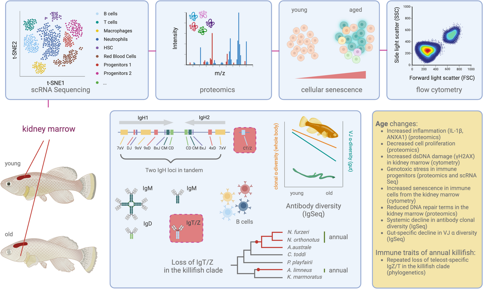
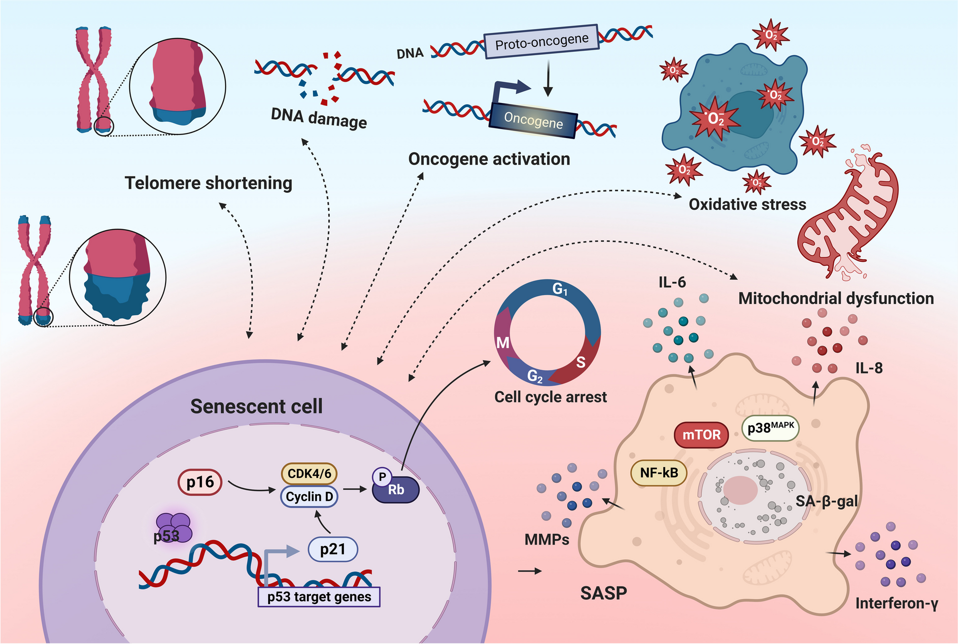
Recent work, still published as a preprint, shows that the main hematopoietic organ of turquoise killifish – the kidney marrow – presents typical vertebrate immune cell lineages, distinct in myeloid and lymphoid lineages [22]. Thanks to the possibility to characterize cell type identity through single-cell RNA Sequencing, it has recently become possible to study cellular and transcriptional changes in aged immune organs, including the kidney marrow and the spleen [22, 23]. The advent of powerful “omics” technologies, including proteomics, has recently demonstrated that immune organs (e.g., the kidney marrow) from young (two-months old) individuals have a proteomic profile compatible with active DNA damage repair and cellular proliferation. On the other hand, immune organs from old (four months old) fish, are characterized by typical inflammation-related terms, such as interleukin-1β synthesis. Plasma proteomics further indicates that plasma from young-adult killifish is characterized by anti-inflammatory terms, such as Annexin 1A, a known NF-kB pathway inhibitor [22]. Conversely, in aged killifish, the plasma reveals marked increases in acute-phase proteins closely associated with inflammatory processes, including elevated levels of clotting factors like FGG and FGB, as well as heightened presence of components from the complement system such as C8G and C8B. These findings collectively underscore the organism's heightened response to wounds and infections. To note, blood from old killifish, compared to blood from young-adult individuals, presents hyperinsulinemia – which is strongly associated with human aging [24] – and higher levels of IGF1.
Both systemic (serum) and organ-specific proteomics in killifish show spontaneous onset of molecular hallmarks of aging associated with inflammation, reduced DNA repair, response to pathogens and metabolic dysfunctions. At the cellular level, cytometric analysis in the kidney marrow of aging killifish indicates that markers of DNA double-strand breaks (γH2AX) and cellular senescence (SA- β-Gal) accumulate in cells from aged killifish, and in particular in progenitor cells [22].
Overall, killifish appear to recapitulate several markers associated with aging during mammalian hematopoiesis, including genotoxic and replicative stress [25].
The killifish IgH locus architecture and evolutionLymphocyte-based adaptive immunity is a distinct trait associated with the evolutionary success of jawed vertebrates [26]. The evolution of adaptive immunity allowed jawed vertebrates to recognize and respond to a vast range of pathogens and parasites. Lymphocyte-mediated adaptive immune recognition enabled for the establishment of long-lasting immunological memory, resulting in effective and precise immune responses upon re-exposures to previously encountered immunological threats. The evolution of this formidable line of defense and immune recognition has also been associated with the establishment of highly complex commensal vertebrate-specific microbial communities [27]. Age-dependent dysfunctions in lymphocyte-mediated adaptive immunity has been demonstrated in humans and mice [28, 29], and has been associated with impaired responses to infectious agents – as well as to vaccines – in the elderly population [30]. A key genomic locus that mediates adaptive immune responses is the Immunoglobulin Heavy Chain (IgH) locus. This locus undergoes somatic recombination during B cell maturation and is responsible for the generation of a vast diversity of antibodies in naïve B cells.
As vertebrates, killifish are equipped with an IgH locus and cellular machinery able to build a humoral immune response against potentially pathogenic antigens. Bradshaw and Valenzano studied the IgH locus architecture in killifish and showed that indeed killifish have two IgH loci in tandem on chromosome 6, able to generate both IgM (secreted and transmembrane) and IgD antibody isotypes [31] (Fig. 1). To note, killifish of the genus Nothobranchius, together with the genus Aphyosemion (a genus of non-annual African killifish) and Austrofundulus (a genus of annual south American killifish), appear to have completely lost a teleost-specific mucosal antibody isotype, i.e., IgT/Z [31] (Fig. 1). The causes underlying the loss of same antibody isotype in several killifish species remains unclear. Consistent with other teleosts, which do not undergo class-switch recombination [32], also killifish do not show IgG/A/Es.
Fig. 1
Multi-dimensional molecular profiling of immune aging in turquoise killifish
The genomic structure of the IgH locus among killifishes is highly heterogeneous. Different killifish species present large variation in the IgH locus, which underwent extensive remodeling and diversification throughout the evolution of this clade.
Nonetheless, the assessment of the IgH locus's products, namely antibodies, cannot rely solely on the examination of the genomic locus; it necessitates the complementary analysis of B cell-specific transcripts.
Aging changes in the killifish immunoglobulin repertoireDuring aging, B cells become less effective in establishing powerful humoral immune responses, contributing to higher titers of autoreactive antibodies, increasing the risk for infections and leading to worse vaccine outcomes. Furthermore, aging B cells undergo clonal expansion even without antigenic challenges, leading to lowered primary B cell diversity [33], which might contribute to defective humoral immunity. Human elderly appear to have more specialized and less plastic B cell responses [29]. Like humans, mice also display age-dependent decline in spleen-specific antibody diversity [28]. What about non-mammalian vertebrates? Do teleosts undergo aging in the B cell compartment? Assembling the genomic sequence of the IgH locus’ constant region in turquoise killifish has been instrumental for characterizing the expressed antibody repertoire. By selective amplification and sequencing of B cell-specific transcripts from the IgH locus via IgSeq [34], it was recently possible to study the expressed killifish immunoglobulin diversity [35]. Expression data via IgSeq, enabled to estimate the entropy of turquoise killifish’s productive IgH repertoire, which amounts to 23 bits [35]. Each adult killifish is therefore able to generate ~ 107 possible unique sequences before selection in the primary lymphoid organ. While 23 bits represents a rather diverse range of possible primary antibodies, the killifish IgH locus has a generative entropy that is 50 times smaller than the human’s [36]. Bradshaw et al. discovered that the entropy of the antibody generative process does not decline during killifish aging, suggesting that killifish might retain the capacity to generate novel naïve B cells throughout life [35]. However, while the primary killifish antibody repertoire appears to be unaffected during aging, the diversity of the largest B cell clones rapidly declines during aging. Furthermore, the sequence diversity (VJ α-diversity) of intestinal B cell antibodies does undergo a dramatic age-specific decline. One possible explanation could be that the constant antigen exposure of the intestinal mucosa might lead to extensive clonal expansion, resulting in a lower relative number of unique VJ combinations in older killifish. Another plausible reason for the age-dependent decline in gut VJ α-diversity could be that the intestinal epithelium has a smaller clone-size distribution than the whole-body repertoire, hence sampling the repertoire diversity towards the larger-size clones [35].
As repertoire diversity declines during aging in killifish, the pairwise inter-individual repertoire diversity (repertoire dissimilarity index, or RDI), increases in aged fish. Hence, young killifish have more similar antibody repertoires, while older killifish have more individualized antibody repertoires, which might reflect the individual-specific exposure to antigens occurring throughout life [35].
To address which immune-system-wide functional terms correlated with high antibody repertoire diversity (from Shannon entropy to diversity measures accounting for relative abundance), Bradshaw et al. conducted a differential expression analysis of total intestinal RNA transcripts, with respect with antibody repertoire diversity, factoring out age [35]. This analysis showed that B cell functional terms, such as “B cell signaling pathway”, and “B cell proliferation”, alongside “microglia cell activation” (possibly enteric glia) and “erythrocyte development” ranked among the top GO terms.
Overall, killifish undergo dramatic antibody diversity loss during aging, and antibody diversity per se strongly correlates with immune terms associated with B cell activation and proliferation. Strikingly, several features of aging in the killifish B cell compartment resemble mechanistic changes identified in human aging, including narrowing of clonotypic diversity [37].
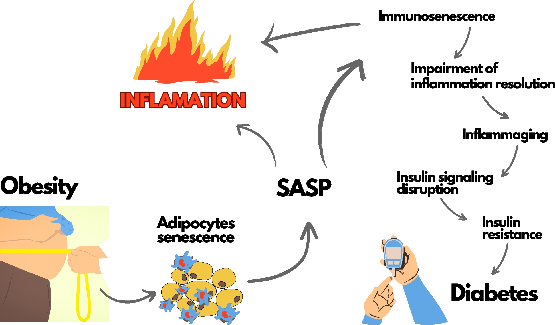
Microbiome age changes and immune cell functionSmith et al., found that young-adult vs. old turquoise killifish have significant differences in their gut microbiome, including declined taxonomic richness, increased prevalence of pathogenic Proteobacteria, and increased pairwise microbial taxonomic diversity [38]. Aging-dependent shifts in microbiome composition have also been reported in Drosophila [39]. Similarly, mice undergo taxonomic and functional changes in the microbiome during aging [40]. In humans, aging has been associated with increased microbiome uniqueness [41, 42]. The age-dependent changes in microbial diversity observed in killifish occur alongside intestinal transcriptional changes associated with increased inflammation, activation of innate immune responses, as well as reduced cell adhesion. In a heterochronic transplantation experiment where gut content from young fish was transplanted into aged killifish, a striking increase in lifespan was observed when compared to mid-age control fish that received gut content from age-matched peers. This effect was accompanied by distinctive transcriptional signatures indicating reduced intestinal inflammation, increased B cell activation (notably POU2AF1), as well as elevated levels of bactericidal lipid-binding serum glycoproteins, such as BPI, and antimicrobial peptides, for instance, HAMP [38]. Together, heterochronic microbiome transfers appears to induce intestinal transcriptional changes associated with lowered inflammation and improved immune protection from pathogens. Recent work in mice suggests that heterochronic stool transplantation in mice ameliorates immune function and brain phenotypes [43, 44]. Furthermore, targeted restoration of the healthy microbiome in humans has been suggested as a promising strategy to promote healthy aging [45].
Cancer in killifishAnnual killifish of the genus Nothobranchius have long been reported to carry a remarkably high incidence of spontaneous neoplastic transformations [9,10,11], which have been associated with thymic involution and malignant changes, including lymphomas. The most affected organs appear to be the liver and the kidney marrow, i.e., the main teleost hematopoietic organ [46]. However, a recent study issues a caveat regarding the reliance on conventional histological methods for the diagnosis of tissue lesions as neoplastic. It underscores that host defense responses to mycobacterial infections can exhibit neoplastic-like characteristics, such as the activation of mononuclear phagocytic processes and the formation of inflammatory granulomas [47]. While responses to mycobacterial infections might be mistaken as neoplasias, their increased occurrence in elderly fish are nonetheless suggestive of age-dependent immunological dysfunctions.
The common platyfish (Xiphophorys maculatus) and the green swordtail (Xiphophorus helleri), which belong to the same order (Cyprinodontiformes) of annual killifish of the genus Nothobranchius, have been extensively studied as models of genetically and environmentally-induced melanoma [48, 49]. Zebrafish and medaka, on the other hand, which are powerful models for experimentally-induced genetic manipulations, have been developed as models for oncogene-induced melanoma (e.g., RAS-RAF) [48, 50]. Whether killifish naturally manifest unequivocal cancerous lesions requires further comprehensive molecular validation. Nevertheless, killifish can become a promising cancer model due to their intrinsic characteristics. Like platyfish, they exhibit spontaneous pigment aberrations [15], while sharing with medaka and zebrafish the amenability to genetic manipulation through transgenesis and genome editing [51,52,53,54]. Furthermore, with a short natural lifespan and an organ-wide emergence of aging-related changes, turquoise killifish offer the opportunity to study the impact of aging on organ-specific tumorigenesis.
Future research will be needed to shed light on the onset of spontaneous malignant transformations in aging killifish.
Opportunities for interventionsThe spontaneous onset molecular, cellular, tissue-specific and systemic changes associated with aging of the immune system, make annual killifish of the genus Nothobranchius an ideal experimental model for immune-targeted interventions aimed at ameliorating health through the aging process.
A recent preprint by Morabito et al., showed that while primary immune cells extracted from the killifish kidney marrow from young vs. old turquoise killifish respond differently to LPS (endotoxin) induction, the application of the senolytic drug fisetin to cells from old killifish donors leads to cellular responses to LPS comparable to those of young cells [22]. Senescent cells appear to dramatically accumulate in aging killifish across organs [55, 56]. The application of senolytics, such as a cocktail of quercetin and dasatinib, led to enhanced neurogenesis after traumatic brain injury [57]. However, whether senolytics lead to improved immune-aging phenotypes also in vivo, is still unclear.
留言 (0)