Administering IR tacrolimus orally directly after LuTx leads to a higher variability in tacrolimus concentrations in comparison to continuous intravenous administration. The oral group had a significantly higher IPV% and lower TTR% than the intravenous group in the first 14 days after LuTx. In general, the occurrence of AKI did not differ between oral and intravenous administration. The occurrence of AKI stage 1 was significantly higher after oral administration of tacrolimus. Keeping the total number of clinically diagnosed cases of acute rejection in mind, the frequency of clinically diagnosed acute rejection in the oral compared to the intravenous group appeared to be 28% higher, without statistical significance. ICU and hospital mortality rate and ICU length of stay were similar for both groups.
To our knowledge, this is the largest study in LuTx providing insightful information on the relationship between the administration route of tacrolimus in the early post-LuTx period and the variability in whole blood tacrolimus concentrations. The higher variability in the oral group may be primarily caused by highly variable bioavailability, which is mainly bypassed by the intravenous administration route. It has been shown that the bioavailability of tacrolimus is the most important factor for fluctuations in whole blood concentrations in orally treated patients and bioavailability may even vary up to 55% shortly after LuTx [11].
Variability in tacrolimus blood concentrations can be substantial among lung transplant recipients, even when adjusting for factors such as age, body weight, and concomitant medications [19]. The genetic polymorphisms of CYP3A enzymes mainly responsible for tacrolimus’ metabolism, particularly CYP3A5*3 and CYP3A4*22, and the transporter ABCB1 variants have been implicated as major contributors to the interindividual variability in tacrolimus PK [19, 20]. Moreover, drug–drug interactions involving CYP3A inducers or inhibitors can significantly alter tacrolimus metabolism, leading to subtherapeutic or toxic blood concentrations, respectively [21]. In addition to interindividual variability, tacrolimus blood concentrations may also vary within the same individual over time, especially shortly after transplantation. Factors such as inflammation, severe bleeding, shock, and organ dysfunction with gut dysmotility and liver dysfunction can influence the IPV% in tacrolimus PK directly after transplantation. Specifically, gut dysmotility is a common complication in the direct postoperative phase with a possible influence on the bioavailability of oral tacrolimus [11].
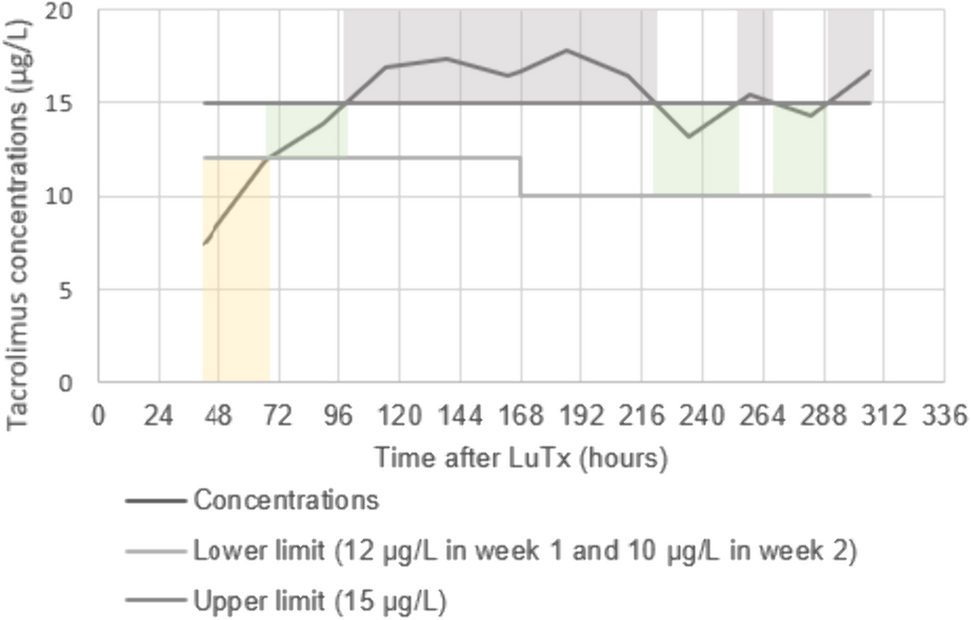
The time that tacrolimus concentrations were within the therapeutic range was shown to be longer with continuous intravenous administration compared to intermittent oral administration. After oral administration of tacrolimus, the TTR% was about 35% when therapeutic ranges of 12–15 μg/L and 10–15 μg/L were applied in the first and second week after LuTx, respectively. Ensor et al., observed a median TTR% of 21% for a therapeutic range of 12–15 μg/L in the first 6 months and 10–12 μg/L up until 1 year after LuTx [3]. In another study in lung transplant recipients, receiving oral tacrolimus in the direct post-LuTx period, a TTR% of 47% was observed, using a therapeutic range of 10–15 μg/L [5]. These are low numbers for a drug for which frequent TDM is conducted. In a recently published cohort study including 67 LuTx recipients receiving tacrolimus continuously intravenously within the first 14 days after LuTx applying a therapeutic range of 10–15 µg/L, the median percentage of tacrolimus TTR% was 35.7% [22]. These findings align closely to our TTR% of 34.6% in the intravenous group, in which a therapeutic range of 12–15 µg/L in the first week and 10–15 µg/L in the second week after LuTx was applied. These studies show the challenges in maintaining whole blood tacrolimus concentrations within the therapeutic range for oral and intravenous administration. Nevertheless, intravenous administration seems to be superior to oral administration.
It is further noticeable that there is no indisputable therapeutic range for tacrolimus after LuTx [23]. In the second consensus report on therapeutic monitoring of tacrolimus, it is stated that the therapeutic range of 15–20 µg/L early after LuTx must be revised [23], because it has been shown that a trough concentration above 15 µg/L has an increased risk of AKI in these patients [9]. Additionally, a subtherapeutic concentration may lead to higher rejection rates in LuTx patients [2]. Since dose adjustments may lead to overcompensation, resulting in more severe toxic or subtherapeutic concentrations, a concentration just beneath or above the therapeutic range is generally accepted, thereby negatively influencing TTR. Moreover, with continuous intravenous dosing, the therapeutic range for trough concentrations should be higher than the therapeutic range for trough concentrations in oral dosing, in order to obtain an area under the curve (AUC) exposure similar to that of intermittent dosing. Nevertheless, in our study, we did not find more adverse effects compared to oral treatment. Currently, no consensus is present regarding the optimal therapeutic range shortly after LuTx. Ultimately, it is of utmost importance to define specific therapeutic ranges for the postoperative LuTx tacrolimus whole blood concentrations for oral and intravenous administration to guide transplant doctors and improve tacrolimus treatment.
A standardized approach to diagnose acute rejection early after LuTx is lacking in both clinical care and in research. In a study conducted by Katada et al., there was no observed correlation between clinically diagnosed acute rejection within 2 weeks after LuTx and tacrolimus TTR [4]. However, they did report TTR to be a predictor for acute rejection after 4 weeks, albeit in a small sample of four patients. Kao et al., correlated routine biopsy-proven acute rejections with tacrolimus TTR [5]. In 157 recipients who underwent routine bronchoscopies, acute rejection was diagnosed in 20.9% and 25.7% of the biopsies, taken at 1 and 2 months post-LuTx, respectively, while no difference was found between TTR for patients with (46%) and without (47%) acute rejection. In the current study, the frequency of clinically diagnosed acute rejection within 6 weeks after LuTx tended to be 28% higher in the oral group than in the intravenous group. While this outcome did not meet the threshold for statistical significance, it may still be of relevance. Acute rejection in LuTx is associated with an unfavorable long-term graft outcome [24, 25]. Consequently, our findings may hold clinical significance. Nevertheless, more research is needed to explore this further, preferably with biopsy-proven rejection data.
There was no difference in the overall rate of AKI stage 1–3. This study only showed an effect of the tacrolimus route of administration on AKI stage 1. This could be due to the higher variability of the tacrolimus blood concentrations in the oral group. However, AKI is multifactorial and not all risk factors have been included in analyses. Moreover, no information was available about pre-LuTx pulmonary hemodynamics/right ventricular function, post-LuTx inflammation status, shock, duration of ischemia of the lung allograft, or the use of concomitant nephrotoxic medication, and were therefore not studied. When comparing our results to a large meta-analysis including > 40,000 LuTx recipients, the AKI incidence (stage 1–3), according to the KDIGO criteria, was lower for both groups in our study (42.6% for the intravenous group and 46.0% for the oral group vs 53%) [7]. In another large multinational study, including 1.800 ICU patients, the AKI incidence (stage 1–3) was 57%, with a severe AKI (stage 2 or 3) incidence of 39% and an incidence of 13.5% for AKI with need for RRT. [26] As seen in this study by Hoste et al., higher stages of AKI are associated with higher mortality rates in ICU patients, the OR for mortality and stage 1 AKI being 2.19 (95% CI 1.44–3.35) compared to no AKI, and could therefore be a clinically significant disadvantage for oral tacrolimus treatment. If we stratified by higher and lower IPV%, patients in the intravenous group with a lower IPV% had a higher AKI rate than patients with higher IPV%. This is a striking result, but could be explained by a higher TAR% in this intravenous group, while AKI is predominantly influenced by AUC.
Identified confounders for AKI were the need for ECLS in the first 14 days post-LuTx and the lowest hematocrit in the first 14 days post-LuTx. Low hematocrit increases the unbound tacrolimus plasma concentration [27,28,29]. Hematocrit in the oral group was higher than in the intravenous group, which could have had an effect on toxicity, such as nephrotoxicity. Additionally, the group without ECLS had a larger proportion of patients with obstructive airway disease as the reason for LuTx. Obstructive airway disease has been found to be predictive for a worse renal outcome [28]. All these factors could explain the higher AKI incidence in this subgroup.
The use of two large cohorts added to the strength of this study. In addition, the LuTx protocols were similar and tacrolimus concentrations were determined with the same analysis method: LC-MS/MS. The latter is in contrast to Gallagher et al., Kao et al., and Katada et al., who used immunoassay to analyze tacrolimus levels, which is known to have cross-reactivity with tacrolimus metabolites [2, 4, 5]. This potentially leads to a higher variability and makes interpretation more difficult. Whereas we compared PK variability between two administration routes, previously mentioned studies have focused on the effects of variability, often defined as the coefficient of variance or SD with only one administration route [2, 5]. These approaches ignore the outliers and may soften the observed effect.
Despite the abovementioned strengths, there are limitations in view of the retrospective nature of this study.
Due to challenges in data collection, information on tacrolimus doses and drug–drug interactions with tacrolimus could not be incorporated into the multiple linear regression model. Consequently, we were not able to determine the length of intravenous administration nor exclude peak concentrations once the switch to oral administration was made. As a result, it was not possible to correct the calculation of IPV% for dose adjustments. Doses are often altered in the early post-transplantation period, and this may in part explain the observed IPV%. Regardless, the switch to oral administration is only made after clinical stabilization, a period in which the gastro-intestinal blood supply and uptake of tacrolimus have stabilized. The contribution of drug–drug interactions to the results is expected to be small because the protocols for administration of co-medication were comparable for both hospitals. It would be desirable to confirm the results from this study in a randomized controlled trial with a direct comparison of oral and intravenous administration.
Further, even though we attempted to exclude tacrolimus peak concentrations, we cannot be certain that we excluded all misclassified ‘supratherapeutic trough’ concentrations, which could have led to a variability higher than the variability in reality. This was only relevant for the oral group and once patients in the intravenous group had switched to oral administration.
Finally, we correlated TTR% within the first 14 days to acute rejection within the first 6 weeks post-LuTx, and did not have information on whether patients had adequate tacrolimus concentrations within the desired therapeutic range from week 2 to 6 after LuTx. This would have been useful additional information.
For future perspectives, the individual tacrolimus concentrations should be corrected for the preceding dose before calculating IPV%. Furthermore, it may be interesting to look at the long-term kidney and graft outcomes.


留言 (0)