
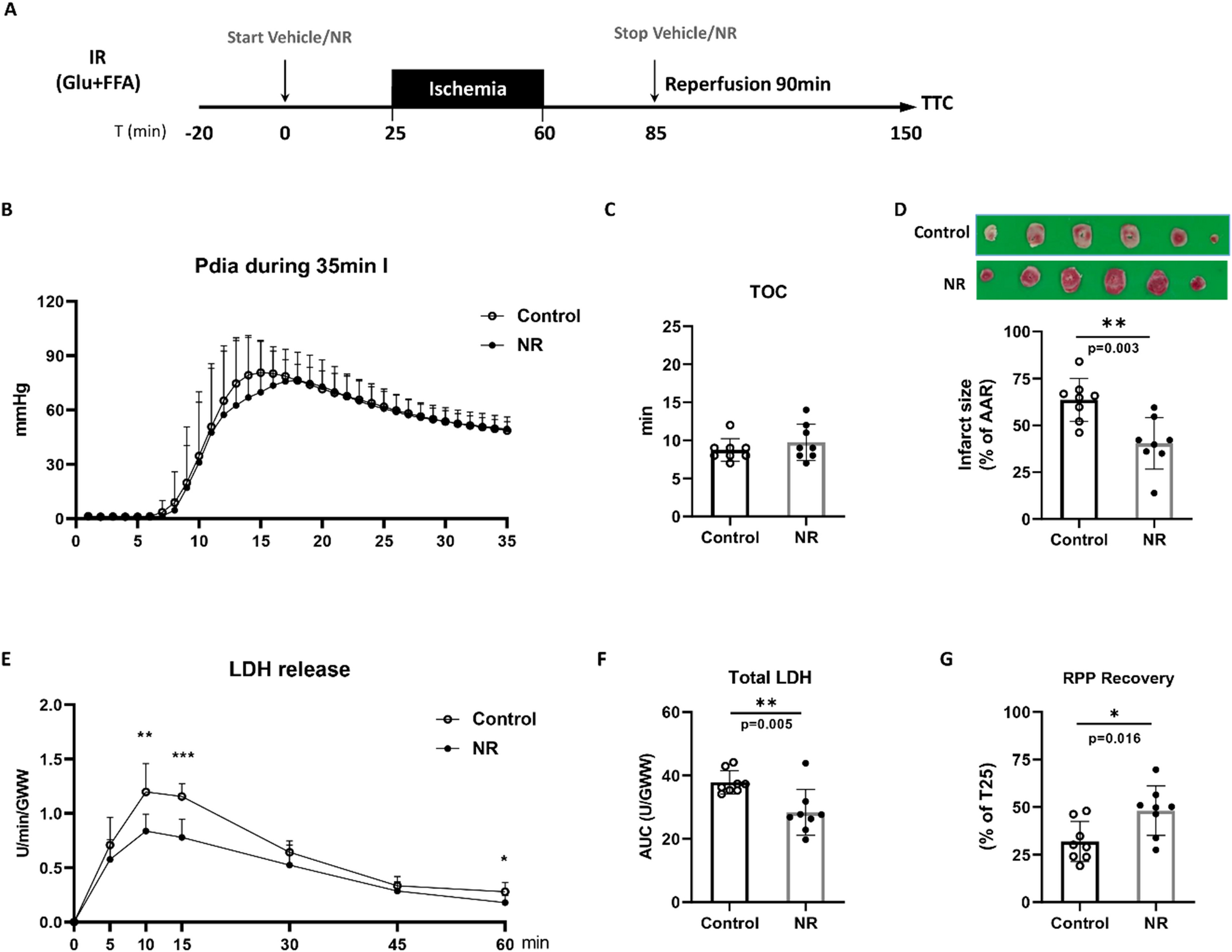
記住我


The endogenous fluorescence properties of the myocardium can be preserved after clarification with aqueous-based and hydrogel embedding solutions. We developed and evaluated a new methodological pipeline, as depicted in Fig. 1a, to determine whether the endogenous fluorescence of the heart could serve as a reliable indicator to measure the degree of infarct injury resulting from ischemia–reperfusion. A mouse model of MI was induced by occluding the left-anterior coronary artery for 1 h, followed by reperfusion for different periods (15 min, 3, and 24 h) until the mice were killed [12] (Supplemental Figure S1). The mice included in the study were categorized into three groups: a negative control group composed of 24-h sham mice, an ischemic group at different times post-reperfusion, and a positive control group for cardioprotection represented by mice undergone ischemia–reperfusion for 24 h with an ischemic post-conditioning (IPoC) at the onset of reperfusion. The hearts of the mice were collected, fixed, and processed for standard X-clarity protocol without bleaching by hydrogen peroxide. Details of the process are provided in the supplemental methods section. Hearts were then imaged using either confocal microscopy or light sheet microscopy, and the resulting images were then processed and analyzed using various software. It is worth noting that while the clearing procedure did not achieve complete transparency of the myocardium, it significantly increased the depth of photon penetration (Fig. 1b). By utilizing multiple lasers with excitation wavelengths of 405, 488, 561, and 633 nm and capturing the resulting light with a spectral detector, specifically a single-photon avalanche photodiode (SPAD), a wide fluorescence bandwidth was detected (Fig. 1c). This endogenous fluorescence of the myocardium provided high-quality macroscopic imaging of myocardium structures and myofibrils (Fig. 1d).
Fig. 1
Cleared mouse hearts conserved endogenous fluorescent emitters in the absence of hydrogen peroxide bleaching. a Methodological pipeline of the study: a mouse model of myocardial infarction, mouse heart before clarification (top), after clarification with the X-Clarity system (bottom right) and after incubation of the cleared heart in the mounting medium (bottom left), fluorescence imaging of unlabeled hearts and image analysis. b A tenfold increase in photon penetration in the depth of the myocardium could be measured in the cleared heart by a mono-photonic confocal microscope under excitation at 488 nm. c A 16-tile image of a cleared myocardium excited by 4 laser lines: 405/488/561/633 nm and acquired with a spectral detector showed endogenous fluorescence over a large wavelength band. Split image (right) and a composite image of merging the 32 channels are shown to the left. d Tiles image of a spectral acquisition acquired tangentially endocardium side of the left ventricle shows the papillary muscles. Inset: magnification of the papillary muscles enables the identification of single fibers
Structure of the endogenous fluorescence signal in cardiomyocytes resembles myoglobin localizationBy focusing on the red wavelength range (bandpass >620 nm), expected to be partly generated by myoglobin fluorescence, and optimizing the resolution using a confocal microscope, we revealed various myocardial structures such as artery layers, cardiomyocytes and interstitial cells (Fig. 2a). In addition to the cellular phenotypes, the endogenous fluorescent signal exhibited a distinct grid-like pattern within the cardiomyocytes (Fig. 2b). This pattern is typically associated with the ultrastructure of cardiomyocytes, including t-tubules, sarcoplasmic reticulum, mitochondria, and myofilaments. We then aimed to investigate if Mb could be responsible for generating this spatial distribution. We have immunostained isolated cardiomyocytes with Mb and GRIM-19, a mitochondrial protein marker (Fig. 2c). Overlay of Mb and GRIM-19 signals revealed that Mb colocalized with GRIM-19 at the periphery of mitochondria and spread away from mitochondria in a well-ordered grid-like pattern resembling the organization of the t-tubules (Fig. 2d). Overall, immuno-detection of Mb exhibited a spatial signature that closely resembled the endogenous fluorescence detected in cleared hearts’ cardiomyocytes. Taken together, our results and previous research support the hypothesis that Mb could be one of the primary sources of endogenous fluorescence in cardiomyocytes, specifically in the red wavelength range.
Fig. 2
Myoglobin localization in cardiomyocytes correlates with the subcellular structures emitting endogenous fluorescence in cleared mouse hearts. a Images of a cleared unlabeled mouse heart taken with a confocal microscope (60× objective, laser source at 633 nm, and SPAD detector) at maximal resolution. Insets 1, 2, and 3 show magnification of cardiomyocytes, aorta, and an interstitial cell. Scale bar: 50 µm. n = 3. b Higher magnification of a single cardiomyocyte section before (left) and after (right) entropic filtering. c Images of adult mouse cardiomyocyte immunolabelled with anti-myoglobin (green channel) and GRIM-19 (red channel). The overlay is shown on the right, and the pixels with both fluorescence signals are white. d Magnification of the image shown in c
The oxidized myoglobin’s spectral signature recapitulates the infarcted area’s spectral signature in failing heartsPeroxynitrite or hydrogen peroxide can efficiently oxidize the iron atom in the heme group of Mb, forming oxidized myoglobin (MetMb). Over-oxidation of MetMb leads to the formation of ferryl myoglobin (ferrylMb), whose absorption spectrum is similar to that of MetMb. We utilized the spectral detector of a confocal microscope to assess how the changes in its oxidation–reduction state modified the fluorescence emission spectrum of pure horse myoglobin. Initially, we corrected the gaps in the fluorescence spectrum due to the dichroic mirrors 405/488/565/647 nm and the glass reflection to prevent the bleed-through phenomenon observed at the peripheries of the 647 nm dichroic mirror (Supplemental Figure S2A). We also conducted a dose–effect and time-effect study using hydrogen peroxide (H2O2) to determine the optimal dose required for MetMb [41]. As displayed in Supplemental Figure S2B, a dose of 0.03% H2O2 was deemed appropriate for oxidizing Mb without inducing any signal bleaching or pigment destruction. As demonstrated in Fig. 3a, enrichment in reduced myoglobin (CarbMb) decreased fluorescence emission intensity, whereas enrichment in MetMb increased it compared to oxygenated myoglobin (OxyMb). Assuming that Mb indeed contributed to the endogenous fluorescence of myocardium, we postulated that alterations in the oxidoreduction state of myocardium would exhibit a comparable signature in myoglobin’s spectra. Pretreatment of control hearts with H2O2, without clearing, revealed a significant increase in the endogenous fluorescence intensity above 600 nm (Fig. 3b). Interestingly, using sodium dithionite to reduce Mb prior to clearing slightly increased endogenous fluorescence intensity around 650 nm, with no effect in greater wavelengths, unlike H2O2. Notably, the observed fluorescent signal around 650 nm was suppressed after heart clearing, suggesting it was likely due to lipids (Fig. 3c). The mean oxidation of myocardium was associated with an increase in endogenous fluorescence intensity above 600 nm, while reduction by sodium dithionite had no significant effect or slightly decreased it. Additional controls assessing treatments’ time and concentration are provided in Supplemental Figures S2C and S2D. In addition, spectral profiles of the fluorescence of individual hearts are given as examples in Supplemental Figure S2E. In order to strengthen our demonstration, we confirmed the involvement of Mb by measuring and comparing the endogenous fluorescence spectrum in both the expected area-at-risk and the healthy area (estimated near to the right ventricle) in confocal transversal slices of wt and KO-Mb hearts. As shown in the Fig. 3d, the endogenous fluorescence intensity dropped by 2.5-fold at all wavelengths in the bandpass 500–730 nm in the KO-Mb hearts in comparison to the wt. These results confirm that myoglobin is a major component of the heart endogenous fluorescence in the red bandpass. After normalization of the fluorescence intensity, we observed an increase in endogenous fluorescence intensity above 600 nm in the area-at-risk of the wt hearts compared to the healthy area (Figure S2F). This red shift signal was similar to the one observed in oxidized myocardium (Fig. 3c) and was absent in the area-at-risk of KO-Mb hearts.
Fig. 3
Myoglobin oxidation and myocardial ischemia–reperfusion share a spectral shift in the red wavelength range. a Fluorescence spectra of pure horse myoglobin measured with the confocal microscope under illumination with laser lines: 405, 488, 561, and 633 nm and detected by a spectral avalanche photodiode. Myoglobin was enriched in either the form bound to CO2 (CarbMg), or the oxidized form (MetMg). b Fluorescence spectra of uncleared heart incubated either in PBS, hydrogen peroxide to induce oxidation (H2O2), or dithionite to induce reduction (NaS2O4) were determined as in a. c Fluorescence spectra of heart incubated either in PBS, H2O2 (oxidation) or NaS2O4 (reduction) before clarification were determined as in a. d Average total intensity fluorescence spectra from heart optical slices of wild type (wt; left inset) and myoglobin knockout (KO-Mb; right inset) mouse hearts in both healthy area (HA) and area-at-risk (AAR). n = 3 each. e Fluorescence spectra of healthy area (labeled by Unisperse blue) and area-at-risk determined as in a. Inset shows a cleared I/R mouse heart labeled with Unisperse blue (healthy area) and contrasting with the brownish area-at-risk. f Normalized fluorescence spectra of healthy volume (HV, blue line) and volume-at-risk (VAR, red line) of a 24-h reperfused heart and a sham heart (black). Value: Mean ± SD; n = 3 Sham and 4 IR. *p < 0.05. g Images of a 24-h reperfused heart by linear unmixing of the mean spectra of sham and VAR presented in f. Confocal slice (top left), 3D rendering volume of healthy volume (bottom left), volume-at-risk (bottom right) and combined volumes (top right). h Heart slice labeled by Unisperse blue (healthy area) shows the distribution of dead tissue (white) and living tissue (pink) in the area-at-risk as labeled by a TTC assay
Altogether, these findings indicate that myoglobin’s oxidation can partly explain the shift in the endogenous fluorescence spectrum and will be referred to as the spectral signature of MetMb. However, the lack of precision delimitation of the area-at-risk and the healthy area, and the analysis of area instead of volume could have slightly modified the interpretation. We thus took advantage of Unisperse blue labeling (Fig. 3e) to selectively extract endogenous fluorescence spectra from both healthy myocardial volume and the whole occluded myocardial volume, defined as “volume-at-risk” (VAR). The control spectrum was retrieved from the heart of a sham mouse without Unisperse blue. Following normalization by the peak value of fluorescence intensity (at 585 nm), the mean endogenous fluorescence spectrum in the VAR exhibited a significant increase of fluorescence intensity above 600 nm, with a maximal difference from sham and healthy volume around 650 nm (Fig. 3f). Except for the specific signature of Unisperse blue in the 500 nm bandpass, sham and healthy hearts showed similar endogenous fluorescence spectra. This increased fluorescence intensity in the bandpass 620–660 nm in the VAR resembles the shift of in vitro spectral signatures between CarbMb and MetMb. While emission spectrum analysis of images is a powerful technique, it is subjected to significant drawbacks when light passes through a diffractive or absorbing media: an in-depth red shift in the light spectrum is anticipated due to the lower penetrance of the shorter wavelengths. In the cleared heart samples, we were able to control for this effect and detected only a slight occurrence on the inner border of the myocardial wall (Supplemental Figure S3A).
We then applied spectral linear unmixing to segment sham-like spectrum and VAR spectrum in all hearts (Supplemental Figure S3B). The spectral signature of VAR, which resembles the spectral signature of MetMb, was detected at the expected position of VAR in the left ventricle. However, it did not propagate transmurally but radiated from the middle of the myocardium wall (Fig. 3g), what was similar to the necrotic area (white area) detected in the TTC assay (Fig. 3h). This finding excluded the possibility that the in-depth red shift phenomenon caused the detected signal due to in-depth photon absorption. In addition, this suggested that the fluorescence spectrum in the VAR was enriched in MetMb spectrum. By contrast, some random noise signal was detected in sham hearts (Supplemental Figure S3C).
Altogether, the spectral features and spatial distribution of the endogenous fluorescence above 600 nm in infarcted mouse hearts fit could be explained by myoglobin’s oxidoreduction state within the infarcted volume. We next aimed to establish a pipeline to quantify the oxidized volume characterized by MetMb fluorescent signature in hearts collected at different reperfusion times and the decrease in the oxidized volume induced by ischemic post-conditioning (IPOC).
The intensity of endogenous fluorescence at 633 nm is associated with reperfusion time, and cardioprotection by IPoC, with a stronger oxidation signal observed 3 h post-reperfusionIn comparison to the z-stacks generated by confocal microscopy, the spatial resolution and imaging depth have been drastically improved using light sheet microscopy (Supplemental Figure S4A), particularly in the Z plane, with a difference of 100 versus 4 µm, for image files of similar size (50–100 Go), with the notion that spectral emission was not possible. All the hearts were imaged using light sheet microscopy at a wavelength of 633 nm, and the light was collected with an extended pass filter above 650 nm. The first step of the analysis involved an anatomical segmentation of the left ventricle. Subsequently, using the strong contrasting effect of the Unisperse blue in the healthy area, we extracted the volume-at-risk (VAR) with intensity-based thresholding (Fig. 4a). Voxels from VAR were then plotted on a distribution histogram (Fig. 4b), which revealed apparent differences between the experimental groups. This difference in intensity distribution among the voxels was accompanied by variations in the spatial organization of the oxidation signal (Fig. 4c). However, robust and objective signal quantification would be required to demonstrate significant and reproducible differences in the oxidized signal between experimental conditions.
Fig. 4
Light sheet imaging at 633 nm reveals hypersignal endogenous fluorescence in the area-at-risk. a Stack of images obtained by an ultramicroscope at 633 nm were segmented as represented: the right ventricle (blue) left ventricle (red) and volume-at-risk (VAR). All hearts except the sham were labeled by Unisperse blue, which was used to retrieve the VAR. An equivalent section was selected in the sham hearts to compare fluorescence intensity on a similar volume. b Distribution histograms of VAR voxels by fluorescence intensity (1 VAR per histogram). c Medial transversal slice of the 3D segmented VAR were colored such as deep blue labeled the minimal fluorescence intensity value of the histogram and red labeled the maximal fluorescence intensity. A representative heart (among 6 per group) is shown for each group: sham heart (no infarct) 24 h after the surgery: sham (24 h), heart after 1 h ischemia: I (1 h), reperfused heart at 15 min, 3 and 24 h: IR (15 min), IR (3 h) and IR (24 h), respectively; and a heart subjected to ischemic post-conditioning and reperfused for 24 h: IR (24 h) + IPoC
There were two major concerns that needed to be addressed in this analysis: (1) variability in the degree of clarity among the hearts, resulting in heterogeneity in the photon absorption along the light spectrum, and (2) heterogeneity in the light path due to differences in myocardium wall length, ventricular lumen, and, excepted in sham hearts, the presence of Unisperse blue in the healthy area (Supplemental Figure S4B). These factors contributed to experimental noise that made it challenging to use an intensity-based segmentation to analyze the oxidation signal accurately. To overcome this, we aimed to develop a more robust analysis flow to compare the intensity distribution of the oxidized signal, defined an objective segmentation threshold and finally quantify the oxidized volume in all hearts, individually. Sham hearts were not included in the first phase because of the lack in Unisperse blue which slightly modifies the distribution of the fluorescence intensity as compared to the end of the ischemic phase (Fig. 4b). To normalize the experimental noise, we deleted low intensities (background noise), removed low-frequency intensities at the foot of the distribution at a fixed threshold of 0.002%, and normalized intensity values between 0 and 1 (where 0 is the lowest intensity voxel and 1 is the highest intensity voxel) (Supplemental Figure S5A-D). A frequency histogram was thus plotted by dividing each column value by the total number of voxels in VAR (Supplemental Figure S5E). These frequency histograms were used to normalize VARs before averaging. Frequency histograms of the same experimental groups were then averaged (Supplemental Figure S5F). Finally, after testing multiple Gaussians fits, we found that the model with two Gaussian best fits the mean frequency histograms (Supplemental Figure S5G and Fig. 5a). As demonstrated in Table 1, the increase in higher fluorescence intensities with reperfusion time was confirmed by a decrease in areas under the curves (AUC) of the left Gaussian fit (lowest fluorescence intensities), an increase in the AUC of the right Gaussian fit (highest fluorescence intensities), and a decrease in the overlap between the AUC of the two Gaussian fits. In the first section of our study, we have demonstrated that the oxidation signal generated by MetMb lied in the increased intensity of endogenous fluorescence in the red wavelengths. As a result, we defined the right Gaussian fit as the best estimator of the shift from CarbMb to MetMb. We conducted a comparison among the different experimental groups. The median of the right Gaussian fit was 0.485 towards the end of the ischemic period; it rose to 0.527 after 15 min of reperfusion and increased to 0.566 after 3 h of reperfusion. Interestingly, the median of the right Gaussian fit at 24 h after reperfusion was similar to that obtained with IPoC (0.490 and 0.477, respectively). In summary, these results suggested that the oxidation intensity increased during reperfusion, reached its maximum by 3 h of reperfusion, and then decreased. This observation correlates with the blood level of Troponin I used in clinical settings to estimate infarct size and evaluate reperfusion lesions (Fig. 5b). The correlation over time between blood troponin level and MetMb fluorescence intensity was almost perfectly linear with different slopes from ischemia to 3 h reperfusion and 3 to 24 h reperfusion (Fig. 5c). Only two median values: IR(3 h) and IR(24 h) of MetMb fluorescence intensity were found above the mean of the median of all experimental groups (0.509) and only the condition IR(3 h) had blood troponin level above the mean value of all experimental groups (37,985). Finally, in addition to measuring the intensity of oxidation, our approach aimed to quantify the oxidized volume.
Fig. 5
Quantification of oxidation intensity at reperfusion by oxidation–reduction imaging. a Mean voxel distribution histogram of fluorescence intensity per experimental group (n = 6 AAR per group) is shown in red. The two Gaussian fits of the mean distribution histogram: left Gaussian fit for the lowest fluorescence intensities and right Gaussian fit for the highest fluorescence intensities model the proportion of CarbMb and MetMb. The green dotted line shows the median of normalized right Gaussian fit. Fluorescence intensity. b Blood troponin I concentration assayed by ELISA (n = 4 per group). c Biplot showing median value of blood troponin concentration in function median value of normalized MetMb fluorescence intensity. Straight line connects time points of the pseudo-kinetic. The yellow area indicates MetMb fluorescence intensity above the mean of the medians of all experimental groups. The green area indicates blood troponin level above the mean of the medians of all experimental groups. The blue area reports junction between yellow and green area
Table 1 Gaussian fit values of the various experimental groupsThe oxidized volume is maximal at 3 h but is sustained up to 24 h post-reperfusion and correlates with infarct volume measured by late gadolinium enhancement MRIAs the median of right Gaussian fit at 1 h ischemia, 24 h reperfusion, and IPoC were similar and all below the mean of the medians, we used their averaged value (0.485) as a segmentation threshold above which the fluorescence intensity most probably come from MetMb molecules. This threshold value was used to segment the oxidized volume within each individual VAR, including those from sham hearts. The right Gaussian fit spanning from 0 to 1 in the normalized histograms, a threshold at 0.485 is equivalent to a threshold at 48.5% of the maximal intensity in intensity histograms. As shown in Fig. 6a, segmentation of sham hearts revealed a patchy signal that may be either attributed to steady-state oxidation level in healthy myocardium, local retention of hemoglobin or statistical errors in the image processing analysis. At the same time, segmentation of the oxidized volume in the VAR of a 24-h reperfused heart resulted in a transmural hypersignal consistent with the shape and localization of the necrotic area observed by TTC assay and shown in Fig. 3h. It was noticeable that the patterns of the oxidized signal in sham and IR (24 h) hearts, respectively, were similar to the ones detected by linear unmixing of myoglobin spectra in confocal images (Supplemental Figure S3C). The mean ratio of oxidized volume normalized by VAR shows a slight increase during ischemia and a significant increase 3 h after reperfusion (Fig. 6b). This value remained stable at 24 h post-reperfusion, supporting that although the peak intensity of oxidation was reached locally at 3 h, the volume of tissue oxidized expanded in the first hours but was kept stable at least for 24 h post-reperfusion.
Fig. 6
Quantification of the oxidized volume extension during reperfusion and its correlation with infarct size measure by MRI. a 3D rendering volumes of sham (top panels) and 24-h reperfused heart (bellow). VAR is shown in a green-fire blue color scale; oxidized voxels are colored with the hot color scale. b Plots represent values for the oxidized volume divided by VAR (top) or left ventricle volume (down) for each heart in each experimental group. Red line: median; blue dotted line: IC 95%; n = 6 per group. Significant difference with Sham (24 h): a significant difference with I (1 h), b was assessed by non-parametric Brown–Forsythe post-test and Welch ANOVA test. c Images acquired by Late Gadolinium Enhancement MRI at 3 and 24 h post-reperfusion in a living mouse and by oxidoreduction imaging post-mortem. d (Top) Correlation plot showing the linear correlation between infarct size (at 3 and 24 h post-reperfusion) and oxidized volume, normalized by left ventricle volume and (down) fit residual; n = 6 per group
Moreover, the emergence of this oxidized myocardial volume was prevented by IPoC. Noteworthy, the mean ratio of oxidized volume normalized by VAR at 3 and 24 h post-reperfusion is consistent with the value measured by TTC in this mouse model. A ratio of oxidized volume normalized by the left ventricle volume was calculated to compare with values obtained from Late Gadolinium Enhancement MRI (Fig. 6b). The late gadolinium enhancement MRI was performed on the same animals subjected to late 3 and 24 h reperfusion (Fig. 6c). The values obtained from ORI and MRI were plotted on the same graph, displaying a good correlation irrespective of the reperfusion time (Fig. 6d). However, while the maximal infarct volume measured by MRI was observed at 24 h post-reperfusion, the maximal oxidized volume was already detectable at 3 h post-reperfusion.
In summary, we demonstrated that the duration of reperfusion is associated with detectable oxidation of Mb in cleared and unlabeled mouse hearts, as determined by measuring the endogenous fluorescence through intensity or spectral imaging. We also established a reliable and robust analytical pipeline to quantify the intensity and volume of oxidized myocardium. We confirmed these values with blood levels of Troponin I and infarct volume determined by MRI.
留言 (0)