
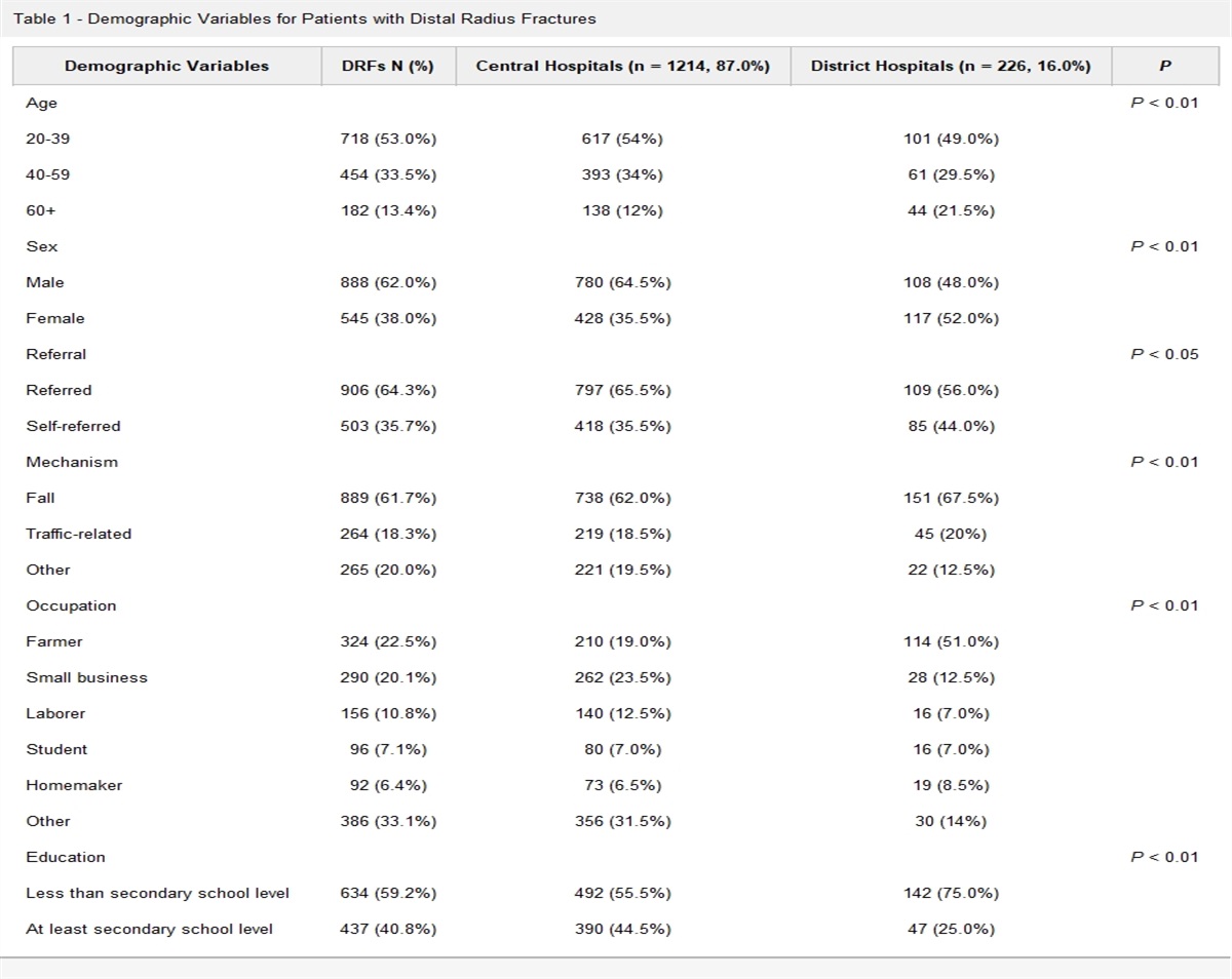
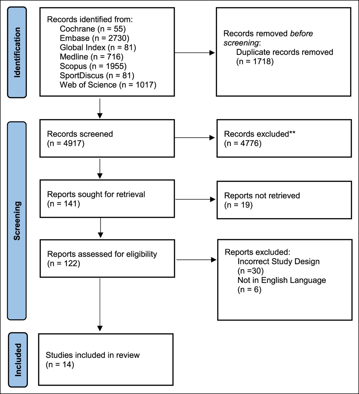
記住我

Periarticular injection (PAI) in total knee arthroplasty (TKA) is a powerful and effective tool to provide perioperative pain control while minimizing motor deficits. It typically involves administering a cocktail of a local anesthetic, an anti-inflammatory, and sometimes a narcotic. Numerous techniques have been described by administering PAI in various areas around the knee for optimal analgesia; however, no single technique has been determined to be superior.1–4
Similarly, an adductor canal block (ACB) is an adjunct method of providing analgesia at the time of TKA. This can be performed by an anesthesiologist before or after the procedure or may be performed intraoperatively by the surgeon. The benefit of an intraoperative surgeon-administered ACB is the avoidance of surgical delays preoperatively and the relative ease of administration. Greenky et al5 found surgeon-administered ACBs to be not inferior to anesthesiologist-administered ACBs. Various techniques have been proposed, and recent cadaver studies have confirmed that intraoperative techniques are able to reliably administer the injectate into the adductor canal. Pepper et al6 described their technique and found a surgeon-administered ACB to be safe with a 1.5-inch needle and to reliably administer the injectant into the canal. Vanamala et al7 conducted a cadaver study, which described an accurate technique with reliably identifiable anatomical landmarks to perform the ACB in cadavers. This technique successfully infiltrated the adductor canal and bathed the saphenous nerve and nerve to the vastus medialis in all cases without evidence of neurovascular injury. They postulated that 10 mL of a local anesthetic is sufficient for an adequate regional block.
Unlike the ACB, PAI sites are not as well described in the literature. In particular, the spread of the local injection is not well defined. The authors of this study performed a PAI in the standard manner that we have used in clinical practice for the past decade with emphasis on a posteromedial injection site as one part of the injection protocol.1 We hypothesize that this injection site will penetrate the adductor canal and posteriorly along the capsule. A posteromedial injection site could simplify the surgeon's workflow and minimize duplicate steps or additional blocks. The purpose of this study was to perform a posteromedial periarticular injection and assess the spread of the injection in both the adductor canal and to the posterior capsule in a cadaver model and compare this with a standard surgeon-administered ACB.
MethodsFour fresh frozen cadavers were used for this study. Adherence to all local, state, and federal laws involving the use of cadaveric specimens was ensured throughout the study. Three specimens were male and one was female.
We sought to compare a standardized protocol for an ACB versus a posteromedial PAI during TKA. In all cadaver specimens, a medial parapatellar approach was performed by a fellowship-trained arthroplasty surgeon. All arthrotomies were extended approximately 3 cm proximal to the superior pole of the patella with the leg bent in 90° of flexion. The distal aspect was extended approximately to the tibial tubercle. The left lower extremity on each of the four cadaver specimens was used to test the ACB, whereas the right lower extremity was used to test the PAI. One type of injection was performed on each side to serve as an internal control for the patient anatomy. On the right lower extremities, additional bony cuts were made to allow the posteromedial injection to be performed in the same location that we use during surgery. For the right lower extremity, a distal femoral cut was made, followed by anterior femur, posterior femur, and finally tibial cuts made using standard mechanical instrumentation. These were all done by the same surgeon for consistency. On the left lower extremities, bony cuts were not necessarily based on the location of the ACB injection.
Next, the injection solution was prepared. This consisted of dilution of 12.5 mL of methylene blue in 237.5 mL of normal saline for 250 mL of total solution for a concentration of 5%.
The ACBs were performed in a standard fashion on the left lower extremities of each of the four cadavers by the same surgeon. We used a validated technique where a 1.5-inch 18-gauge needle was directed posteriorly at the level of the adductor tubercle in the supracondylar region, angled approximately 15° medial in relation to the sagittal plane, with the needle buried until the syringe hub met resistance.6 20 mL of the methylene blue solution was then injected7 (Figure 1).
 Figure 1:
 Photograph showing adductor canal block demonstration.
The posteromedial PAI was performed as a single injection site into the posteromedial capsule. A 1.5-inch 18-gauge needle was at the midpoint of the posteromedial femoral condyle, midway between the ends of the femoral and tibial bone cuts while the knee was bent to 90°. The needle was parallel to the cut surface of the tibia, direct 10° medial, and advanced until the needle and syringe were hubbed typically on the bone (approximately 1 inch of the needle). 20 mL of the methylene blue solution was injected at this location based on our standard intraoperative protocol (Figure 2).
 Figure 2:
 Photograph showing posteromedial injection demonstration.
After injection of all specimens, dissection was performed on the medial thigh to evaluate the proximity of each injection to (1) the adductor canal and (2) the posterior capsule. In addition, the most proximal distance of dye spread was measured from the tip of the needle placement after injection. The tip of the needle injection site to the most proximal distance of dye spread was measured with a ruler and rounded to the nearest 0.25 cm.
ResultsEight total dissections were performed on four cadavers as described earlier. The right lower extremities of the cadavers were dissected medially and posteriorly to identify the spread of the posteromedial injection in the adductor canal and the posterior capsule. A similar dissection was performed on the left lower extremities after the ACBs.
The dye was successfully injected into the adductor canal in all four ACB specimens (100%) and three of four posteromedial PAI specimens (75%). Figures 3 and 4 demonstrate an ACB with proximal dye spread. Figure 5 demonstrates the dye spread in a specimen after posteromedial PAI. Methylene blue was found bathing the posterior capsule in three of four of the posteromedial PAI specimens (75%) and 0 of four in the ACB specimens (0%). Figure 6 is a representative photograph of the dye bathing the posterior capsule, but away from the tibial nerve and popliteus artery after posteromedial PAI.
 Figure 3:
 Photograph showing left lower extremity dissection demonstrating dye spread into the adductor canal after an adductor canal block.
 Figure 4:
 Photograph showing left lower extremity dissection demonstrating the proximal extent of dye spread into the adductor canal after an adductor canal block.
 Figure 5:
 Photograph showing right lower extremity dissection demonstrating dye spread into the adductor canal after posteromedial periarticular injection. The saphenous nerve is indicated by the instrument.
 Figure 6:
 Photograph showing a prone view of the posterior capsule bathed in dye after posteromedial periarticular injection to a right knee. The instrument is indicating the tibial nerve and popliteus artery.
The proximal dye spread from the needle tip was also assessed. The mean proximal dye spread in the adductor canal group was 10.125 cm compared with 6.5 cm in the posteromedial injection group (P = 0.04). The distance of proximal dye spread in the adductor canal group was farther in all but one specimen. The results are summarized in Table 1.
Table 1 - Comparison of Dye Spread in adductor canal block (ACB) versus posteromedial periarticular injection (PAI) Proximal Dye Spread from the Needle Tip (cm) Dye Injected into the Adductor Canal? Dye Surrounding the Posterior Capsule? Cadaver # ACB PAI ACB PAI ACB PAI 1 9 6 Y Y N Y 2 14 6 Y N N N 3 7 8 Y Y N Y 4 10.5 6 Y Y N Y Average 10.125 6.5ACB = adductor canal block, PAI = periarticular injection
PAIs and ACBs are both excellent options to provide analgesia without causing motor deficits in patients undergoing TKA. To our knowledge, there have been no studies conducted evaluating the specific location of injection spread with a PAI. This study demonstrates that there may be benefit in performing a posteromedial PAI over ACB because of the dissemination into the posterior capsule and the adductor canal.
PAIs and ACBs have been compared clinically in numerous studies with some mixed results. Kulkarni conducted a single-blinded randomized controlled trial comparing surgeon-administered ACB versus PAI. They demonstrated superior pain control at 6, 12, and 24 hours postoperatively in the PAI group.4 By contrast, Tong et al8 compared a PAI administered by the surgeon consisting of ropivacaine 150 mg, ketorolac 30 mg, morphine 10 mg, and epinephrine 200 µg in 75 mL total volume to a 30 mL ACB of 0.5% ropivacaine. They found lower total morphine consumption in the first 24 hours in the ACB group, but no difference in functional outcome including total morphine consumption in the first 48 hours, pain scores, quadriceps strength, Timed Up and Go test, 30 s Chair Stand Test, or length of hospital stay. Goytizolo et al3 evaluated the addition of an ACB to PAI and found that patients had lower maximal pain and greater pain relief at 24 hours after anesthesia with the addition of an ACB. However, there was no difference in opioid consumption, opioid-related adverse effects, and Numeric Rating Scale pain scores between groups. A prospective randomized trial by Grosso9 randomized patients to three groups: ACB alone (15 mL of 0.5% bupivacaine), PAI alone (50 mL of 0.25% bupivacaine with epinephrine), and ACB + PAI. Interestingly, patients had markedly higher pain scores and opioid consumption after TKA was done with an ACB, suggesting that ACB alone is inferior for perioperative pain control to PAI alone.
PAIs incorporating a posteromedial injection site seem to have the benefit of bathing the posterior capsule. This was true in three of four of the cadaver specimens. This would theoretically provide enhanced anesthesia to the posterior capsule, much like an iPACK block,10 potentially providing additional pain relief in the posterior knee. The iPACK block (infiltration between the popliteus artery and the capsule of the knee) improves postoperative pain control as an adjunct to ACB in several studies.10–12 Based on the results of our study, using 20 mL of a PAI cocktail in the posteromedial knee allows for penetration into the adductor canal and the posterior capsule, which could perform similarly to the combination of an ACB and iPACK block. The clinical ramifications of this have not been studied; however, the goal would be to improve postoperative mobilization and decrease narcotic usage. The iPACK + ACB has been previously compared in a randomized controlled trial with PAI + ACB, and no difference in visual analog scale pain scores was found.13 If the posteromedial PAI functions similarly to ACB + iPACK, then this may eliminate the need for these additional procedures.
Despite having better posterior capsular coverage, the PAI was less reliable in achieving injectate into the adductor canal (75% versus 100%). The single PAI that missed the adductor canal was in a very thin female cadaver, and the needle was positioned too deep and exited the adductor canal (Figure 7). One should be cautious of this in the clinical setting as well. In addition, the ACB group did have more proximal dye spread (10.125 cm versus 6.5 cm). The clinical benefit of additional spread is unclear, and it is unknown whether this correlates with improved pain control in TKA patients. The injectate spread of both the PAI and ACB may be sufficient to provide similar anesthesia around the knee, but this has not been sufficiently studied. Another clinical implication of this study is potentially eliminating the need for separate surgeon-administered or additional perioperative ACB performed by an anesthesiologist, saving both time and additional cost. Reducing the additional surgical or perioperative blocks would be useful, especially as surgeons move toward higher efficiency ambulatory surgery centers.
 Figure 7:
 Photograph showing dissection of cadaver 2 after posteromedial periarticular injection, where no dye infiltrated the adductor canal and the injection was placed too superficially.
There are several limitations to this study. First, cadaveric tissues may not precisely replicate live tissues and may affect the spread of the injection. Cadaver tissues and tissue planes likely have different dye spread patterns based on tissue pliability, but fresh frozen cadavers were used to minimize this effect. Second, our sample size of cadaveric specimens is small and may be underpowered for absolute comparisons. Furthermore, this small sample does not account for all anatomic variation in a patient population. Next, we used methylene blue to visualize the spread of the injectate, and this may possess a different viscosity and diffusion patterns than local anesthetics and analgesics, especially in live tissues compared with cadaver specimens, where tissue planes may not be as intact. Finally, the volume of injection selected was 20 mL based on our intraoperative protocol. Numerous protocols have used different volumes and may vary based on medication concentrations. Prior studies have demonstrated that even less volume of ACB injection may be effective, however.7
ConclusionsA posteromedial injection as part of a PAI may provide additional analgesia for patients undergoing TKA by bathing the adductor canal and the posterior capsule. By contrast, an isolated surgeon-administered ACB did not provide any spread of injectate to the posterior capsule but did allow more proximal spread in the adductor canal. Incorporating PAI into a routine protocol may theoretically have clinical benefits.
References 1. Spangehl MJ, Clarke HD, Hentz JG, Misra L, Blocher JL, Seamans DP: The chitranjan ranawat award: Periarticular injections and femoral & sciatic blocks provide similar pain relief after TKA: A randomized clinical trial. Clin Orthop Relat Res 2015;473:45-53. 2. Dalury DF: Periarticular injection technique to enhance pain relief after knee arthroplasty. JBJS Essent Surg Tech 2014;4:e7. 3. Goytizolo EA, Lin Y, Kim DH, et al.: Addition of adductor canal block to periarticular injection for total knee replacement: A randomized trial. J Bone Joint Surg Am 2019;101:812-820. 4. Kulkarni MM, Dadheech AN, Wakankar HM, Ganjewar NV, Hedgire SS, Pandit HG: Randomized prospective comparative study of adductor canal block vs periarticular infiltration on early functional outcome after unilateral total knee arthroplasty. J Arthroplasty 2019;34:2360-2364. 5. Greenky MR, McGrath ME, Levicoff EA, et al.: Intraoperative surgeon administered adductor canal blockade is not inferior to anesthesiologist administered adductor canal blockade: A prospective randomized trial. J Arthroplasty 2020;35:1228-1232. 6. Pepper AM, North TW, Sunderland AM, Davis JJ: Intraoperative adductor canal block for augmentation of periarticular injection in total knee arthroplasty: A cadaveric study. J Arthroplasty 2016;31:2072-2076. 7. Vanamala R, Hammer N, Kieser D: Anatomical landmarks for intraoperative adductor canal block in total knee arthroplasty: A cadaveric feasibility assessment. Arthroplasty Today 2021;10:82-86. 8. Tong QJ, Lim YC, Tham HM: Comparing adductor canal block with local infiltration analgesia in total knee arthroplasty: A prospective, blinded and randomized clinical trial. J Clin Anesth 2018;46:39-43. 9. Grosso MJ, Murtaugh T, Lakra A, et al.: Adductor canal block compared with periarticular bupivacaine injection for total knee arthroplasty: A prospective randomized trial. J Bone Joint Surg Am Volume 2018;100:1141-1146. 10. Mou P, Wang D, Tang XM, et al.: Adductor canal block combined with IPACK block for postoperative analgesia and function recovery following total knee arthroplasty: A prospective, double-blind, randomized controlled study. J Arthroplasty 2022;37:259-266. 11. Vichainarong C, Kampitak W, Tanavalee A, Ngarmukos S, Songborassamee N: Analgesic efficacy of infiltration between the popliteal artery and capsule of the knee (iPACK) block added to local infiltration analgesia and continuous adductor canal block after total knee arthroplasty: A randomized clinical trial. Reg Anesth Pain Med 2020;45:872-879. 12. Wang Q, Ma T, Hu J, Yang J, Kang P: Minimum effective volume of ropivacaine for ultrasound-guided adductor canal + IPACK block in total knee arthroplasty: A double-blind, randomized dose-finding trial. J Orthop Surg (Hong Kong) 2023;31:10225536231161873. 13. Kertkiatkachorn W, Kampitak W, Tanavalee A, Ngarmukos S: Adductor canal block combined with iPACK (interspace between the popliteal artery and the capsule of the posterior knee) block vs periarticular injection for analgesia after total knee arthroplasty: A randomized noninferiority trial. J Arthroplasty 2021;36:122-129.e1.
留言 (0)