Nanoparticles are currently utilized in every branch of science and in commercial applications to make products cleaner, lighter, stronger, more precise, more efficient, and more aesthetic [5]. Among them, AuNPs are highly remarkable with their unique functional properties and easy synthesis, allowing them to be used in a wide range of medical applications, including biosensing, photothermal therapy, photodynamic therapy, radiotherapy, X-ray imaging, computed tomography, and gene and drug delivery [14]. Due to the fact that NPS may negatively affect male reproductive organs, spermatogenesis, and hormone levels, reproductive toxicity caused by NP exposure is regarded as an essential topic to be researched in general toxicology [16].
This study aimed at studying the toxic effects of AuNPs on the reproductive system of adult male Albino rats and assessing their reversibility after 30 and 60 days of withdrawal. To the best of the authors' knowledge, there are no available studies investigating the reversibility of AuNPs-induced reproductive toxicity.
In this experimental study, sixty adult male Albino rats were divided into four groups (fifteen rats each): the control group, the test group, withdrawal group I, and withdrawal group II. Control group rats received deionized water daily through the intraperitoneal route for 28 days. Test group and withdrawal groups I and II rats received 570 μg/kg of AuNPs (13 ± 4 nm) daily through the intraperitoneal route for 28 days. Then, withdrawal groups I and II continued for another thirty and sixty days, respectively, with free access to tap water and food to assess the recovery.
Several researchers have already utilized the animal model used in this study to evaluate the AuNPs induced reproductive toxicity [10, 13, 35]. Regarding the AuNPs dose used in the current study, Zhang et al. [41] have used an equivalent dose in mice, and this dose was calculated in rats according to Nair and Jacob [27] conversion tables. The same dose was also used in rats by Velikorodnaya et al. [35] to evaluate AuNP-induced reproductive toxicity. The intraperitoneal route was preferred due to the dense blood vessels and lymph in the peritoneum, which allow good and rapid drug absorption [41].
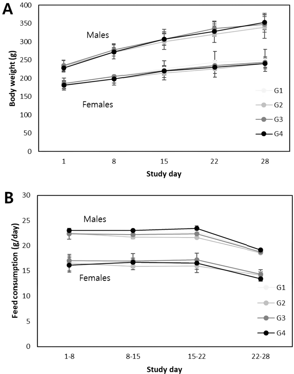
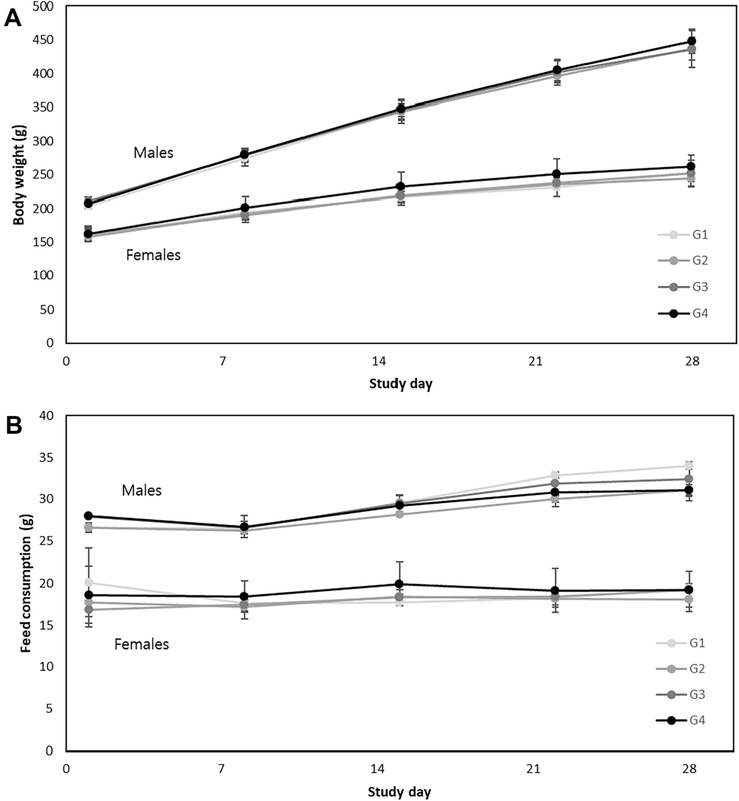
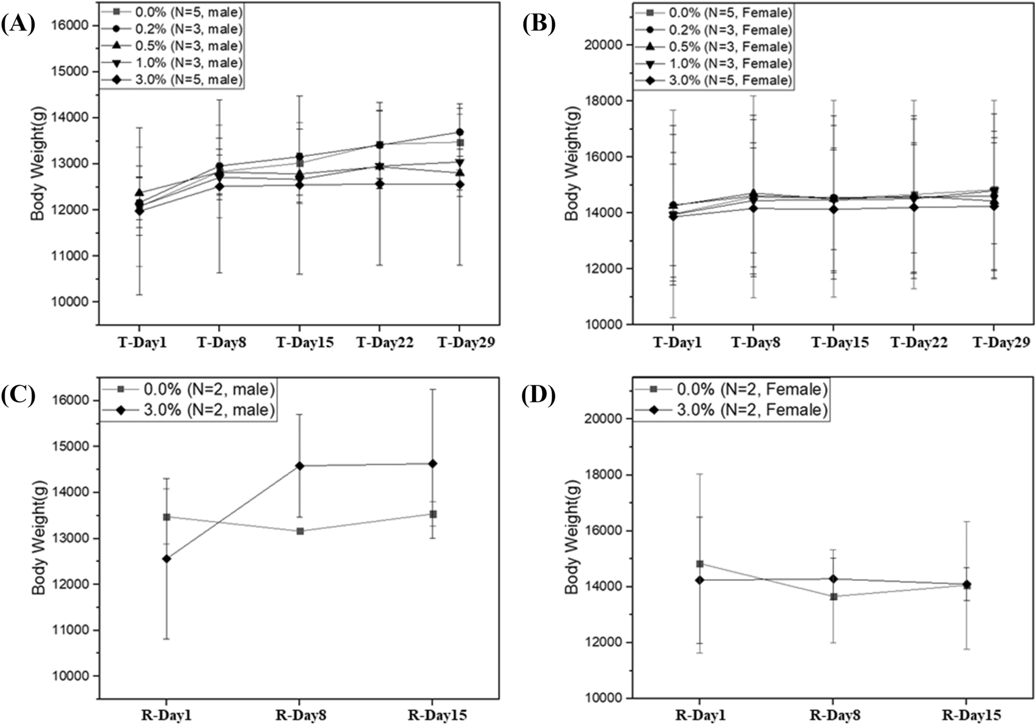
In the present study, there was statistically significant decrease in final body weights in animals of the test group compared to animals of the control group. Zhang et al. [41] obtained a similar result following daily IP injection of mice with AuNPs (13.5 nm) for 28 days. The metabolic effects of AuNPs, which have been reported by Chen et al. [6], could be the cause of this outcome. They found that IP injection of AuNPs (20–30 nm) into mice improved lipid and glucose metabolism, decreased fat mass, and aided in weight loss. However, compared to the test group, withdrawal groups I and II showed statistically non-significant and statistically significant increases in final body weights, respectively. When the final body weights of both withdrawal groups were compared, withdrawal group II showed a statistically significant increase. This could be attributed to the gradual recovery from AuNPs-induced toxicity after AuNPs clearance from the body.
Furthermore, the test group showed a statistically significant decrease in absolute testicular weights compared to the control group. Meanwhile, withdrawal groups I and II showed statistically non-significant and statistically significant increases in absolute testicular weights compared to the test group, respectively. The toxic effects of NPs on germ cell mass could be the cause of the test group's decreased absolute testicular weight [11]. While, its increase in withdrawal groups I and II indicated gradual regeneration of germ cells and gradual recovery from AuNPs induced spermatogenic defects.
Regarding serum testosterone hormone levels, they were statistically decreased in the test group compared to the control group. This result was in agreement with Behnammorshedi et al. [4] who observed that after daily IP injection of different doses of AuNPs for ten days in rats, the mean testosterone level decreased with increasing AuNPs dose. Similarly, Liu et al. [21] suggested that after daily intravenous injection of AuNPs (5–10 nm) for 14 days in mice, the testosterone production in Leydig cells reduced due to down regulating of the expression of 17α hydroxylase enzyme, which has crucial importance in androgen synthesis and degeneration of Leydig cells which are responsible for testosterone production.
Meanwhile, withdrawal groups I and II showed statistically significant increases in serum testosterone hormone levels compared to the test group. This result may be attributed to regaining of Leydig cells mitochondrial secretory activity after resolving their degeneration.
As regards semen analysis, the test group displayed statistically significant decreases in sperm count and percentage of motile sperms and a statistically significant increase in the percentage of sperms with abnormal morphology compared to the control group. These results were in accordance with Wiwanitkit et al. [36], who reported the effect of mixing AuNPs (9 nm) with a fresh semen sample from a healthy human male. They observed that after 15 min of exposure to AuNPs, the motility was lost in 25% of the sperms, and some human sperms were clumped and fragmented with an accumulation of AuNPs in the sperm tails and heads. Another study conducted by Taylor et al. [32] on bovine spermatozoa reported detrimental effects on sperm motility, morphology, and fertilizing capability after mixing with AuNPs (10.8 nm in average).
As well, Nazari et al. [28] reported a significant decrease in sperm motility and an increased number of abnormal spermatozoa after repeated IP injection of AuNPs (10–30 nm) in mice. In addition, Liu et al. [21] reported sperm malformations (including small heads, large heads, double heads, double tails, and coiled tails) after daily intravenous injection of AuNPs (5–10 nm) for 14 days in mice.
These detrimental effects of AuNPs on sperm quantity and quality were explained by the induction of reactive oxygen species (ROS), resulting in oxidative stress and mitochondrial damage with subsequent metabolic dysfunction [11].
Furthermore, withdrawal groups I and II showed statistically significant increases in sperm count and percentage of motile sperms and decrease in the percentage of sperms with abnormal morphology compared to the test group. This result reflected the gradual reversibility of AuNPs-induced damage to the epididymal sperms.
Concerning light microscopic results, the test group showed statistically significant decreases in JTBS for spermatogenesis and the mean thickness of the epithelial lining of the tubules compared to the control group. The negative effect of AuNPs on spermatogenesis in the current study may be attributed to their ability to produce ROS, which leads to formation of oxidative stress and disruption of cellular metabolism associated with inducement or exacerbation of the NPs-related inflammatory response [38], as well as induction of oxidative DNA damage that leads to cell cycle arrest and cytotoxic effects on male germ cells. This explains why sperms and spermatids were rarely seen in the test group [11].
Regarding other light microscopic results, the test group showed statistically significant disruption of seminiferous tubules, detachment of spermatogenic cells, interstitial inflammation and edema, congestion of vessels, and degeneration of Sertoli and Leydig cells compared to the control group. The mean thicknesses of tunica albuginea and blood vascular walls and the mean diameter of the seminiferous tubules showed statistically significant increases compared to the control group.
These toxic testicular histopathological changes could be explained by the intracellular leaching of gold ions from AuNPs and their effects on the surrounding biomacromolecules. Consequently, these ions strongly inhibit mitochondrial membrane depolarization and/or inactivation of mitochondrial enzymes, rendering direct or indirect mitochondrial damage, leading to alteration of cellular redox balance and promoting cell necrosis or apoptosis [34].
Furthermore, testicular interstitial inflammation and edema generated in the current study could be explained by AuNPs activation of inflammatory mediators' synthesis by disturbing the normal mechanisms of cell metabolism [17]. Additionally, congestion of blood vessels and interstitial edema could be attributed to the induction of nitric oxide production, which is an endothelial relaxing factor [23].
Moreover, the thickening of tunica albuginea, basal lamina, and blood vascular walls observed in the test group of the current study can be attributed to increased production of glycosaminoglycans and proteoglycans, a mechanism that is considered a defense reaction against the damaging activity of the probably induced ROS [3].
Although the mean thickness of the epithelial lining of seminiferous tubules was decreased in the test group of the current study, their mean diameters were increased. This could be explained as a result of the detachment of spermatogenic cells into the lumen, leading to blocking of the efferent ducts with subsequent impairment of seminiferous tubule fluid passage from the testis to the epididymis, resulting in increased seminiferous tubule diameter [25].
Regarding the electron microscopic results of the test group, most of the intratubular and interstitial testicular changes could be explained by lipid peroxidation of the cell membranes and organelles. It also destroys the structure of the spermatozoal lipid matrix, which can be associated with loss or affect sperm motility [7].
Cytoplasmic vacuolization of testicular cells in the test group of the current study might have arisen from lysosomal membrane damage induced by ROS with subsequent release of lysosomal hydrolases into the cytosol, uncontrolled extra lysosomal proteolysis, and tissue destruction [15]. The clear vacuoles within the cytoplasm might represent distended and pinched-off segments of the endoplasmic reticulum. The cellular swelling might occur as a result of failure of energy-dependent sodium–potassium ion pumps in the plasma membrane, leading to intracellular accumulation of sodium and progressive changes in osmolarity with consequent entry of water into the cells. This pattern of injury could be referred to as hydropic change [19].
The accumulation of AuNPs in Sertoli cells, spermatogenic cells, and Leydig cells in the current study was strong evidence of their ability to cross the blood testis barrier (BTB). AuNPs accumulation in Sertoli cells and its associated Sertoli cell degeneration would have altered the structural and functional integrity of testicular tissues, with subsequent disruption of the Sertoli-germ cell interaction leading to the detachment of spermatogenic cells from the seminiferous epithelium. Moreover, Sertoli cell degeneration would have impaired the production of growth factors and nutrients, which would have a harmful impact on the normal maturation of spermatogenic cells at various stages [33]. In addition, AuNPs accumulation inside the interstitial Leydig cells and its subsequent degeneration explain the decrease in testosterone levels and impaired sperm production and maturation of the test group rats [21].
Mohamed et al. [26] also noted the presence of intercellular gaps between the spermatogenic cells as found in the test group of the current study. They attributed this to the disruption of tight junctions in BTB upon exposure to the ROS, leading to the entry of excess water and toxic agents between the spermatogenic cells and widening of intercellular spaces.
Both light and electron microscopic results of the test group came in accordance with Gupta et al. [10], who stated that after oral exposure of mice to AuNPs (15 nm) for 90 days, there was considerable accumulation of AuNPs in the testes, degeneration of testicular tissues, detachment of germinal epithelium from the basement membrane, and a reduction in the population of germ cells. Also, these results came in agreement with Liu et al. [21], who performed combined in vitro and in vivo studies on Leydig cells and mice and recognized that after AuNPs (5 nm) internalization into Leydig cells lysosomes, they induced the formation of autophagosomes, increased the production of ROS, and arrested the cell cycle in S phase, resulting in concentration-dependent cytotoxicity and DNA damage with a significant reduction of testosterone production. Additionally, after daily intravenous injections of AuNPs for 14 days in mice, they accumulated and were retained in the testes in a dose-dependent manner.
However, the results of the current study were in disagreement with Leclerc et al. [20], who reported that after daily intramuscular injection of AuNPs (70 nm) for 45 days, there were neither testicular histopathological toxicity signs nor AuNPs testicular accumulation. This inconsistency of results could be due to different sizes of AuNPs and different routes of their administration.
Both light and electron microscopic results of withdrawal groups I and II of the present study reflected the gradual reversibility of AuNPs-induced damage to the testicular tissues. Similar reversible effects for the damage of the testicular tissues were detected by Bai et al. [2], who used carbon nanotubes, Ren et al. [30], who used silica nanoparticles,and Nirmal et al. [29], who used graphene oxide nanoparticles.
In this study, the reversibility of AuNPs-induced male reproductive toxicity may be attributed to: (A) exocytosis of NPs as suggested by Sakhtianchi et al. [31], (B) clearance of AuNPs from a variety of body organs as investigated by Han et al. [12], (C) deoxyribonucleic acid repair process as detected by Xu et al. [37], and (D) subsidence of AuNPs-induced oxidative stress and lipid peroxidation as concluded from other nanoparticle reversibility studies [2, 30].


留言 (0)