Clinically, patients with cutaneous adverse drug reactions induced by ABC commonly develop systemic symptoms, such as fever and general malaise [3, 4]. Although the pathological changes in the skin of patients with drug eruptions are well-characterized, the details of what occurs in the body during the development of drug eruptions remain unclear [18]. This study showed that weight loss and thymic atrophy preceded skin eruption in ABC-administered B*57:01-Tg, but not in LM. These results suggest that some HLA genotype-dependent stress is induced by medications even before the onset of skin eruptions. In addition, inflammatory cell infiltration occurred in various organs simultaneously with skin eruption in ABC-administered B*57:01-Tg, suggesting that HLA-mediated ABC-induced effects occurred systemically.
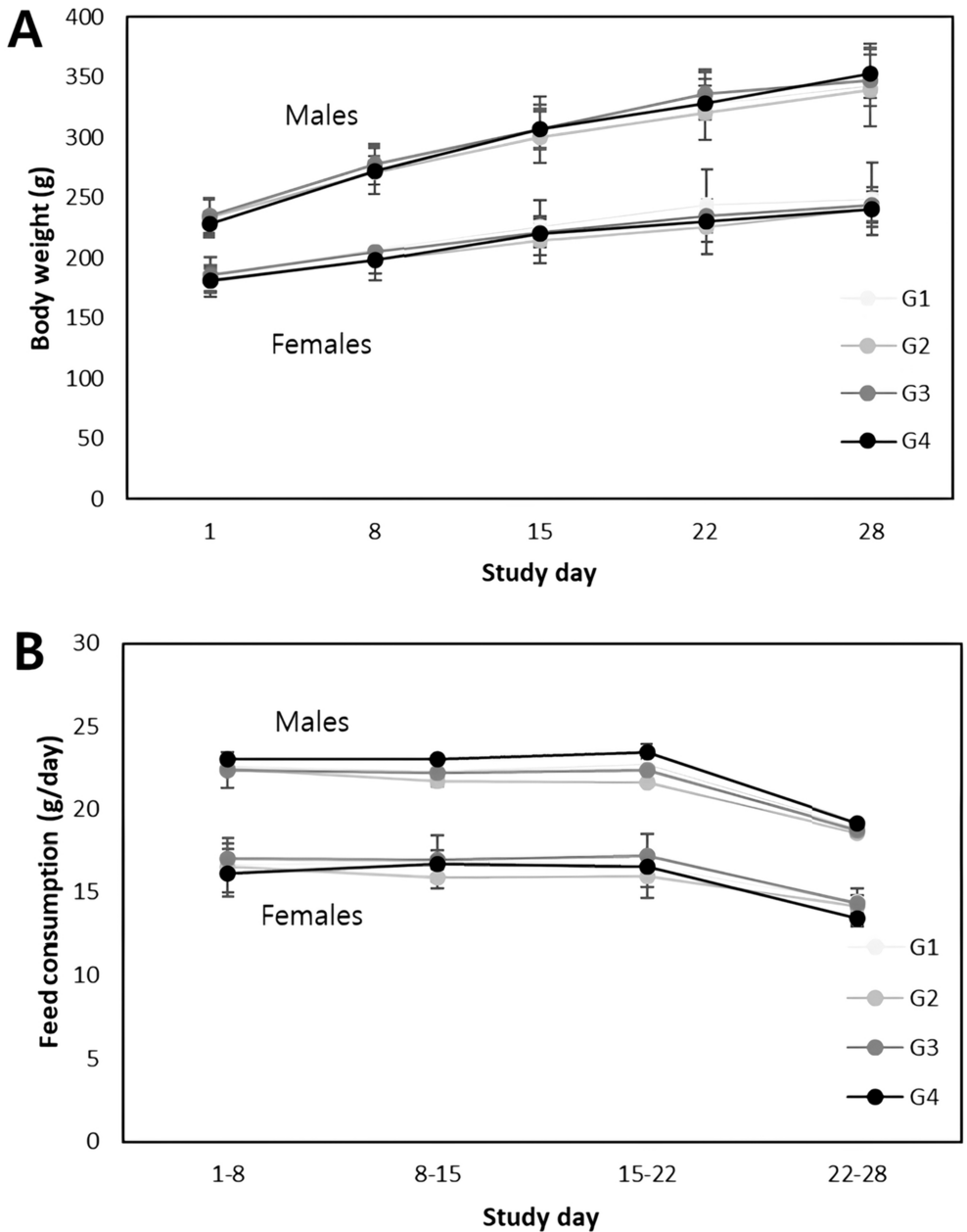
We have previously reproduced abacavir-induced skin rash using B*57:01-Tg and obtained findings regarding cutaneous manifestations such as rash, infiltration of CD8+ T cells into skin tissues, and elevated serum CCL17 levels[15, 16]. In those experiments, we also encountered a situation in which many mice died during ABC dosing. The infiltration of inflammatory cells into skin tissues was so mild that it was unlikely that the mice died from skin inflammation, thus, we wondered if there was also an HLA-B*57:01-dependent effect of abacavir on tissues other than skin. Therefore, in this study, we expected that the pathological analysis of various organs of this mice model yield interesting insights into understanding the pathogenesis of deadly drug eruptions. We attempted to analyze the pathology of various organs and observed the following sequential changes. On day 1, weight loss was observed, suggesting that a stress response may have occurred immediately after administration (Fig. 1D). On day 3, thymic atrophy occurred, and increase in serum IFN-γ and decrease in serum ALB were recorded but the differences were not statistically significant, suggesting a systemic effect (Figs. 4D, 5G, 6A). On day 5, body temperature rose and skin eruption occurred (Fig. 1B, E). Furthermore, on day 5, inflammatory cell infiltration was observed in the skin, liver, kidney, and lungs, and increases in serum levels of GLB and various cytokines and chemokines were also observed, indicating systemic inflammation (Figs. 2, 3, 5H, 6A–E). On day 7, the AST level was markedly elevated, and a slight increase in ALT was also observed, suggesting that cellular injury may have occurred in various tissues, including the liver, in association with inflammatory cell infiltration (Fig. 5A, B). The ALT level was elevated by ABC administration while the ALP level was not (Fig. 5B, C), suggesting hepatocellular injury rather than cholestasis in the liver. The serum level of CK, a myocardial deviation enzyme, was not increased and no finding related with ABC administration was observed in the heart in the histopathological examination (Fig. 5D, Table 1), suggesting no significant myocardial damage. Even though serum BUN levels were elevated on day 7, the histopathological examination of the kidneys showed no obvious damage, suggesting an extrarenal regulation of BUN level (Fig. 5E).
B*57:01-Tg, but not LM, showed body weight loss and thymic atrophy. Notably, these were observed immediately after the administration of ABC (Figs. 1D, 4D). Clinical case reports of AHS patients indicated that some experienced symptoms, such as myalgia, before the onset of skin eruptions [6]. Taken together, these clinical case reports and our observations in mice suggest that HLA genotype-dependent systemic signs may precede the appearance of skin eruptions in the pathogenesis of AHS. HLA genotype-dependent drug hypersensitivity is considered to be caused by acquired immunity, which requires a specific timeframe [19]. In addition to antigen presentation by HLA, various co-stimulatory factors, such as activation of innate immunity, signals from barriers including the epidermis, and stress responses, are considered necessary to induce acquired immunity [20,21,22]; HLA genotypes are thought to have little effect on these factors. Therefore, an HLA genotype-dependent response immediately after ABC administration was unexpected. Although the detailed mechanism underlying this phenomenon is currently unknown, previous studies have suggested that innate immunity and stress response activation are involved in the pathogenesis of AHS. Martin et al. [23] showed that HSP70-induced innate immunity may be involved and that ABC induces HSP70 expression in the endoplasmic reticulum of PBMCs from patients with AHS. As HSP70 is induced by an unfolded protein response [24], HLA-B*57:01 may form abnormal HLA complexes upon exposure to ABC, causing an unfolded protein response. Shirayanagi et al. [25] reported an increased formation of aberrant HLA complexes in HLA-B*57:01-expressing cells upon exposure to ABC. Therefore, we hypothesized that the aberrant HLA complex induces HSP70 by triggering an unfolded protein response and that the downstream immune response is involved in the early pathogenic process of AHS. However, further studies are needed to confirm this hypothesis.
Biomarkers that predict the occurrence of idiosyncratic adverse drug reactions, such as drug eruptions, including AHS, have not yet been established, making it challenging to avoid side effects [18]. Idiosyncratic adverse drug reactions are exceptionally rare, and comprehensive examinations are seldom conducted before significant symptoms arise [26]. Animal models can be valuable in identifying biomarkers that can effectively predict the occurrence of drug eruptions. The mice treated daily with ABC showed elevated serum IFN-γ levels and decrease serum ALB levels in some individuals before the onset of skin eruptions and inflammatory cell infiltration into multiple organs (Figs. 1B, 2, 3, 4, 5G, 6A). These findings again indicate that there can be signs of systemic inflammation during the process that leads to AHS. The increased IFN-γ may have induced subsequent increases in CCL17 and CXCL10 levels [27, 28], which may have been involved in the induction of skin inflammation. Gao et al. [16] reported that CCL17 is involved in T-cell infiltration into the skin. Consistent with the previous observation, elevated serum CCL17 levels were observed after day 5 (Fig. 6D), which coincided with skin eruption and CD8+ T cell infiltration (Figs. 1B, 2B). The involvement of CCL17 in the infiltration of CD8+ T cells into other tissues is unlikely [16], and further studies are required to clarify the underlying mechanism. Notably, on day 5 (Fig. 6), some individual mice exhibited markedly elevated levels of IFN-γ, TNF-α, and IL-6, although these levels appeared to decrease on day 7. Tracking the same individuals throughout the study period was not feasible as blood and organs were collected from each mouse at each time point. This implies that mice with markedly elevated cytokines on day 5 might have subsequently progressed to a cytokine storm-like syndrome [29] and died before day 7. However, due to the unavailability of data immediately before death, establishing a definitive relationship between the cause of death and cytokine levels was challenging. At present, the current study has not identified any biomarkers that predict the onset of skin eruption, i.e., biomarkers prior to day 5, and the relationship between cause of death and cytokines. A more comprehensive analysis that follows the same individual mouse from before the onset of hypersensitivity to just before death is needed.
In our mouse model, the fatality rate on day 7 was 70%, which is considerably higher than what is observed in clinical practice [3, 4]. This may be due to the loading of extreme conditions that readily induced immune activation, such as PD-1 knockout and CD4+ T-cell depletion. The infiltration of inflammatory cells into each organ, including the skin, was very mild, making it unlikely that the mice died from inflammation in the skin, liver, kidneys, or lungs. Systemic inflammation leading to death is believed to be the result of the cumulative inflammatory and immune responses in multiple organs. In fact, patients with AHS often exhibit elevated levels of serum markers associated with liver or kidney injury, including AST, ALT, ALP, lactate dehydrogenase, and creatinine [4, 6, 30, 31]. Therefore, patients with AHS may have lesions in several tissues, such as liver and kidneys, even if they do not show any noticeable symptoms. Moreover, some patients go into shock and die; however, they rarely show significant damage to specific organs [4, 31, 32]. While direct extrapolation of the results to clinical practice is challenging owing to variations in specific backgrounds of the mice, the findings imply that when administering ABC to HLA-B*57:01 carriers, it is crucial to consider early systemic inflammation and its impact on organs beyond the skin.
In conclusion, the histopathological examination of a mouse model of ABC-induced skin eruption shows that disorders in various organs other than the skin should be considered and provides insights into the unexpected early systemic responses dependent on HLA-B*57:01.


留言 (0)