Test items
HemoHIM, an extract of a triple herbal combination of Angelica gigas Nakai, Cnidium officinale Makino, and Paeonia lactiflora Pallas, contains chlorogenic acid (25–60 mg/100 g), paeoniflorin (200–400 mg/100 g), and nodakenin (50–150 mg/100 g). The sources are purchased from GEUNONONGLIM Agricultural Co. (Yeoju, Korea). It was manufactured by Kolmar BNH Co. Ltd. (Sejong, Korea). HemoHIM was prepared following the method detailed in our previous report [5]. Briefly, Angelica gigas, Cnidium officinale, and Paeonia lactiflora were extracted in equal amounts for 4 h in boiling water to obtain the extract. Half of this extract was then precipitated with ethanol to yield a water-soluble polysaccharide fraction. Subsequently, the HemoHIM was obtained by adding this polysaccharide fraction to the remaining half of the extract, which was concentrated to a solid content of 30% ± 3%. The product was then lyophilized using a freeze-dryer (FDU-1110, EYELA, Japan) and stored in a refrigerator at 4 ℃. No additional additives were introduced during this process. The formulation was prepared immediately before administration, specifically on the day of administration.
Animals and husbandry
All rats (N Tac: SD, 7 weeks old) for acute and 28-day repeated dose oral toxicity studies were obtained from Vivo Bio tech Ltd. (Hyderabad, Telangana, India). Environmental conditions in the animal room were maintained as follows: temperature = 19.6–23.6 °C, relative humidity = 50–62%, air exchange rate 14 changes/h, and light/dark cycle = 12 h/12 h. Variations in these conditions had no effect on the study outcomes. All rats (Crl:CD(SD), 6 weeks old) for 13-week repeated dose oral toxicity study were obtained from Orientbio Inc. (Seongnam, Korea). Environmental conditions in the animal room were maintained as follows: temperature = 19–25 °C, relative humidity = 30–70%, air exchange rate 10–15 changes/h, and light/dark cycle = 12 h/12 h. Variations in these conditions had no effect on the study outcomes. All mice (Swiss albino, 8–10 weeks old) for in vivo micronucleus test were obtained from Mahaveera Enterprises (Hyderabad, Telangana, India). Environmental conditions in the animal room were maintained as follows: temperature = 19.6–22.8 °C, relative humidity = 52–61%, air exchange rate 14 changes/h, and light/dark cycle = 12 h/12 h. Variations in these conditions had no effect on the study outcomes.
Acute oral toxicity
An acute oral toxicity study was conducted in accordance with the Organization for Economic Cooperation and Development (OECD) Guideline 423 [18], and OECD principles of Good Laboratory Practice (GLP) [C (97) 114/Final]. The acute toxicity of HemoHIM was assessed in male and female Sprague–Dawley rats via oral gavage, with the test item suspended in water, and administered up to the OECD dose limit (2,000 mg/kg of body weight). All animals were fasted overnight (14 h) (water ad libitum with no feed) prior to the administration of the test item. All animals were observed for mortality, morbidity, and signs of toxicity (clinical signs) at 30 min, and 1, 2, 3, and 4 h after dosing on day 0 and once daily thereafter for 14 days. Body weights were recorded prior to dosing on days 0, 7, and 14. At the end of 14 days observation period, necropsy and gross pathological examinations were performed.
28-Day repeated dose oral toxicity
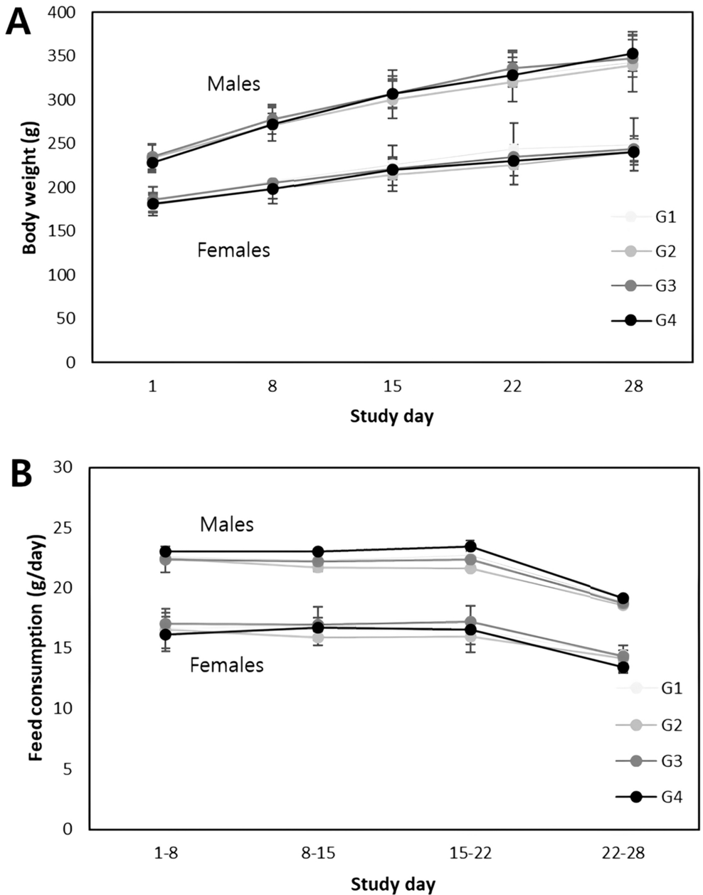
A repeated-dose oral toxicity study was conducted in accordance with the OECD Guideline 407 [19], and OECD principles of GLP [C (97) 114/Final]. The doses were administered orally to Sprague–Dawley rats for 28 consecutive days, followed by a 14-day recovery period to assess the reversibility of any toxic effects. The test item was weighed, suspended in water, and administered to rats through the oral (gavage) route using a disposable syringe with a rat intubation cannula at dose levels of 500 mg/kg/day for low-dose (G2), 1,000 mg/kg/day for mid-dose (G3), 2,000 mg/kg/day for high-dose (G4), and high-dose recovery (G4R) groups. Rats in the control (G1) and control recovery (G1R) groups received only water. The administered dose volume was 10 mL/kg/day. Each group comprised five rats of each sex. Vehicle or test formulations were administered to each rat group once daily for 28 consecutive days. After 28 days of treatment, the administration of vehicle and test item dose preparation to the control recovery (G1R) and high-dose recovery (G4R) groups was discontinued, and the potential reversibility or persistence of any toxic effects was observed for 14 days. The animals were observed twice daily for mortality/morbidity and once daily for cage-side clinical signs. Detailed clinical examinations were performed once prior to the initiation of treatment and thereafter at weekly intervals and at the end of the treatment and recovery periods. The rats were observed once per week for changes in body weight and feed consumption. Hematological and clinical chemistry investigations, and measurement of organ weight were performed at the end of the treatment and recovery periods. Animals were subjected to detailed necropsy at the end of the treatment and the recovery periods and the organs specified in the study plan were collected and weighed. The relative organ weights were calculated as percentage of body weight. Histopathological examination was carried out on all collected organs from control (G1) and high dose (G4) groups.
13-Week repeated dose oral toxicity
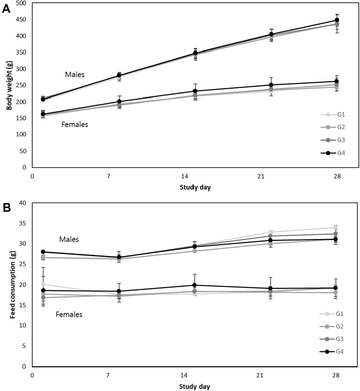
A repeated dose 13-week oral toxicity study was conducted in reference to OECD Guideline 408 [20], and in accordance with OECD principles of GLP (as revised 1997) ENV/MC/CHEM(98)17. The doses were administered orally to Sprague–Dawley rats for 13 consecutive weeks. The test item was weighed, suspended in water, and administered to rats through the oral (gavage) route using a disposable syringe with a rat intubation cannula at graduated dose levels of 500 mg/kg/day for low-dose (G2), 1,000 mg/kg/day for mid-dose (G3), and 2,000 mg/kg/day for high-dose (G4). The rats in the control (G1) received only water. The administered dose volume was 10 mL/kg/day. Each group comprised 10 rats of each sex. Vehicle or test formulations were administered to each rat group once daily for 13 consecutive weeks. During the observation period, clinical and detailed clinical signs, measurement of body weight and food consumption, ophthalmological examinations, and urinalysis were observed. At the end of the observation period, hematological and clinical chemistry examinations, observation of the estrus cycle, organ weight measurements, gross postmortem examinations, and histopathological examinations were performed. Histopathological examination was performed as follows: Organs and tissues from all animals of the control group (G1) and high dose group (G4); and organs and tissues from dead animal in the low dose group. Histopathological examination was performed on the following organs: brain; Pituitary gland; thyroid gland; parathyroid gland; thymus; lung with bronchus; heart; aorta; trachea; spleen; liver; adrenal gland; kidney; salivary gland (submandibular, sublingual, parotid); esophagus; stomach; duodenum; jejunum; ileum with Peyer’s patch; cecum; colon; rectum; pancreas; testis; epididymis; prostate gland; seminal vesicle; coagulating gland; ovary; uterus with cervix; vagina; urinary bladder; sciatic nerve; mesenteric lymph node; submandibular lymph node; eye; optic nerve; Harderian gland; skin (inguinal); mammary gland (inguinal); bone (sternum, femur); bone marrow (sternum, femur), skeletal muscle (biceps femoris); spinal cord (cervical, lumbar, thoracic).
Bacterial reverse mutation assay
An in vitro bacterial reverse mutation assay was conducted in accordance with the OECD Guideline 471 [21]. In the preliminary cytotoxicity assay, TA100 of Salmonella typhimurium was treated with the test item at 156.3, 312.5, 625.0, 1250.0, 2500.0, and 5000.0 μg/plate with (5% v/v, S9) or without metabolic activation. Vehicle and positive controls were maintained concurrently with the treatment groups. Based on the results observed in the preliminary cytotoxicity assay, 5000.0 μg/plate was selected as the highest concentration for the mutagenicity assay. Mutagenicity assays were performed using the TA1537, TA1535, TA98, and TA100 strains of S. typhimurium and WP2uvrA of Escherichia coli. The bacterial strains were treated with the test item at 312.5, 625.0, 1250.0, 2500.0, and 5000.0 μg/plate with (5% v/v, S9) or without metabolic activation. HemoHIM showed no genotoxic activity in this assay.
In vitro mammalian chromosomal aberration assay
An in vitro mammalian chromosomal aberration assay was conducted in accordance with the OECD Guideline 473 [22]. Based on the results of the preliminary cytotoxicity assay, a chromosome aberration assay was conducted using test item concentrations of 156.25, 312.5, and 625 μg/mL with or without metabolic activation. Cyclophosphamide (with metabolic activation S9) and ametycin (without metabolic activation S9) were employed as clastogenic positive controls. Human blood lymphocytes were cultured using RPMI 1640 medium supplemented with 10% fetal bovine serum (FBS), 1% penicillin–streptomycin, and 2% phytohemagglutinin in a CO2 incubator at 37 ± 1 ℃ and 5 ± 0.5% CO2. These cultures were exposed to different test item concentrations for the short-term (4 h) and continuous exposure (22 h) groups. In short term exposure, after 4 h of treatment, the culture media with the test item was replaced with RPMI 1640 complete medium and incubated for 18 h at 37 ± 1 ℃ and 5 ± 0.5% CO2. For continuous exposure, cultured cells were treated with different test item concentrations for 22 h. After 22 h, cultures from the short-term and continuous exposure groups were harvested and processed for slide preparation. The slides were stained with Giemsa stain (5% v/v). The slides were evaluated for the Mitotic Index (% of cells in metaphase).
Mammalian bone marrow erythrocyte micronucleus assay
An in vivo mammalian bone marrow erythrocyte micronucleus assay was conducted in accordance with the OECD Guideline 474 [23]. The micronucleus test was conducted at doses of 500, 1,000 and 2,000 mg/kg. The dose levels for the micronucleus test were selected based on a dose range-finding study. In the micronucleus test, HemoHIM was orally administered to Swiss albino mice (five animals/sex/group) at a dose volume of 10 mL/kg for 2 days, with an interval of approximately 24 h. Animals in the positive control group received a single dose of cyclophosphamide monohydrate intraperitoneally at 40 mg/kg a day before bone marrow collection. Approximately 24 h after dosing, all animals were euthanized and both femur bones were collected from each animal. The bone marrow was collected using 100% FBS. After collection, all samples were centrifuged and the supernatant was discarded, leaving a small amount of an FBS cell pellet. Smears were prepared on slides using the cell pellet. The slides were air-dried, fixed with absolute methanol, and stained with 5% Giemsa stain.
Statistical method
The study data was tabulated in MS Excel and subjected to statistical analysis using Graph Pad Prism Software. The data of body weight, weight gain, feed consumption, organ weight (absolute and relative), hematological and clinical chemistry estimations, were subjected to statistical analysis. The data is checked for normality and homogeneity prior to statistical comparisons. All the normal and homogenous data is analyzed using One way ANOVA followed by Dunnett’s multiple comparisons in main groups and Student’s t-test in recovery groups whereas non-normal and/or non-homogenous data is analyzed using Kruskal-Walis test followed by Dunn’s multiple comparisons in main groups and Mann–Whitney U-Test in recovery groups respectively. All analysis and comparisons were evaluated at the 95% level of confidence (p < 0.05).


留言 (0)