Antifungal agents
The test substance used in this study was ATB1651 gel, a prospective antifungal agent produced by MedPharm Ltd., a Good Manufacturing Practice (GMP) facility in the UK. Preparation analyses including stability, homogeneity, and concentration analyses were also conducted.
Test guidelines for dermal toxicity study
Dermal administration of ATB1651 gel to minipigs (Yucatan) was performed at the Contract Research Organization (CRO). This study was conducted in compliance with Good Laboratory Practice (GLP) regulations, the Ministry of Food and Drug Safety Notification No. 2017–71 Test Guidelines for Safety Evaluation of Drugs and Annex 2 Repeated Dose Toxicity Study, and the ICH Harmonized Tripartite Guidelines M3 (R2) Guidance on Nonclinical Safety Studies for the Conduct of Human Clinical Trials and Marketing Authorization for Pharmaceuticals.
Animals
All experimental procedures were approved by the Institutional Animal Care and Use Committee of the Korea Institute of Toxicology (KIT-2112-0092). Animal management and care were in accordance with the guidelines of the Association for the Assessment and Accreditation of Laboratory Animal Care (AAALAC). Yucatan minipigs (Sus scrofa) weighing 10–12 kg (3–5 months old) were housed individually in stainless-steel slide bottom cages during the study period to avoid cross-contamination of the test items and application sites. The animal house was maintained under the following controlled conditions: temperature, 19–27 °C; relative humidity, 30–70%; ventilation, 10–20 times per hour; and ~ 12-h light cycle at 300–700 lx. The minipigs were fed gamma irradiation-sterilized diets (~ 300 g per head) once daily.
Determination of body surface area (BSA)
The dosing site was determined as 10% of body surface area (BSA) according to the OECD test guideline No. 410. First, total body surface was determined using the following formula: BS (m2) = (70 × BW0.75)/1000 for pigs weighing 6–30 kg [25], where BW was the maximum body weight for each sex. The 10% BSA was recalculated based on the recent body weight measured once per week. To avoid the spine, the 10% BSA was split into 2 areas: 5% BSA on the left and 5% BSA on the right side of the spine, with ~ 4 cm distance between both application sites. Prior to dermal application, hair at the dosing sites was clipped or shaved using a clipper, and each corner of the dosing site was tattooed. The procedures were performed under sedation using ketamine and xylazine. During the administration period, hair at the administration site was gently removed to maximize ATB1651 gel absorption.
Dermal administration
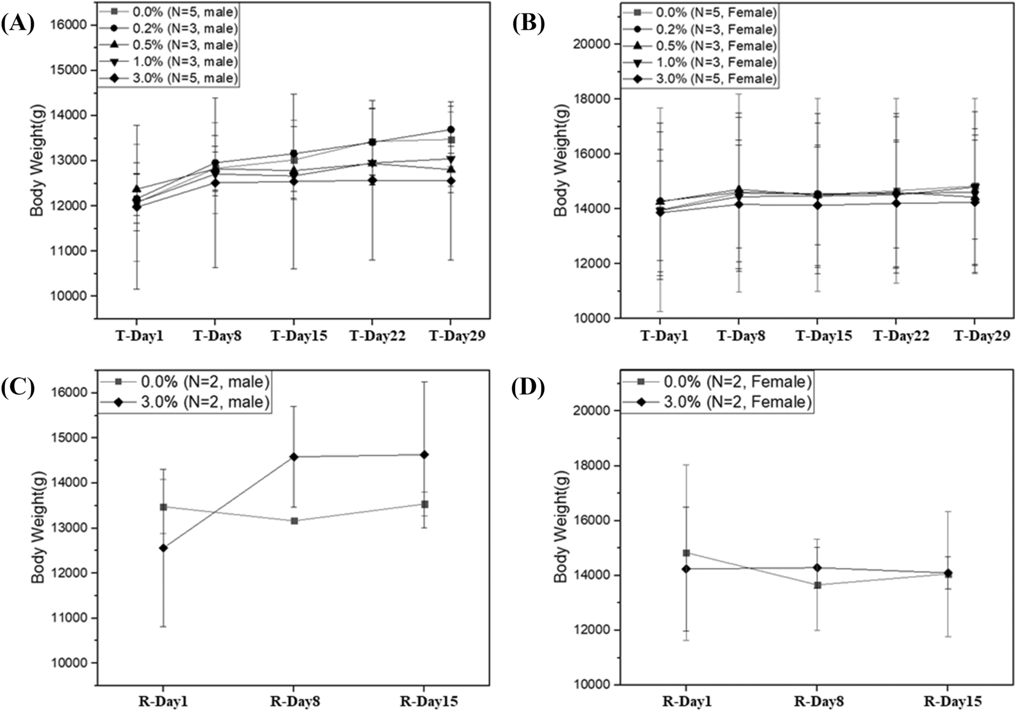
The experimental period comprised 7–8 days of pretreatment to obtain initial data, a 4-week treatment period, and a 2-week recovery period. Yucatan minipigs (n = 38) were randomly assigned to 1 control group (0% ATB1651 gel) and 4 treatment groups (0.2, 0.5, 1.0, and 3.0% ATB1651 gel). Each group comprised 3 males and females except the control and 3.0% groups, which contained an additional 2 males and 2 females to evaluate reversibility during the recovery period. In a previous 4-week dermal administration study of liquid formulation in minipigs, the systemic no-observed-adverse-effect level (NOAEL) was determined at 2.0%. It was considered that the turns from liquid to gel type of the test item formulation due to the change in vehicle control item would weaken dermal toxicity, so the high dose level was determined to be 3.0% (1.0% higher than in the previous study). Therefore, the high-dose level used in the present study was 3.0%, the middle-dose levels were 1.0% and 0.5%, and the low-dose level was 0.2% (no expected toxicity).
Both control and test formulations were gently and uniformly applied to the open application sites (10% BSA) and administered dermally once daily for 4 weeks (dose volume of at least 5 μL/cm2) using a soft spatula. Administration was performed in the cages without animal restraint. The exposure time for the control and test formulations was ~ 6 h; application sites were subsequently rinsed with clean water to remove any residual formulations and dried with porous gauze. The administration site was not occluded with gauze during formulation exposure, as the formulation absorption time was short (as determined through our own preliminary tests).
Observation and measurements
Most experimental measurements and observations, including body weight, food consumption, clinical signs, ophthalmic examination, electrocardiogram, and dermal scoring, were recorded using Pristima XD software (Version 7.4. Xybion Corporation; Lawrenceville, New Jersey, USA).
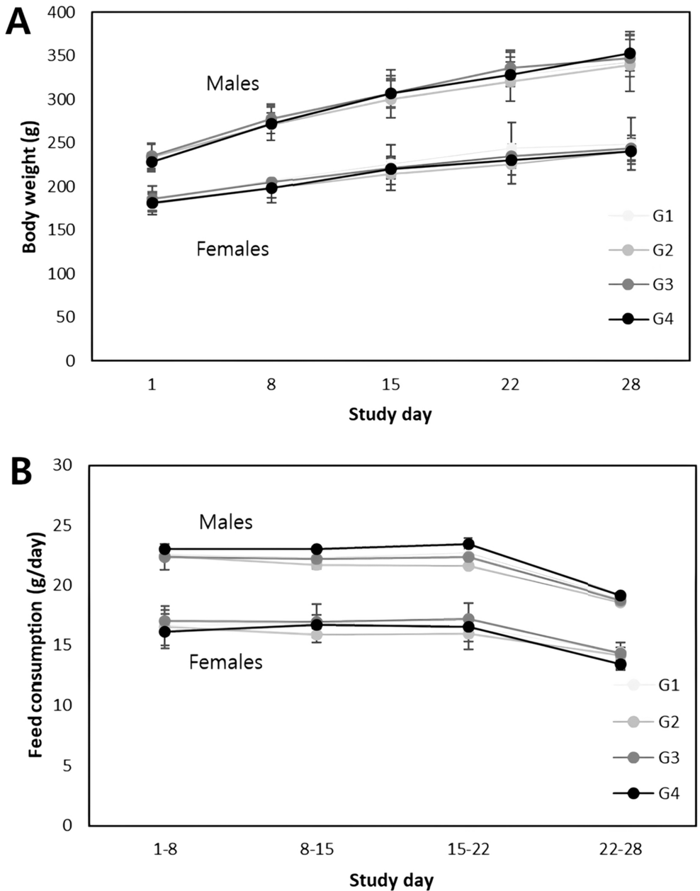
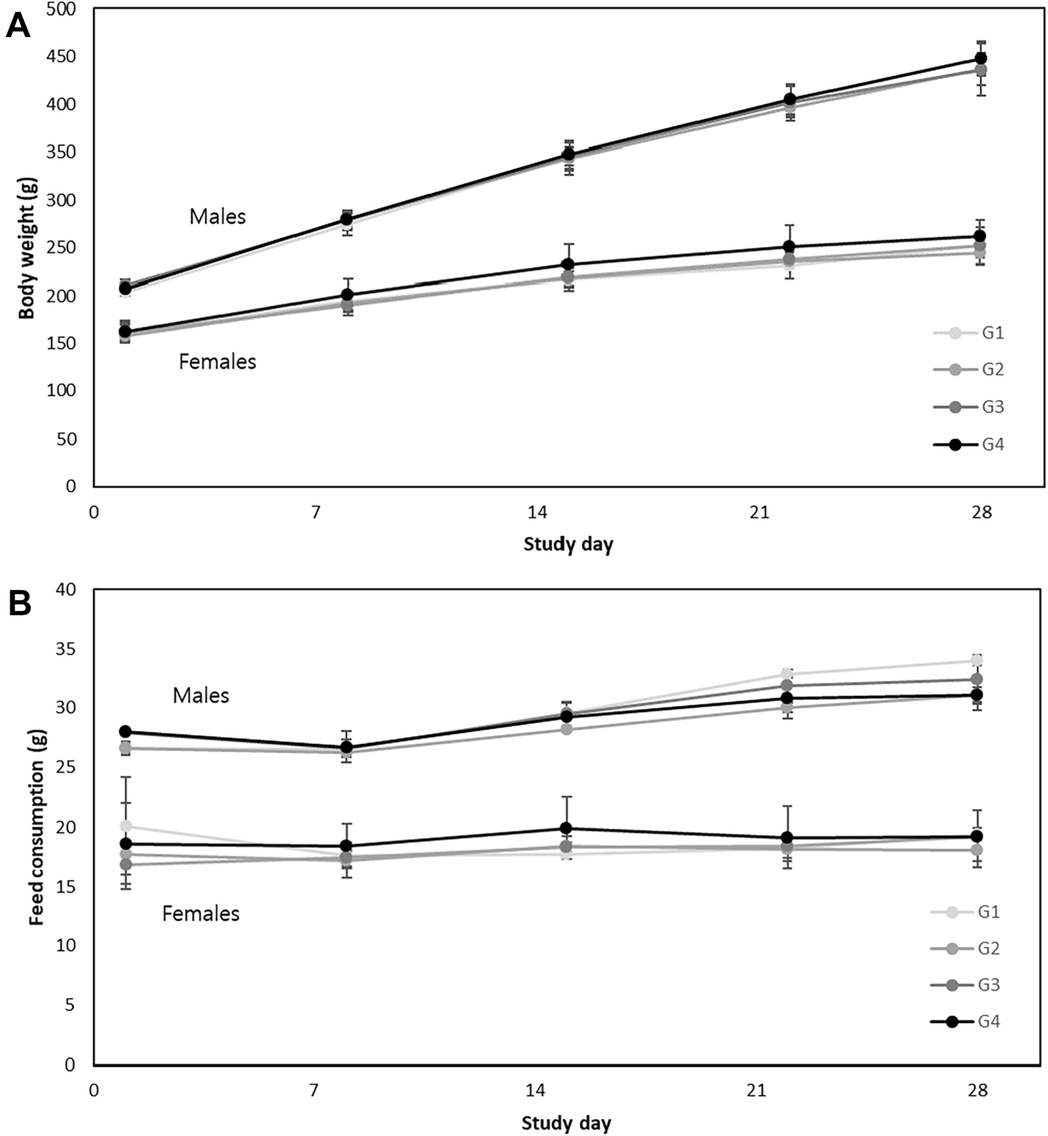
Mortality and clinical signs, such as moribund state, general appearance, and behavioral changes, were observed once daily during the entire experimental period and twice daily during the treatment period. Body weight and food consumption were measured weekly during the entire study period. Ophthalmological and electrocardiographic examinations were conducted by a veterinarian under sedation with ketamine (11–12 mg/kg) and xylazine (2–3 mg/kg) for all live animals during pretreatment, test treatment (day 24), and the recovery period (day 9 or 10). A slit lamp (XL-1, Ohira Co., Ltd., Japan) and binocular indirect ophthalmoscope (Vantage Plus Digital, Keeler Ltd., England) were used for ophthalmic examination of animals after receiving 1–2 drops of a mydriatic agent (Mydriacyl Oph Soln 1%, Alcon, Geneva, Swiss) to both eyes. Cardiac electrocardiogram intervals (QT, QTc, PR, and QRS) and heart rate were measured using an electrocardiograph (Cardio XP; Bionet Co., Ltd., Korea). QTc at each time point was obtained using Bazett’s formula [26, 27]. During treatment and recovery periods, the administration site of each minipig was observed in detail once a week before dosing for erythema, edema, and eschar formation. Dermal scores were recorded according to Draize [28].
Clinical pathology examination
Blood obtained from the jugular vein or vena cava was collected in EDTA-2K tubes, 3.2% sodium citrate tubes, and serum-separating tubes to assess hematology, coagulation, and clinical chemistry, respectively. Clinical pathology examination was performed 2–3 times during the entire study period on the following days: one week before administration, day 29 for all animals, or day 43 for recovery animals. All animals were fasted for ~ 16 h before blood sampling and provided with water ad libitum.
Hematological parameters, including total white blood cell (WBC) count, total red blood cell (RBC) count, hemoglobin, hematocrit, mean corpuscular volume, mean corpuscular hemoglobin, mean corpuscular hemoglobin concentration, platelet count, reticulocyte count absolute, reticulocyte count relative, neutrophil count absolute, neutrophil count relative, eosinophil count absolute, eosinophil count relative, basophil count absolute, basophil count relative, monocyte count absolute, monocyte count relative, lymphocytes absolute, lymphocytes relative, large unstained cells absolute, and large unstained cells relative, were measured using an ADVIA2120i hematology analyzer (Siemens, USA). Coagulation parameters, including prothrombin time and activated partial thromboplastin time, were measured using an ACL Elite Pro coagulation analyzer (Instrumentation Laboratory, Italy). Clinical parameters, including glucose, BUN, creatinine, total protein, albumin, albumin/globulin ratio, total cholesterol, triglyceride, phospholipid, AST, ALT, total bilirubin, AP, gamma-glutamyl transpeptidase, creatine phosphokinase, calcium, inorganic phosphorus, sodium, potassium, and chloride, were measured using a TBA 120FR chemistry analyzer (Toshiba Co., Japan).
Urine was collected from the cage pans of all animals on the day before administration, terminal sacrifice (day 29), and recovery sacrifice (day 43) for analysis. Before urine collection, the animals were fasted overnight; however, drinking water was provided. Urine volume was recorded using a measuring cylinder, and the following parameters were measured using a Cobas U411 urine analyzer (Roche, Switzerland) with a urine reagent strip: color, clarity, pH, specific gravity, bilirubin, protein, urobilinogen level, nitrite level, glucose level, erythrocyte count, ketone, leukocyte count, urine potassium, urine chloride, urine sodium, urine cast, epithelial cell count, WBC count, and RBC count.
Necropsy and histopathological examination
All animals were anesthetized intravenously via the ear vein using thiopental sodium (75–80 mg/kg) after intramuscular sedation with ketamine (11–12 mg/kg) and xylazine (2–3 mg/kg) on the day of terminal sacrifice (day 29) and recovery sacrifice (day 43), and then euthanized by exsanguination. Tissue samples collected from all animals at the terminal and recovery sacrifices were examined macroscopically by a veterinary pathologist. The brain, adrenal glands, pituitary gland, testes, liver with gall bladder, epididymides, spleen, lung with bronchi, heart, thyroid, thymus, uterus and cervix, salivary glands, ovaries with oviducts, seminal vesicle, prostate, and kidneys were weighed. All organs were preserved in 10% neutral buffered formalin, except the eyes (with optic nerve), which were fixed in Davidson’s fixative and the testes and epididymides, which were fixed in Bouin’s fixative for ~ 24 to 72 h and transferred to 70% ethanol. Formalin was infused into the lungs via the trachea and urinary bladder. Subsequently, the sections were embedded in paraffin, sectioned (section thickness: 2.5 μm), stained with hematoxylin and eosin (H&E), and examined under a microscope (BX53, Olympus, Tokyo, Japan). Images were obtained at × 200 magnification using a microscope and evaluated microscopically by a veterinary pathologist. The following tissues were examined and analyzed in this study: abnormal lesions, adrenal glands, animal ID, aorta (thoracic), brain, cecum, colon, duodenum, epididymis, esophagus, eyes with optic nerve, femur with marrow, heart, ileum, jejunum, kidneys, liver with gall bladder, lung with bronchi, mammary gland, uterus with cervix, vagina, injection sites, pancreas, prostate, pituitary gland, rectum, salivary glands, sciatic nerve, seminal vesicles, skeletal muscles, skin, thoracic spinal cord, spleen, sternum with marrow, stomach, testes, thymus, thyroids, tongue, trachea, urinary bladder, mesenteric lymph node, ovaries, and mandibular lymph nodes.
Statistical analysis
All data were analyzed using Prism (Version 8; GraphPad Software, San Diego, CA, USA) and Pristima software (Version 7.4; Xybion Medical Systems Corporation, Lawrenceville, New Jersey, USA). Data were analyzed for homogeneity of variance using Bartlett’s test. Homogeneous data were analyzed using analysis of variance (ANOVA), followed by Dunnett’s test for multiple mean comparison. Heterogeneous data were analyzed using a Kruskal–Wallis test, followed by Dunn’s rank sum test to compare the control and experimental groups. After performing an F test to assess homogeneity of variance between the control and recovery groups, a Student’s t test was conducted to analyze significant differences between the homogeneous data groups. The Wilcoxon rank-sum test was used to assess differences between the heterogeneous data groups. Statistical significance was set at p < 0.05.


留言 (0)