
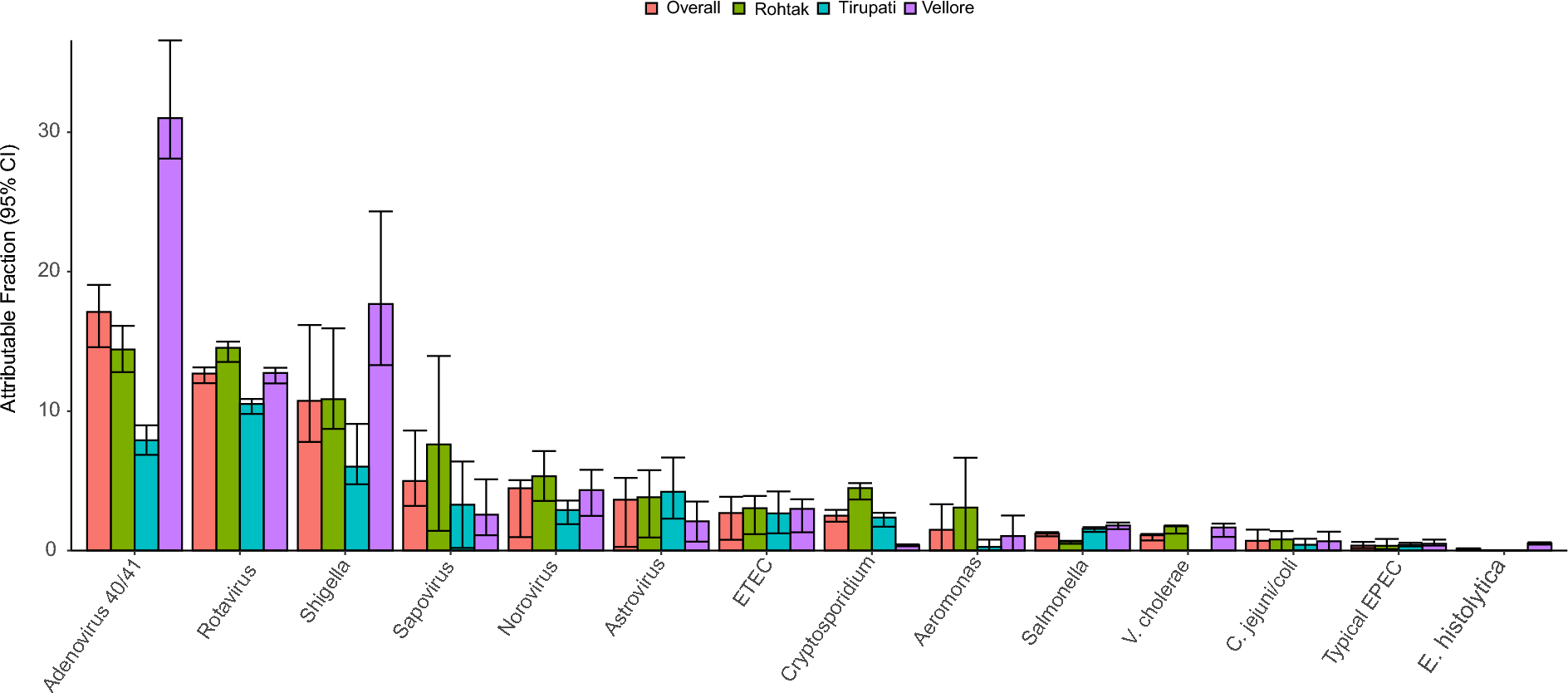
記住我


To explore the potential relationship between F. nucleatum and ALF, we used FISH to detect the abundance of F. nucleatum in 15 liver tissues of ALF patients and 15 normal liver tissues. The liver histological score and the abundance of F. nucleatum detected in liver tissues of ALF patients was significantly higher than that of normal liver tissues (Fig. 1a, b). Then we analyzed the metabolic patterns of liver tissues of ALF patients and normal controls. Compared with the normal control group, the liver tissue of ALF patients showed inhibition of energy metabolism (Fig. 1c). As ATP and NAD+ have an excitatory effect on immune cells and adenosine has an anti-inflammatory effect on immune cells, NAD+ is an important coenzyme that mediates redox reactions [24]. We have detected that compared with normal liver tissue, liver NAD+ levels in ALF patients are significantly lower (Fig. 1d). According to reports, lack of NAD+ can cause moderate hepatic inflammation and damage [25]. We speculated that in the process of ALF, NAD+ biosynthesis was impaired. To this end, we tested the expression levels of major enzymes that control NAD+ biosynthesis, NAMPT and indolamine 2,3-dioxygenase (IDO) are rate-limiting enzymes for NAD+ salvage and de novo biosynthesis pathways respectively [26]. Interestingly, we found that the expression of NAMPT in liver tissues of ALF patients decreased. At the same time, the expression level of IDO had not changed much (Fig. 1e, f). We then evaluated the relationship between the abundance of F. nucleatum and clinicopathological features as shown in Table 1. The abundance of F. nucleatum was positively associated with the clinical course and refractory behavior (P < 0.05). These results showed that the NAD+ salvage synthesis pathway controlled by NAMPT might be an important factor in the consumption of NAD+ in the process of ALF, and the infection of F. nucleatum might be an important link, while the NAD+ de novo synthesis pathway might be an adaptive response.
Fig. 1
Fusobacterium nucleatum is Associated with ALF activity. a The representative images of HE staining and FISH of liver in each group to assess the amount of F. nucleatum in ALF and healthy control tissues. FUS664 (green) is a FITC-conjugated F. nucleatum-specific oligonucleotide probe; EUB338 (red) is a Cy3-conjugated universal bacterial oligonucleotide probe. Magnification, 200×. b The liver histological score of liver in each group (*P < 0.05; unpaired Student’s t-test; the error bars indicate the SDs). c Liquid Chromatography–Mass Spectrometry (LC–MS) Metabolomics of liver in each group. d NAD+ level in liver tissues obtained by hepatectomy from patients. e, f Immunohistochemistry analysis on the protein levels of NAMPT and IDO in liver tissues obtained by hepatectomy from patients. *P < 0.05; unpaired Student’s t-test; The error bars indicate the SDs for triplicate samples
Table 1 Clinicopathologic characteristics in F. nucleatum-negative vs. F. nucleatum-positive ALF Fusobacterium nucleatum aggravated the degree of inflammatory damage in ALF model and regulated the expression of NAMPTWe hypothesized that the infection of F. nucleatum might aggravated the degree of inflammatory damage during ALF. To tested this hypothesis, we established the model of ALF. Compared with the normal control group, the mice treated with F. nucleatum or E. coli alone did not change significantly. However, mice treated with ALF + F. nucleatum exhibited more severe liver inflammation symptoms compared with the ALF group and E. coli + ALF group, including massive hemorrhagic necrosis, liver lobule structure disorder, and obvious inflammatory cell infiltration and higher histological scores (Fig. 2a, b). The 24 h survival rate of mice was observed in each group. The results showed that 40.0% of mice survived in E. coli + ALF group and ALF group, whereas only 16.0% in F. nucleatum + ALF group (Additional file 1: Fig. S1a). In addition, we evaluated the liver NAD+ levels and plasma levels of liver enzymes in each group. Consistent with previous observations, ALF mice showed lower NAD+ levels after the pretreatment of F. nucleatum (Fig. 2c). The levels of ALT and AST in the ALF group increased significantly and F. nucleatum treatment could significantly aggravate this abnormal increase (Fig. 2d). In addition, we tested the expression levels of NAMPT and IDO in vivo and in vitro respectively. Immunohistochemical staining showed that the expression level of NAMPT in mice interfered with F. nucleatum decreased, while that in ALF mice decreased significantly, and the expression level decreased further after the intervention of F. nucleatum. During this period, the expression level of IDO did not decrease but increased slightly (Fig. 2e). In vitro we incubated L02 cells with F. nucleatum or E. coli in a time-dependent manner. Western blotting results showed that the level of NAMPT was positively correlated with the time of F. nucleatum intervention, while the level of IDO did not change significantly. In addition, the intervention of E. coli had no effect on the expression of NAMPT and IDO (Fig. 2f, g). We also tested the level of NAD+ at the cellular level, and the results showed that with the intervention of F. nucleatum, the level of NAD+ gradually decreased, while the intervention of E. coli had no effect (Fig. 2h, i). These results showed that F. nucleatum might affect NAD+ by regulating the expression level of NAMPT, and further promote the progress of ALF.
Fig. 2
Fusobacterium nucleatum aggravated the degree of damage in ALF model and regulated the expression of NAMPT in vitro and in vivo. a Mice (n = 5–7 per group) were administered F. nucleatum, E.coli or PBS for 4 weeks and treated with LPS and D-gal for another 24 h. The representative images of FISH to assess the amount of F. nucleatum in livers of each group. Representative images of histological analyses are shown in (a) and quantified in (b) (200 × magnification). c NAD+ level in liver tissues obtained from mice. d The plasma levels of ALT and AST were measured in each group. e Immunohistochemistry analysis on the protein levels of NAMPT and IDO in liver tissues obtained from mice. f, g Western blotting was performed to measure the expression of NAMPT and IDO in L02 cells cocultured with F. nucleatum, E. coli or PBS (Control, Con) and quantified. Data shown are means ± SD of three separate experiments. *P < 0.05; one-way ANOVA combined with Bonferroni's post hoc test; the error bars indicate the SDs for triplicate samples. h, i The NAD.+ content of L02 cells treated by F. nucleatum or E.coli with a time gradient. *P < 0.05; unpaired Student’s t-test; The error bars indicate the SDs for triplicate samples
Fusobacterium nucleatum aggravated macrophages infiltration and pro-inflammatory response in ALF modelSince endotoxins derived from the intestine can not only directly destroy liver tissues, but also induce local non-specific hypersensitivity reactions in the liver, induce macrophages to release a large amount of cytokines, and further produce natural immune cascades, resulting in a "second blow", and aggravated the damage of liver cells in the process of ALF. Therefore, we tested the level of macrophages infiltration and the degree of inflammatory response. As showed in Fig. 3a, the result of immunofluorescence revealed that the number of macrophages within liver tissues was increased in ALF model and infection with F. nucleatum enhanced this effect, but there was no similar macrophages infiltration in E. coli pretreated mice (Fig. 3a, c). Besides, the expression of TNF-α, and IL-1β in the liver tissues of ALF model were up-regulated and pretreatment with F. nucleatum enhanced this effect. At the same time, the structural damage of the liver lobules was more obvious and E.coli pretreated mice had no such effect (Fig. 3b, d). We found the same phenomenon in vitro. As the infection time of F. nucleatum increased, the expression levels of TNF-α and IL-1β also increased (Fig. 3e-g). Not only that, the protein level of anti-apoptotic protein Bcl-2 decreased, and the level of pro-apoptotic protein Bax increased (Additional file 1: Fig. S1c). These results indicated that the infection of F. nucleatum might aggravate macrophages infiltration and inflammation in the ALF model, and promote hepatocyte apoptosis.
Fig. 3
Fusobacterium nucleatum aggravated macrophages infiltration and pro-inflammatory response in vitro and in vivo. a Representative images and quantitative analysis (c) of infiltrated monocytes by F4/80 (left panel) and CD68 (right panel) immunofluorescence staining in liver tissues of each group. *P < 0.05. n = 6 for each group. b Immunohistochemistry analyses of the pro-inflammatory factors TNF-α and IL-1β in liver tissues and quantitative analysis (d). *P < 0.05. n = 6 for each group. (e–g) real-time PCR and immunofluorescence staining were performed to measure levels of pro-inflammatory cytokines in L02 cells infected by F. nucleatum with a time gradient. *P < 0.05 vs. Data were analysed by Student’s t-test. The error bars indicate the SDs for triplicate samples
Fusobacterium nucleatum inhibited the antioxidant capacity of the ALF modelNext, we investigated the molecular mechanisms of the pro-inflammatory effect of F. nucleatum on the ALF model. Oxidative stress has been shown to promote inflammation during ALF [27]. Therefore, we evaluated the antioxidant capacity of the ALF model 24 h after the infection of F. nucleatum. As showed in Fig. 4a, b, the level of ROS stimulated in ALF model was significantly enhanced by F. nucleatum. Compared to the control group, the activities of malondialdehyde (MDA) were increased with the intervention of F. nucleatum, while superoxide dismutase (SOD) and glutathione peroxidase (GSH‐Px) were opposite, indicating that the antioxidant activity was decreased (Fig. 4c-e). The involvement of SIRT1 in the process of anti-oxidation and anti-inflammatory is well known. According to reports, NAD+ can affect the activity of SIRT1 and its expression level [28]. Our experiments showed that the expression of SIRT1 in the ALF model was reduced, and further decreased after the infection of F. nucleatum (Fig. 4f, g). These results showed that F. nucleatum might have the ability to inhibit the antioxidant capacity in the ALF model.
Fig. 4
Fusobacterium nucleatum inhibited the antioxidant capacity of L02 cells. a ROS productions were detected by DHE staining. Representative images of the DHE staining in different groups. b ROS productions were evaluated by quantification of mean fluorescence intensity in DHE staining. c–e Levels of Malondialdehyde (MDA), Superoxide dismutase (SOD) and Glutathione peroxidase (GSH‐Px) in L02 cells. f Immunoblotting analysis on the protein levels of SIRT1 and quantified in (g). L02 cells stimulated with TNF-α (100 ng/mL) and D-Gal (44 μg/mL) were treated with F. nucleatum for 24 h. Data shown are means ± SD of three separate experiments. *P < 0.05; one-way ANOVA combined with Bonferroni's post hoc test; the error bars indicate the SDs
NAD+ supplementation reversed the inhibition of F. nucleatum on the antioxidant capacity of ALF modelsSince SIRT1 is the most studied NAD+-dependent effector [29], we measured SIRT1 expression in L02 cells stimulated with F. nucleatum, as showed in Fig. 5a, administration of different concentrations of NAD+ in L02 cells could increase the expression of SIRT1 in a concentration-dependent manner even with the intervention of F. nucleatum, but the effect was not so obvious when the NAD+ concentration exceeds 1000 nM. Next, we used 1000 nM of NAD+ to intervene in L02 cells and detected the level of ROS production. Consistent with the previous results, NAD+ intervention could alleviate the generation of ROS in L02 cells stimulated by F. nucleatum (Fig. 5b, c). Besides, the activities of SOD and GSH‐Px were significantly enhanced with the intervention of NAD+, while MDA levels were reduced (Fig. 5d-f). These results showed that the supplementation of NAD+ might reverse the inhibition of F. nucleatum on the antioxidant capacity of ALF models.
Fig. 5
NAD+ supplementation reversed the inhibition of F. nucleatum on the antioxidant capacity of L02 cells. a Immunoblotting analysis on the protein levels of SIRT1 in L02 cells treated with different concentrations of NAD.+ and stimulated with TNF-α (100 ng/mL), D-Gal (44 μg/mL) and F. nucleatum for 24 h. b ROS productions were detected by DHE staining. Representative images of the DHE staining in different groups. c ROS productions were evaluated by quantification of mean fluorescence intensity in DHE staining. d–f Levels of Malondialdehyde (MDA), Superoxide dismutase (SOD) and Glutathione peroxidase (GSH‐Px) in L02 cells. Data shown are means ± SD of three separate experiments. *P < 0.05; one-way ANOVA combined with Bonferroni's post hoc test; the error bars indicate the SDs
Supplemented the natural NAD+ precursor NR corrected the progression of ALF induced by F. nucleatum infection significantly instead of overexpression of SIRT1Next, we verified the effect of supplementing NAD+ on the ALF model of F. nucleatum infection in vivo. Mice were pretreated with F. nucleatum and NAD+ precursor NR, a widely-used NAD+ precursor for increasing NAD+ content [30]. Consistent with in vitro experimental results, compared with the ALF model of F. nucleatum infection, NR treatment could reduce the histological score of the ALF model (Fig. 6a, d), reduce the infiltration of macrophages (Fig. 6a, e) and the expression of inflammatory factors (Fig. 6b, f). The 24 h survival rate of mice was observed in each group. The results showed that 66.6% of mice survived in NR treatment group and ALF group, whereas only 16.0% in F. nucleatum + ALF group (Additional file 1: Fig. S1b). In addition, we investigated whether the loss of SIRT1 caused by NAD+ pool consumption could totally explain the susceptibility of F. nucleatum infection to the ALF model. In vivo, we injected the adenovirus carrying SIRT1 (Ad-SIRT1) from the tail vein to increase the protein expression level of SIRT1 in the liver of mice in the ALF model (Fig. 6c). As showed, SIRT1 overexpression significantly reversed the histological score in the ALF model and 33.3% of mice survived (Additional file 1: Fig. S1b), but the effect was not as obvious as the NR pretreatment group (Fig. 6a, d). The results of immunofluorescence and immunohistochemistry also demonstrated that SIRT1 overexpression could inhibit macrophages infiltration and the expression of inflammatory factors at a certain extent, but the effect was insufficient obviously compared with the NR pretreatment group (Fig. 6a-f). These results showed that the supplementation of NAD+ precursor NR corrected the progression of ALF induced by F. nucleatum infection significantly instead of overexpression of SIRT1.
Fig. 6
NR replenishment, but not SIRT1 overexpression, completely corrected the progression of ALF induced by F. nucleatum infection. a Representative images of HE staining and infiltrated monocytes by F4/80 and CD68 immunofluorescence staining in liver tissues of each group and quantified in (d, e). *P < 0.05. n = 6 for each group. b Effect of SIRT1 overexpression or NR on expression of the pro-inflammatory factors TNF-α and IL-1β in liver tissues and quantified in (f). *P < 0.05. n = 6 for each group. c Western blotting was performed to measure the expression of SIRT1 in liver tissues treated with SIRT1 overexpression or NR in mice infected with F. nucleatum and quantified. Data shown are means ± SD of three separate experiments. *P < 0.05; one-way ANOVA combined with Bonferroni's post hoc test; the error bars indicate the SDs
Fusobacterium nucleatum inhibited SIRT1 expression in a NAMPT/NAD+ dependent manner in the ALF modelMany factors in the cell can regulate the content of NAD+ through different ways. Among the most important factors, besides NAMPT, AMPK increases the content of NAD+ by increasing mitochondrial β-oxidation [31]. Therefore, we tested the effect of F. nucleatum on the expression level of p-AMPK in the ALF model in vivo and in vitro. The results showed that the intervention of F. nucleatum could reduce the expression of p-AMPK indeed, and it gradually decreased in L02 cells in a time-dependent manner (Fig. 7a, b). Next, we wanted to figure out the main reason for the decrease of NAD+ and SIRT1 in F. nucleatum infection. We constructed the NAMPT overexpression plasmid and pretreated the cells with AMPK activator AICAR. As showed in Fig. 7c, western blotting results showed that the expression of SIRT1 was up-regulated after overexpression of NAMPT or activation of AMPK under the intervention of F. nucleatum, but the overexpression of NAMPT was significantly more effective than the activation of AMPK. In the case of supplementation of NAMPT, it was more effective after the activation of AMPK. We also found the same results in the NAD+ test (Fig. 7d). We continued to compare the levels of ROS in L02 cells after different treatments. Compared with activating AMPK, the overexpression of NAMPT could clear ROS in the ALF model infected by F. nucleatum significantly more effectively (Fig. 7e, f). In addition, immunofluorescence detected the expression level of inflammatory factors, and the inflammatory response after activating AMPK was still more severe than the overexpression of NAMPT, but the inflammatory response was significantly weaker when the overexpression of NAMPT combined with the activation of AMPK (Fig. 7g, h). These results showed that the inhibition of F. nucleatum on NAD+ and SIRT1 was mainly through the salvage synthesis pathway of NAD+. In addition, after treatment with metronidazole, Western bolt results showed that the protein level of SIRT1 increased, and the levels of Bax and inflammatory factors decreased, suggesting that the levels of inflammation and apoptosis decreased (Additional file 1: Fig. S1d).
Fig. 7
Fusobacterium nucleatum inhibited NAD+ via the salvage synthesis pathway of NAD+. a Western blots analysis of p-AMPK in mice treated with LPS (100 μg/kg) and D-gal (400 mg/kg) or infected with F. nucleatum and quantified. b Western blots analysis of p-AMPK in L02 cells stimulated with TNF-α (100 ng/mL), D-Gal (44 μg/mL) and infected by F. nucleatum with a time gradient and quantified. c, d The cells were transfected with NAMPT plasmid or treated with AICAR for 24 h and then stimulated with TNF-α (100 ng/mL), D-Gal (44 μg/mL) and infected by F. nucleatum for 24 h. Western blotting was performed to measure the expression of SIRT1 and the NAD+ content of cells were measured in each group and quantified. e Representative images of the DHE staining in different groups and evaluated by quantification of mean fluorescence intensity (f). g immunofluorescence staining were performed to measure levels of pro-inflammatory cytokines and quantified in (h). Data shown are means ± SD of three separate experiments. *P < 0.05; one-way ANOVA combined with Bonferroni's post hoc test; the error bars indicate the SDs
留言 (0)