Isolation and characterization of human NK cellsNK cell enrichment and expansion
All manufacturing and product testing procedures for the generation of human NK cells were performed under GMP conditions (NKMAX Co., Ltd, Seongnam, Korea). Peripheral blood mononuclear cells (PBMCs) were isolated from healthy donors by leukapheresis, and NK cells were expanded as described previously [13], with some modifications. Briefly, CD56+ cells were isolated from PBMCs using CliniMACS CD56 microbeads (Miltenyi Biotech GmbH, Galdbach, Germany) according to the manufacturer’s instructions. Isolated CD56+ cells were then cultured in RPMI-1640 medium (WELGENE Inc., Gyeongsan, Korea) supplemented with 10% fetal bovine serum (FBS; Hyclone, Tauranga, New Zealand), 20 µg/mL gentamicin (GIBCO, Grand Island, NY), g-irradiated (100 Gy) KL-1 and LCL feeders, 500 IU/mL IL-2 (PROLEUKIN®; Norvatis, Basel, Switzerland), and 50 ng/mL IL-21 (NKMAX Co., Ltd., Seongnam, Korea). Growing NK cells were sub-cultured every 3–4 days with fresh RPMI-1640 medium containing IL-2. After 17–18 days of culture, cells were harvested and washed.
Immunostaining and flow cytometric analysis
The following monoclonal antibodies were used to stain NK cells: anti-CD56-FITC, anti-CD3-PE, anti-CD20-PerCP-Cy5.5, and anti-CD14-APC (BD Biosciences). NK cells were stained with the antibodies for 30 min at 4 °C. Sample data were acquired on a FACS flow cytometer (BD FACSDiva™) and analyzed using BD FACSuite v1.2 software.
Cytotoxicity assay
The cytotoxicity of NK cells against K562 cells was assessed using a fluorometric cytotoxicity assay. K562 cells were stained with 4 mM calcein-AM solution (Sigma) for 30 min at 37 °C. NK cells and target cells were mixed at an E:T ratio of 10:1 and cocultured in 96-well U-bottom plates. After 4 h of incubation, 80 µL of the supernatant was transferred to a new 96-well flat-bottom plate. Fluorescence signals were determined using a SpectraMax M2 microplate reader (Molecular Devices, San Jose, CA, USA) and excited at 485 nm, and emission was detected at 525 nm. The percent specific lysis was calculated using the following formula: [(test release − spontaneous release)/(maximum release − spontaneous release)] × 100.
Experimental designsExperimental animals and husbandry
Male and female SCID mice (5–6 weeks old) were purchased from Charles River Laboratories Japan (Kanagawa, Japan). Animals were selected for use in the study on the basis of adequate body weight and freedom from clinical signs of disease or injuries. The mice were inoculated with human NK cells at approximately 6–7 weeks after the acclimation period. The animals were assigned to treatment groups in a stratified manner, using the Pristima System (Version7.2, or 7.3 Xybion Medical System Co., USA), based on the most recent body weight. The animal room environment was automatically controlled (target range: temperature 23 ± 3 °C, relative humidity 30–70%, approximately 12 h light cycle with 150–300 lx, and ventilation 10–20 times/h). A standard mouse pellet diet was provided ad libitum. Microbial monitoring for diet was performed, and a certificate of analysis for the diet was provided by the source. The animals had ad libitum access to filtered, ultraviolet light-irradiated municipal tap water at all times. All animal experiments were conducted under good laboratory practice conditions and reviewed and assessed by the Institutional Animal Care and Use Committee of the Korea Institute of Toxicology (Table 1).
Table 1 Hematology of male and female mice after administration of NKMAX Cell Therapy ProductSingle dose toxicity study
Both male and female mice were randomly assigned to the following four groups, consisting of five animals/group: vehicle control (VC), low-dose (5 × 10⁶ cells/head), medium dose (1 × 10⁷ cells/head), and high-dose (2 × 10⁷ cells/head) groups. Mice in the VC group were inoculated with Hartmann solution, containing IL-2. Mice were inoculated intravenously once via the tail vein, and mortality, clinical signs, body weight, and macroscopic observations were evaluated for 2 weeks following a single intravenous injection.
Eight weeks repeated dose toxicity study
A preliminary study (2 weeks repeated dose toxicity study) was conducted to investigate the approximate toxicity of human NK cells after 10 repeated intravenous administrations, five times a week for 2 weeks, and to evaluate the reversibility for 4 weeks. Considering cell viability and potency, human NK cells can be formulated to contain up to 1 × 10⁸ cells/mL. Therefore, 2 × 10⁷ cells/0.2 mL/head was selected because it is the maximum dose that can be intravenously injected into mice. For 8 weeks repeated dose toxicity study, ten male and ten female animals were assigned to the main group, and six male and six female animals were assigned to the recovery group. At a dose levels of 0 (VC group), 5 × 10⁶ and 2 × 10⁷ cells/head were administered three times a week for 8 weeks. The animals were observed for mortality, clinical signs, body weight, food consumption, ophthalmology, hematology, clinical chemistry, urinalysis, macroscopic findings, organ weights, and microscopic findings.
Real-time polymerase chain reaction (qPCR) method of validation for biodistribution
A qPCR method designed to detect human-specific Alu gene for biodistribution study was validated. The validation parameters studied according to the test guidelines of the Ministry of Korean Food and Drug Safety were linearity, specificity, sensitivity (limit of detection), accuracy, and precision.
Biodistribution analysis
A biodistribution study was conducted to measure the human-specific Alu gene of human NK cells using qPCR analysis and was performed according to the validated method. Five male and five female animals were intravenously administered single or multiple doses (once a week for 6 weeks) of human NK cells (2 × 10⁷ cells/head) and were maintained until the scheduled sacrifice day [day 0 (1 h after administration), day 1, day 7, day 14, day 28, day 35, day 42, day 49, and day 63]. After all animals were necropsied, the major organs (heart, lung, liver, spleen, kidney, testis/ovary, epididymis, uterus with cervix, brain, mesenteric lymph node, skeletal muscle, and adrenal gland) were harvested, frozen in liquid nitrogen, and ground in an automatic homogenizer Precellys 24 (Bertin Technologies, Aix en Provence, France Chiba, Japan) for DNA extraction, using a Maxwell 16 instrument (Promega, WI, USA). DNA was extracted on the day of euthanasia. The concentration of DNA was measured using a SPECTROstar Nano spectrophotometer (BMG Labtech, Offenburg, Germany). The Alu gene was amplified from the extracted DNA using qPCR, with the following primers: sense-GTCAGGAGATCGAGACCATCCC, and anti-sense-TCCTGCCTCAGCCTCCCAAG [14]. qPCR was performed using the QuantStudio 5 Real-Time PCR System and ViiA7 Real-Time PCR System (Thermo Fisher Scientific, MA, USA), using the following conditions: 95 °C for 2 min followed by 35 cycles of 95 °C for 15 s, 68 °C for 30 s, and 72 °C for 30 s. A melt curve was obtained after each qPCR to ensure the precise amplification of the amplicons. This consisted of 20 s at 72 °C, followed by a stepwise increase of 1 °C with a 5-s hold at each step.
Animal observationMortality and clinical signs
Mortality and moribundity were observed twice a day, and clinical signs, including general appearance and behavioral changes, were recorded.
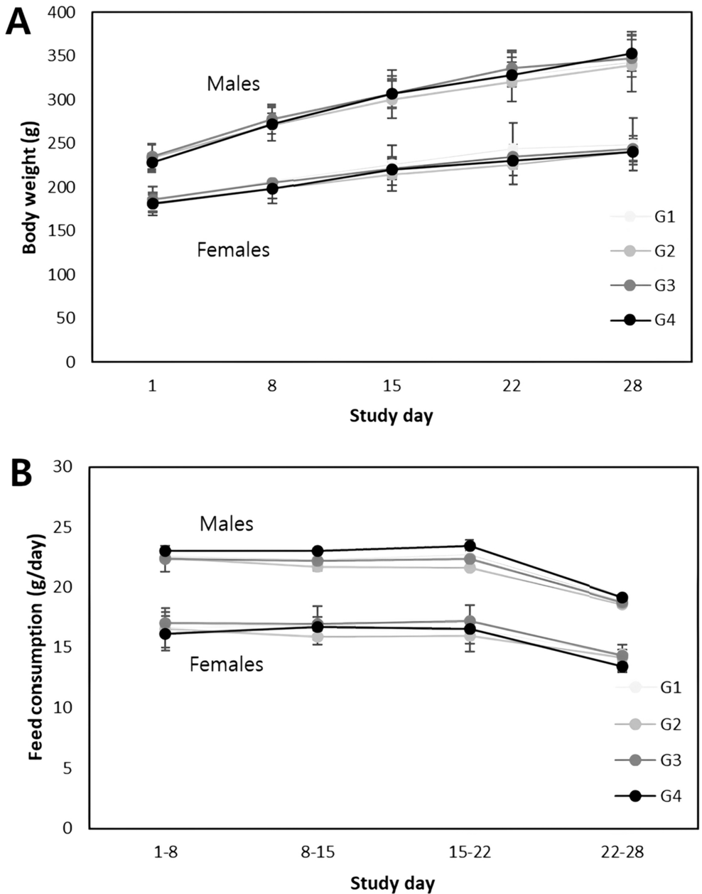
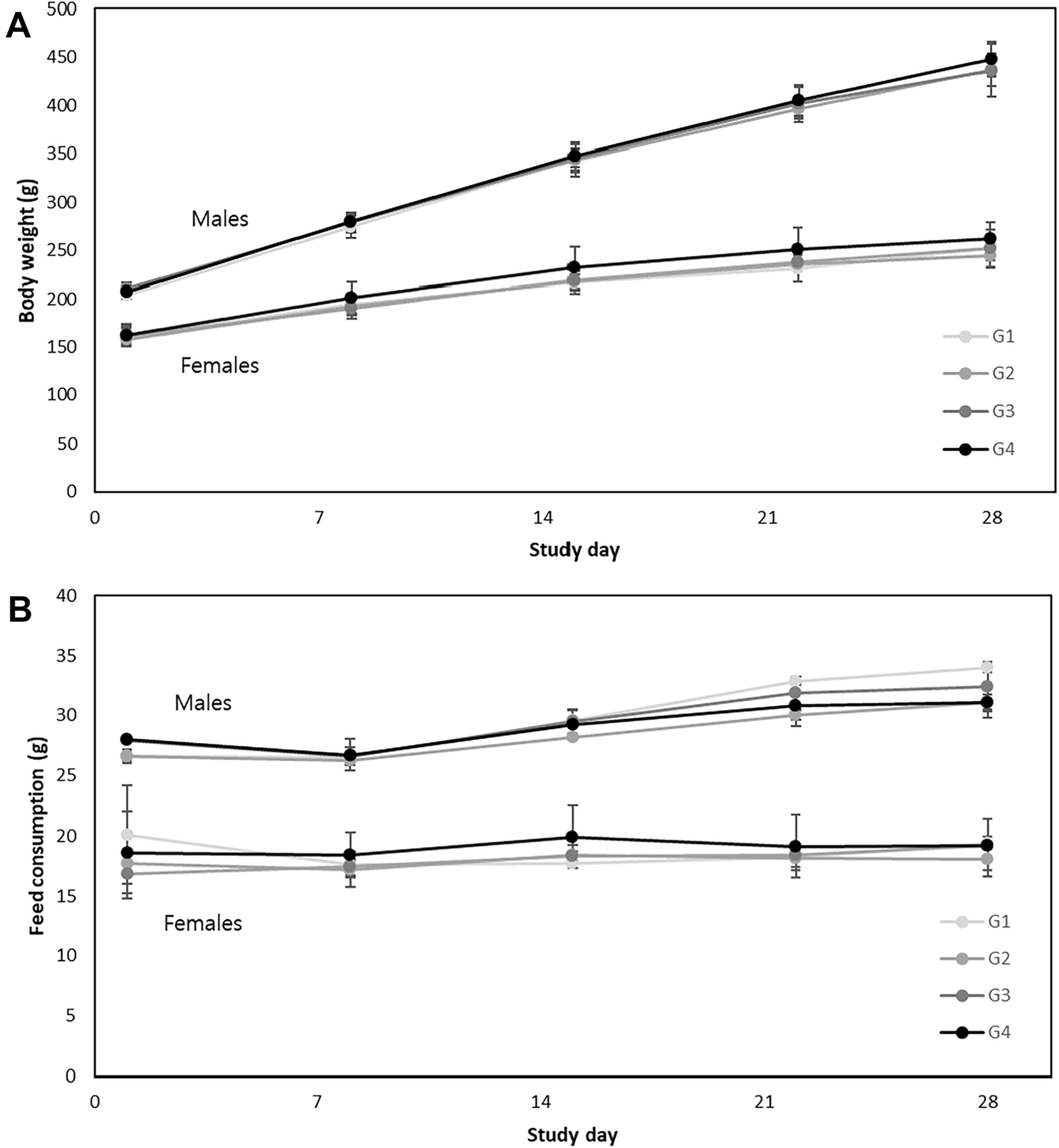
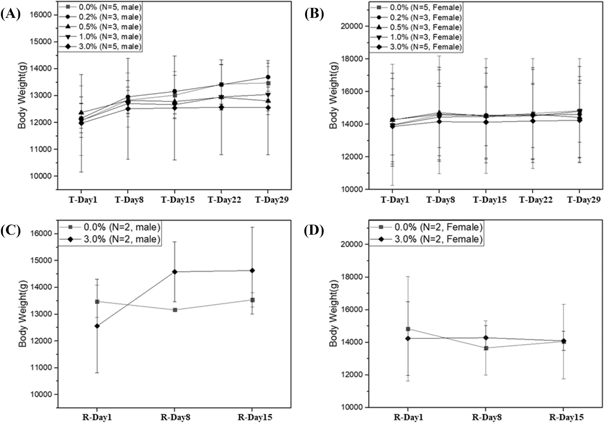
Body weight and food consumption
Body weight of the mice was measured at receipt, grouping, and approximately once a week during the treatment period. The amount of food in each cage was measured. The quantity of food consumed by every animal in each cage, 2 days during the whole week (5–7 days), was measured and presented as g/animal/day.
Ophthalmological examinations
Ophthalmological examinations were performed on all animals in the repeated toxicity studies, using binocular indirect ophthalmoscopy (Vantage Plus Digital, Keeler Ltd., England), after the animals were treated with mydriatics (Mydrin-P, Santen Pharmaceutical Co., Japan).
Laboratory investigationsHematology
Animals were fasted overnight prior to necropsy and sacrificed after blood collection, under isoflurane anesthesia, and EDTA-2 K was used as an anticoagulant. The following parameters were analyzed: white blood cell count, red blood cell count, hemoglobin concentration, hematocrit, mean corpuscular volume, mean corpuscular hemoglobin, mean corpuscular hemoglobin concentration, platelets, neutrophils, lymphocytes, monocytes, eosinophils, basophils, large unstained cells, and reticulocytes. This was done using a hematological autoanalyzer (ADVIA2120i Hematology analyzer, Bayer, USA).
Serum biochemistry
Serum was isolated and used to determine the levels of glucose, alanine aminotransferase, blood urea nitrogen, aspartate aminotransferase, creatinine, total bilirubin, total protein, alkaline phosphatase, albumin, gamma glutamyl transpeptidase, albumin/globulin ratio, creatine phosphokinase, total cholesterol, calcium, triglyceride, and inorganic phosphorus; this was done using a clinical chemistry auto-analyzer (200FR NEO, Toshiba Co., Japan).
Urinalysis
Urine samples were collected at approximately 3–4 h within 1 week prior to necropsy. The following parameters were measured during urinalysis: color, clarity, glucose, pH, erythrocyte, specific gravity, ketone, bilirubin, leukocytes, protein, nitrite, and urobilinogen. All parameters except for urine color were analyzed using Combur 10 TM urine sticks (Roche, Germany) and a Cobas U411 urine analyzer (Roche, Germany).
Necropsy
Animals were sacrificed via exsanguination of the aorta and posterior vena cava under isoflurane anesthesia. Complete necropsy examinations were performed on all animals that were found dead or were terminally sacrificed under the direct supervision of a veterinary pathologist. After blood sampling, the animals were subjected to necropsy and carefully examined for external abnormalities (Table 2).
Table 2 Biochemistry of male and female mice after administration of NKMAX Cell Therapy ProductOrgan weights
The weights of the brain, pituitary gland, liver (with gall bladder), spleen, heart, salivary glands (submandibular and sublingual), seminal vesicles (with coagulation glands), prostate, kidneys, adrenal glands, testes, epididymides, lungs, thyroids (with parathyroids), thymus, uterus (with cervix), and ovaries were measured for all animals at terminal and recovery sacrifice, and organ/brain and body weight ratios (using the terminal body weight obtained prior to necropsy) were calculated. Bilateral organs were measured together.
Histopathology
Tissue samples from adrenal glands, aorta (thoracic), brain, cecum, colon, duodenum, esophagus, femur with marrow (F-T joint), heart, ileum, jejunum, kidneys, liver (with gallbladder), lung (with bronchi), mammary gland (female only), mandibular lymph node, mesenteric lymph node, ovaries, pancreas, pituitary gland, prostate, rectum, salivary glands (submandibular and sublingual), sciatic nerve, seminal vesicles (with coagulation gland), skeletal muscle, skin (inguinal), spinal cord (cervical, thoracic, lumbar), spleen, sternum (with marrow), stomach, thyroids (with parathyroids), thymus, tongue, trachea, urinary bladder, uterus with cervix, vagina, and injection site (tail) were preserved in 10% neutral buffered formalin, while the eyes (with optic nerve) were fixed in Davidson’s fixative and the testes and epididymides were fixed in Bouin’s fixative. The tissues were placed in the appropriate fixative for approximately 48 h and then transferred to 70% ethanol. Formalin was infused into the lungs via the trachea and into the urinary bladder. Abnormal lesions and all tissues collected from animals at sacrifice, animals in the high-concentration and VC groups, and animals found dead or accidentally killed (tissue integrity permitting) were further processed to slides, stained with hematoxylin and eosin, and examined microscopically.
Statistical analysis
Multiple comparison tests were conducted for the different dose groups. The homogeneity of variance was examined using the Bartlett’s test. Homogeneous data were analyzed using analysis of variance, and the significance of inter-group differences was analyzed using Dunnett’s test. Heterogeneous data were analyzed using the Kruskal–Wallis test, and the significance of inter-group differences between the control and treatment groups was assessed using Dunn’s Rank Sum Test.
To compare the control and recovery groups, the data were analyzed for homogeneity of variance using the F test. Homogeneous data were analyzed using the t test, and the significant difference between the control and recovery groups was assessed using Dunnett’s test. Heterogeneous data were analyzed using the Kruskal–Wallis test, and significant differences between the control and recovery groups were assessed using Dunn’s Rank Sum Test (Table 3).
Table 3 Microscopic observation results of male and female mice after administration of human NK cells 


留言 (0)