
記住我
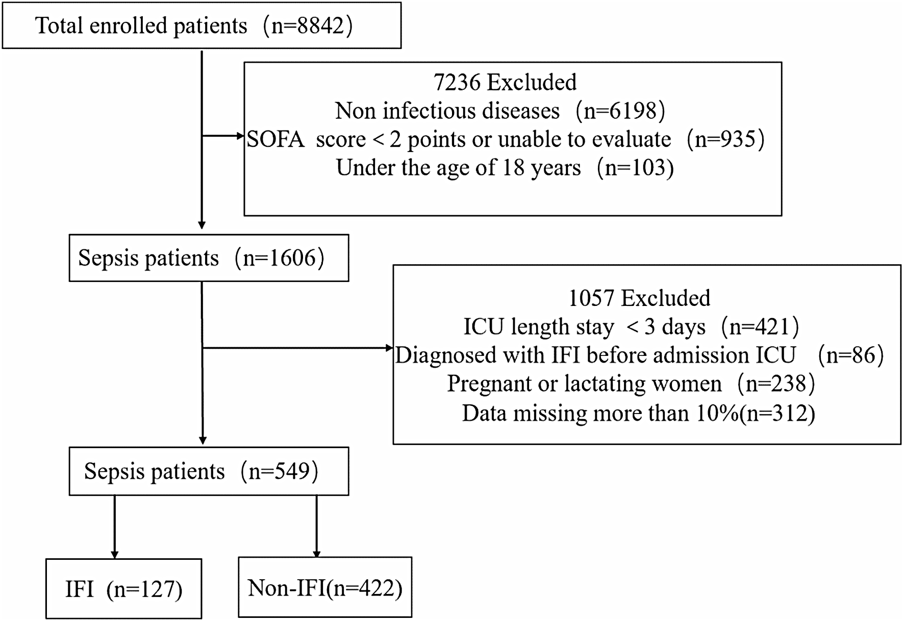
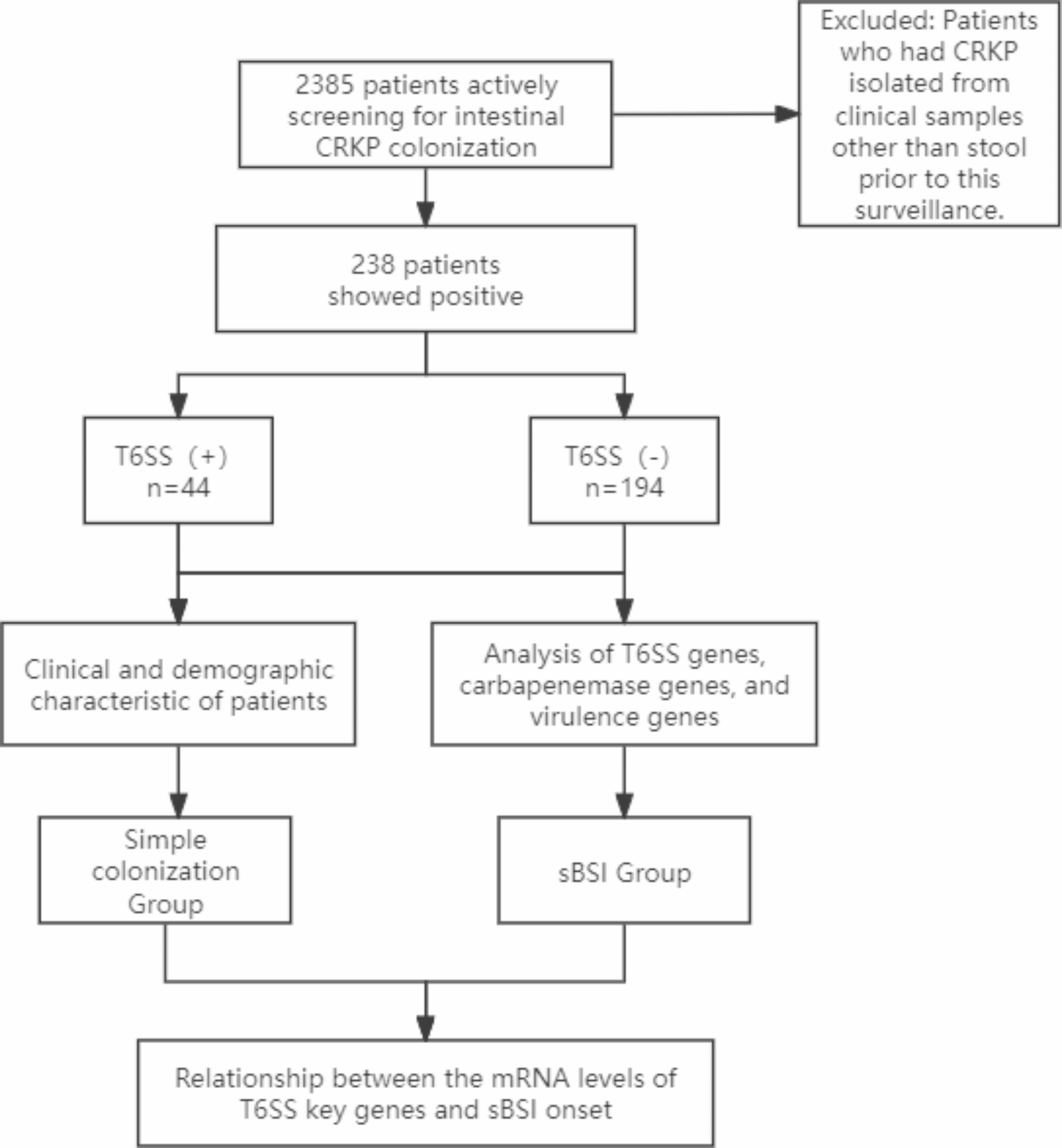
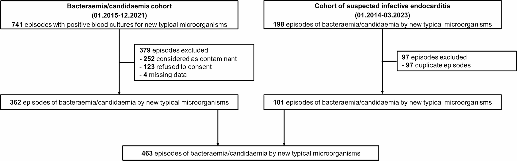
A total of 2,385 patients expected to remain in the ICU for at least three days were selected as subjects for the study from the ICU of the First Affiliated Hospital of Sun Yat-sen University, between January 2020 and 2022. Rectal swabs or stool samples were obtained from all newly admitted patients to proactively screen for CRKP. The sampling frequency was determined as follows: specimens were collected within 2 h of admission, then subsequently every 3 days until a positive result was obtained from the screening, and finally on the day of discharge. Patients from whom CRKP was isolated from clinical samples other than stool prior to this surveillance were excluded from the study.
In this investigation, we performed a follow-up tracking on patients harboring intestinal CRKP colonization, dividing them into two distinct groups: the simple colonization group and the subsequent bloodstream infection (sBSI) group. This classification was based on the occurrence of CRKP-sBSI within six months following the initial detection of CRKP colonization. The flow chart of this study is shown in Fig. 1.
Procedures followed ethical guidelines of the Declaration of Helsinki and obtained written consent from participants. The study was approved by the Clinical Research and Ethics Committee of the First Affiliated Hospital of Sun Yat-sen University, ensuring its ethical and scientific validity.
Fig. 1 Identification of bacterial strains and determination of drug susceptibility
The identification of Klebsiella pneumoniae strains was carried out by matrix-assisted laser desorption ionization-time-of-flight mass spectrometry (MALDI-TOF MS) (bioMérieux, Marcy l’Etoile, France). The antibiotics susceptibility of these strains was tested using Gram-negative susceptibility (GNS) cards on the Vitek-2 system (bioMérieux, Marcy l’Etoile, France). The interpretation of the susceptibility testing results was based on the guidelines recommended by the Clinical and Laboratory Standards Institute (CLSI, 2024). For quality control purposes, E. coli ATCC 25,922 and K. pneumoniae ATCC 700,603 were employed as reference strains during the susceptibility testing.
Pulsed-field gel electrophoresis (PFGE)This study conducted PFGE experiments on CRKP strains isolated from both the patients’ intestine and from blood cultures, confirming their genetic similarity. It was performed as described by Tenover et al. [17] with the Xbal restriction endonuclease (Takara Bio Inc., Otsu, Japan) and the Fingerprinting II Informatix software package system (Bio-Rad Laboratories, Hercules, CA). The similarity of the PFGE banding patterns was interpreted using the Dice coefficient, and the analysis of the acquired data was conducted through unweighted pair group method with arithmetic average clustering, employing the Pearson correlation coefficient.
Detection of carbapenemase genes, virulence genes and T6SS genes by PCRBacterial DNA was isolated from CRKP isolates through boiling. PCR amplification of carbapenemase genes (blaKPC, blaNDM, blaIMP, blaVIM, and blaOXA−48−like), virulence genes (rmpA, rpmA2, and iucA), and T6SS key genes (hcp, vgrG, and icmF), was conducted using TaKaRa Ex Taq reagents (Takara Bio Inc., Otsu, Japan). The primers utilized in this study are detailed in Table 1. Each PCR reaction contained 1 µl of DNA template, 17 µl of nuclease-free water, 1 µl of forward primer, 1 µl of reverse primer, 0.5 µl of Taq enzyme, and 2 µl of dNTP Mixture. The PCR cycling conditions were as follows: an initial denaturation step at 94℃ for 5 min, followed by 25 cycles of denaturation at 94℃ for 30 s, annealing at 58℃ for 30 s, and extension at 72℃ for 1 min. Finally, the PCR products were incubated at 72℃ for 10 min and stored at 4℃ for further analysis. The presence of PCR products was confirmed through the utilization of a 2% agarose gel.
Table 1 Primer sequences of the target genesQRT-PCR analysis of mRNA expression of key genes related to T6SSThe mRNA expression levels of the key genes of the T6SS, including Hcp, VgrG, and icmF, were measured. Total RNA was extracted from the isolates using the TRIzol method (manufactured by Invitrogen, Carlsbad, California, United States), strictly adhering to the recommended protocol. The relative expression levels were determined by real-time PCR using the SYBR® Premix Ex TaqTM reagent (Takara BioInc. Otsu, Japan) on the Applied Biosystems® 7500 Fast Dx Real-Time PCR Instrument (Life Technologies Corporation, Foster City, California). The RT-PCR reaction mixture, with a total volume of 20µL, was composed of 3µL of cDNA, 10µL of 2×SYBR® Premix Ex Taq II (Tli RNaseH Plus), 0.4µL of 50×ROX Reference Dye or Dye II, and 0.8µL of each primer. The PCR reaction proceeded under the following conditions: an initial cycle of 30 s at 95 °C, followed by 40 cycles of denaturation at 95 °C for 30 s, annealing at 53 °C for 30 s, and elongation at 72 °C for 30 s. The reaction concluded with a final cycle of 5 min at 72 °C. Reactions were repeated in triplicate and the fold changes in expression of these genes were calculated relative to the level of housekeeping gene 16 S rDNA using the comparative CT method (2–ΔΔCT method).
Multilocus sequence typing (MLST)A homology analysis of CRKP was conducted utilizing housekeeping genes: rpoB, gapA, mdh, pgi, phoE, infB, and tonB. The PCR-amplified products were subsequently sequenced, and the resulting sequences of the seven housekeeping genes were submitted to the MLST analysis website (http://www.mlst.net) to determine the ST typing of Klebsiella pneumoniae.
Statistical analysisAll data above were processed by Graphpad prism 8.0. Categorical variables were analyzed using the χ2 test or Fisher’s exact test. All tests with a p-value < 0.05 were taken as significant.
留言 (0)