Cell Culture and Bacterial Culture
OCCM-30, a murine cementoblast cell line, was cultured in Dulbecco’s modified Eagle’s medium (Hyclone) supplemented with 10% fetal bovine serum (FBS, Every Green) in an environment with 5% CO2 at 37 °C. Cementogenesis was induced in medium consisting of 5% FBS, 50 μg/mL ascorbic acid (Sigma), and 10 nM Naβ-glycerophosphate (Sigma) was utilized. The P. gingivalis (strain ATCC 33277) used in this study was grown in trypticase soy broth containing 0.1% yeast extract and 1 g/mL menadione in anaerobic incubators (80% N2, 10% CO2, and 10% H2) at 37 °C. A single clone was transferred into the liquid medium and allowed to grow to the logarithmic phase. The OD600 nm was used to determine bacterial concentrations (1 OD = 1 × 10⁹P. gingivalis/mL). Following centrifugation, the bacteria were resuspended in OCCM-30 mineralization medium.
Animal Model
Periapical lesions were induced in 8-week-old C57BL/6 male mice, which were divided into a control group and an AP group (n = 6). After administering anesthesia, a controlled rotation handpiece with a #1/4 round bur was used to open the pulp chambers of the mandibular first molars of mice under the guidance of an operating microscope. The exposed molars were treated with P. gingivalis in 2% carboxymethylcellulose and left uncovered for 21 days. We also used C57BL/6 mice of three different ages, 3-week-old, 6-week-old, and 6-month-old (n = 6 per group), to detect gene expression during cementogenesis. All animal experiments were approved by the Ethics Committee of School and Hospital of Stomatology, Wuhan University, and complied with the updated ARRIVE 2.0 guidelines for preclinical animal studies.
Plasmid Construction and sh-RNA Transduction
For Dnmt3a overexpression, Dnmt3a expression plasmids (Miaoling Biotech) or their negative controls (NCs) were transfected into OCCM-30 cells using TurboFect (Thermo Scientific). After 8 h, mineralization medium with or without P. gingivalis was added to the cells. Dnmt3a knockdown was achieved using lentivirus-mediated delivery of a shRNA (sh-Dnmt3a) with the sequence 5ʹ-CCAGATGTTCTTTGCCAATAA −3ʹ. OCCM-30 cells were incubated with the prepared virus for 24 h and screened with 2 μg/mL puromycin for 1 week.
Chemical Treatment
OCCM-30 cells were exposed to 10 nM Decitabine (MedChemExpress) in mineralization medium for 7 and 14 days. OCCM-30 cells transfected with sh-Dnmt3a were exposed to 30 μM Pifithrin-α (PFT-α) (MedChemExpress) in mineralization medium for 7 days. The chemicals were dissolved in dimethyl sulfoxide (DMSO) and the control group was administered an equal volume of DMSO.
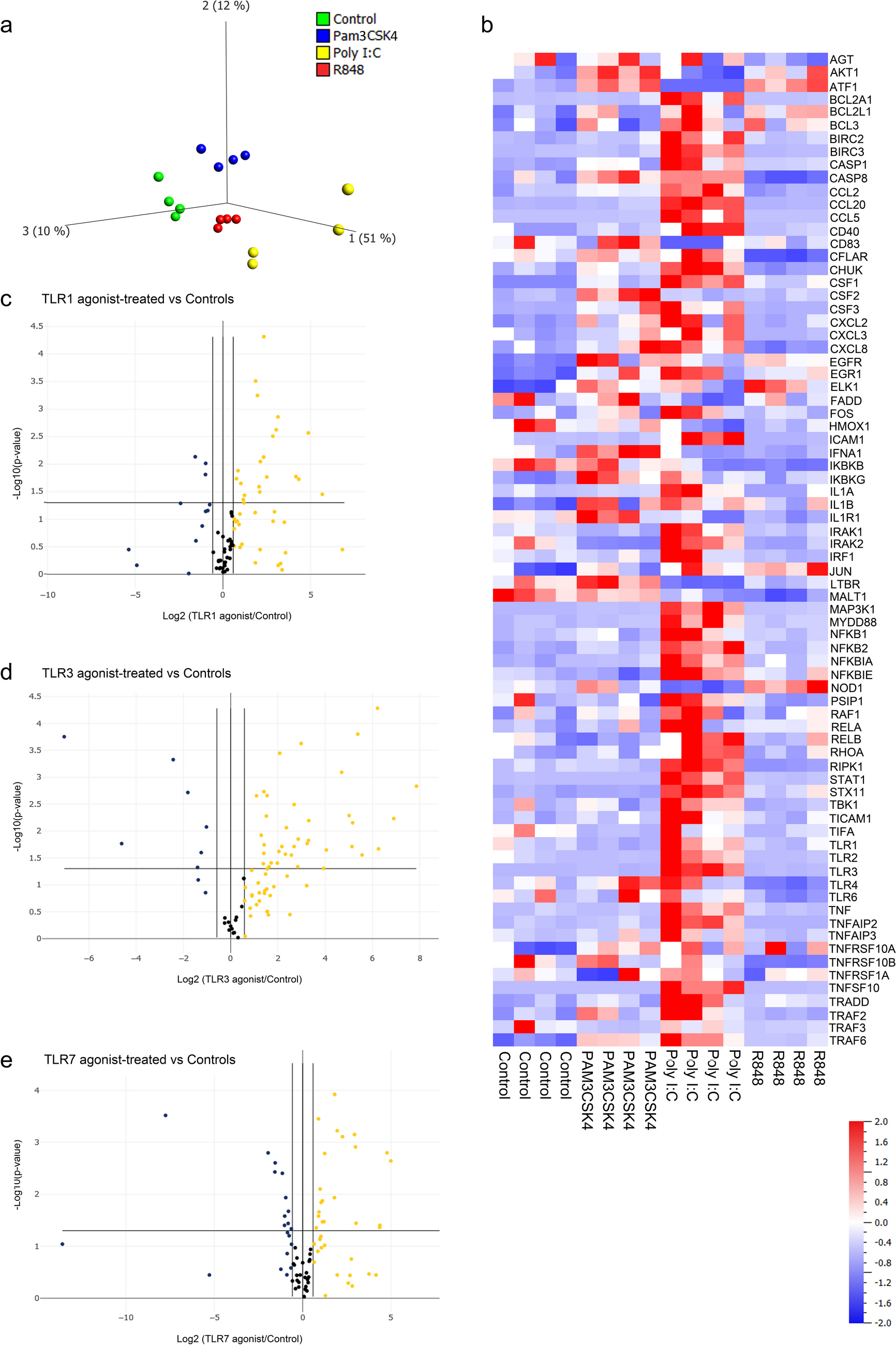
High-throughput RNA Sequencing (RNA-seq)
After 0 and 7 days of osteogenic induction in OCCM-30, RNA was harvested using TRIzol, sample quality control, next-generation sequencing, data acquisition, processing, and analysis were performed by ANOROAD.
For RNA harvested from OCCM-30 cells transfected with sh-NC or sh-Dnmt3a, BGI Tech provided technical support.
Quantitative Real-Time Polymerase Chain Reaction(qPCR)
Total RNA was extracted using the TRIzol reagent (TaKaRa) and reverse transcribed using the PrimeScript RT reagent Kit (TaKaRa). qPCR was performed on a Biosystems QuantStudio 6 with SYBR qPCR Master Mix (Vazyme). Relative expression levels were calculated using the 2−ΔΔCT method and normalized to β-actin. All the primers shown in Appendix Table 1 were synthesized by Sangon Biotech.
Western Blotting
Total protein was extracted from confluent cells using M-PER mammalian protein extraction reagent (Thermo Scientific). Protein concentrations were determined by BCA assay. Equal amounts of protein (25–35 μg) were separated by 10% SDS–PAGE, transferred to PVDF membranes (Millipore), blocked in 5% skimmed milk for 2 h at room temperature, and then incubated with primary antibodies overnight at 4 °C. After washing, the membranes were incubated with the corresponding secondary antibodies for 1 h at room temperature. The blots were visualized on an Odyssey LI-COR scanner (BD Biosciences). Information relating to the primary and secondary antibodies used in the study is presented in Appendix Table 2.
DNA Extraction and Global DNA Methylation Assay
Genomic DNA was extracted from both treated and untreated cells using the Universal Genomic DNA Extraction Kit v.5.0 (TaKaRa). The levels of global DNA methylation in 100-ng DNA samples were analyzed using the MethylFlash Global DNA Methylation (5-mC) ELISA Easy Kit (EpiGentek); the absorbance at 450 nm was determined using a microplate reader. All procedures were performed following the manufacturer’s instructions.
Immunofluorescence Staining of Tissue Sections
Tissue was collected from 3-week-old, 6-week-old, and 6-month-old mice as well as AP model mice, fixed in 4% paraformaldehyde, desalted in 10% EDTA for 30 days, embedded in paraffin, and cut into 5-μM-thick sections. The sections were then incubated first with an anti-Dnmt3a primary antibody (ABclonal) overnight at 4 °C and then with an anti-rabbit Cy3 secondary antibody (1:200; Beyotime) for 1 h. Nuclei were counterstained with DAPI (ZhongShan Jinqiao Biotechnology). Immunofluorescence images were captured under a microscope.
Alkaline Phosphatase (ALP) and Alizarin Red Staining
ALP staining was performed on cells mineralized for 7 days with NBT/BCIP (Beyotime). On day 14, cells were treated with 1% alizarin red solution, pH 4.2. After imaging, the mineral nodules were dissolved using a 10% solution of cetylpyridinium chloride, and their absorbance was measured at 562 nm.
Cell Viability Assay
A Cell Counting Kit-8 (CCK − 8; Biosharp) was used to assess the effect of decitabine on cell viability. Cells seeded in 96-well plates were treated with decitabine for 24, 48, and 72 h, after which the medium was removed, and 100 μL of 10% CCK-8 reagent was added to each well. After incubation for 1 h in the dark at 37 °C, the absorbance of each well was measured at 450 nm using a microplate reader.
Flow Cytometric Analysis for the Detection of Apoptosis
Apoptosis was detected using an Annexin V-PE/7-AAD apoptosis kit (Elabscience). Briefly, the cells were gently digested with EDTA-free trypsin, washed in cold PBS, and centrifuged at 300 × g. The samples were then resuspended in Annexin binding buffer and incubated with Annexin V-PE reagent and 7-AAD reagent for 20 min shielded from light. The results were collected by flow cytometry and figures were generated in CytExpert.
Analysis of Mitochondrial Reactive Oxygen Species (ROS) and Mitochondrial Membrane Potential (MMP)
To detect ROS, OCCM-30 cells were seeded in 96-well plates. After treatment, the cells were incubated with a solution of 5 μM MitoSOX Red Mitochondrial Superoxide Indicator (Yeasen) and Hoechst 33,342 staining solution (Beyotime) for 10 min at 37 °C away from light. For MMP detection, following treatment, OCCM-30 cells were incubated with 50 nM fluorescent dye TMRM Perchlorate (MedChemExpress) and Hoechst 33,342 (Beyotime) at 37 °C for 30 min in the dark. Images were captured and analyzed under a fluorescence microscope.
Statistical Analysis
A minimum of three independent experiments were conducted to obtain the data. GraphPad Prism 8 was used, and Unpaired t-tests and one-way ANOVAs were performed to compare the results. Statistical significance was indicated with *p < 0.05, **p < 0.01, and ***p < 0.001.


留言 (0)