
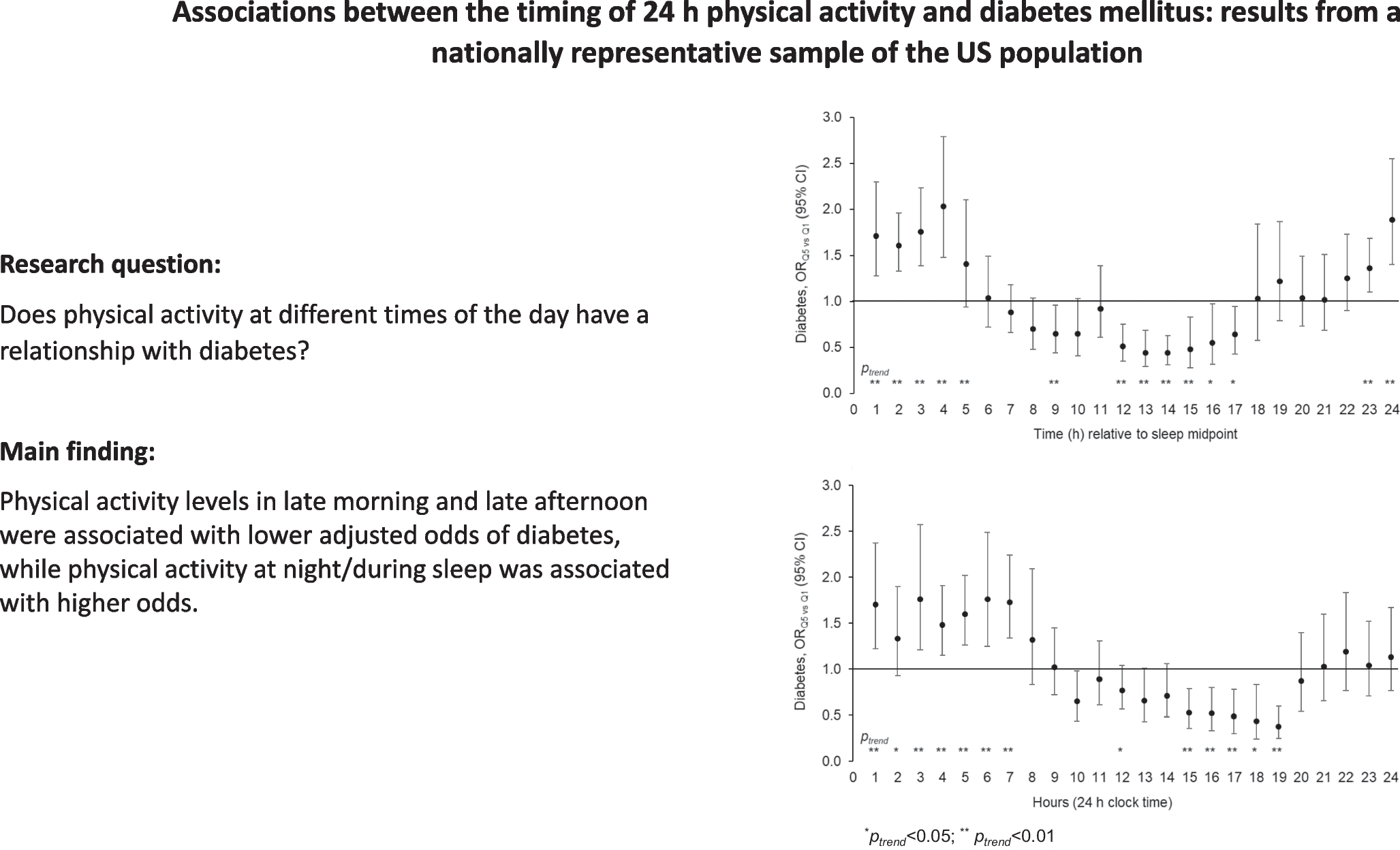
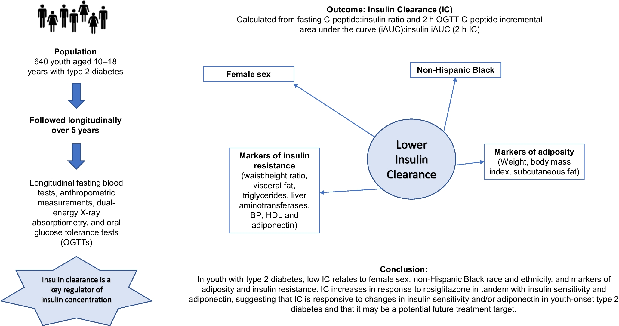
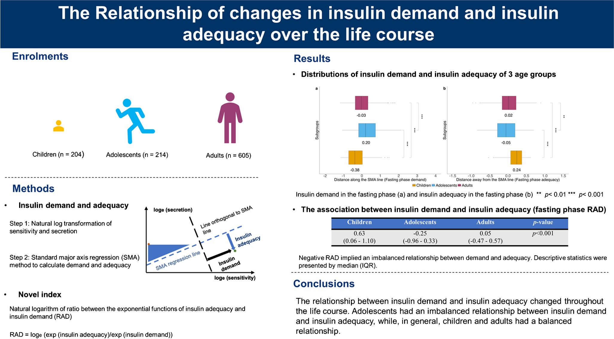
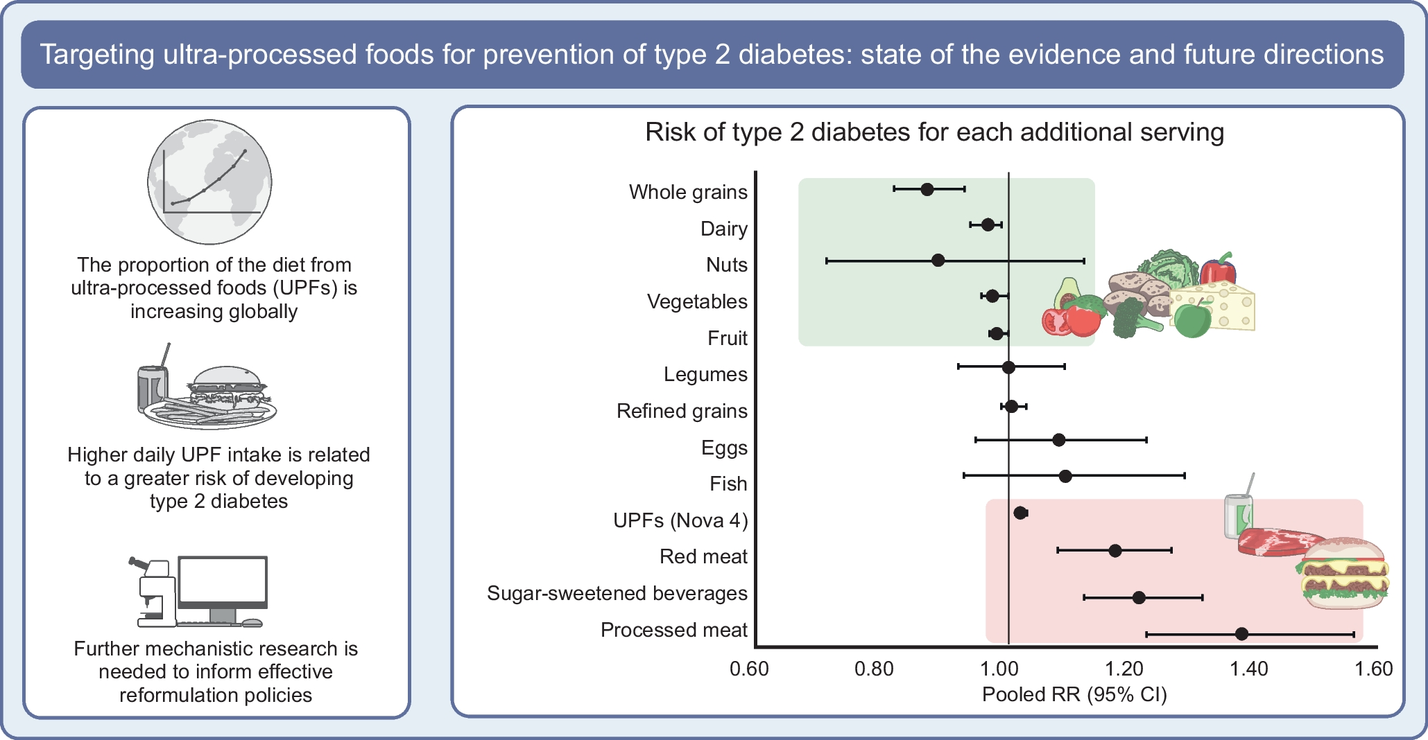
記住我
First, we explored the association between body composition and glucose metabolism using multiple linear regression analyses in participants with the G-A haplotype and in those without (non-G-A). The 5 year change in HOMA2-IR was positively associated with visceral fat score in both groups, and inversely associated with ASM/Wt only in the G-A haplotype (ESM Table 2).
To find clinical subgroups where the G-A haplotype strongly impacts the deterioration of insulin resistance indices, we subsequently categorised participants by the combination of sarcopenic obesity defined by VFA≥100 cm2 and ASM/Wt Q1, and the G-A haplotype. We analysed the association between either serum resistin or HOMA2-IR and low ASM/Wt and high visceral fat, individually or in combination (ESM Fig. 2). Participants were divided into four groups: group 1, VFA<100 cm2 and ASM/Wt Q2–Q4; group 2, VFA<100 cm2 and ASM/Wt Q1; group 3, VFA≥100 cm2 and ASM/Wt Q2–Q4; and group 4, VFA≥100 cm2 and ASM/Wt Q1. Serum resistin and HOMA2-IR were highest in group 4, after adjustment for age and sex (ANCOVA, p=2.06 × 10⁻⁸ and p=1.74 × 10⁻¹², respectively).
We next assessed blood glucose and insulin during OGTT in the same four groups (ESM Table 3). After adjusting for age and sex, differences in insulin at fasting, 1 h, and 2 h were observed among the four groups (ANCOVA, p=3.00 × 10⁻¹⁴, 3.51 × 10⁻⁶ and 1.18 × 10⁻⁹, respectively). In post hoc tests, insulin at fasting, 1 h, and 2 h in group 4 was higher than that in the other three groups. Differences were also found in fasting and 2 h serum glucose among the four groups (ANCOVA, p=0.0098 and p=0.0054), with group 4 having the highest values. Differences in fasting and 2 h serum glucose were observed only when comparing group 4 with group 1. No difference was found in 1 h serum glucose among the four groups (ANCOVA, p=0.0972).
Therefore, participants with both low ASM/Wt and VFA≥100 cm2 showed the highest serum resistin and insulin resistance.
The G-A haplotype, when accompanied by latent skeletal muscle loss and visceral obesity, showed the highest serum resistin and insulin resistanceWe further examined the association between either serum resistin or HOMA2-IR and the G-A haplotypes defined by SNP-420 and SNP-358 (ESM Fig. 3). Since A at SNP-358 was required for G at SNP-420 to confer the highest serum resistin, major haplotypes around RETN were able to be defined by these two SNPs due to almost complete linkage disequilibrium [16]. Participants were divided into three groups based on the G-A haplotype: */*, G-A/* and G-A/G-A, where * represents the C-G or G-G haplotype. Serum resistin was highest in G-A homozygotes after adjustment for age, sex and BMI (ANCOVA, p=1.30 × 10−85) (ESM Fig. 3a). In contrast, HOMA2-IR was not associated with this group (ESM Fig. 3b).
We further examined the association between serum resistin, HOMA2-IR, HOMA2-B or glucose AUC in OGTT, and G-A haplotypes defined by SNP-420 and SNP-358, and low ASM/Wt and high visceral fat, individually or in combination (ESM Fig. 4). The G-A/* and G-A/G-A haplotypes were combined into a single subgroup, and participants were divided into four groups: group 1, VFA<100 cm2 and ASM/Wt Q2–Q4 with */*; group 2, VFA<100 cm2 and ASM/Wt Q2–Q4 with G-A/* and G-A/G-A; group 3, VFA≥100 cm2 and ASM/Wt Q1 with */*; and group 4, VFA≥100 cm2 and ASM/Wt Q1 with G-A/* and G-A/G-A. Serum resistin was highest when low ASM/Wt and visceral obesity were accompanied by the G-A haplotype (G-A/* and G-A/G-A) (group 4) after adjustment for age and sex (ANCOVA, p=2.06 × 10−52) (ESM Fig. 4a). Either HOMA2-IR or HOMA2-B was also highest in group 4, whereas glucose AUC appeared to be highest in group 4 (ESM Fig. 4b–d).
We further assessed blood glucose and insulin during OGTT in the four groups (Table 1). Fasting insulin was highest in group 4, after adjustment for age and sex (ANCOVA, p=1.11 × 10⁻¹²; p for interaction=0.0188). Either 1 or 2 h insulin, or 2 h blood glucose, was also highest in group 4.
Table 1 The G-A haplotype, when accompanied by latent skeletal muscle loss and visceral obesity, showed the highest insulin resistanceTherefore, the G-A haplotype, when accompanied by latent sarcopenic obesity, showed the highest serum resistin and insulin resistance.
The increase in insulin resistance was highest when the G-A haplotype was accompanied by low ASM/Wt and visceral obesity over a 5 year periodWe further analysed changes over 5 years in serum resistin, HOMA2-IR, HOMA2-B or glucose AUC during OGTT in the four groups (Fig. 1). Glucose metabolism was evaluated at baseline and 5 year follow-up, and body composition was assessed at 5 year follow-up. The increase in HOMA2-IR was highest when low ASM/Wt and visceral obesity (defined as sarcopenic obesity) were accompanied by the G-A haplotype (group 4), after adjustment for age and sex (ANCOVA, p=1.96 × 10⁻⁵; p for interaction=0.0005) (Fig. 1b). Similar associations were found for HOMA2-B and glucose AUC (Fig. 1c, d). In contrast, no changes were found for serum resistin (Fig. 1a), suggesting that, in addition to the G-A haplotype as a genetic factor, sarcopenic obesity, as a time factor, could be required for the deterioration of insulin resistance.
Fig. 1
The increase in insulin resistance was highest when the G-A haplotype was accompanied by low ASM/Wt and visceral obesity over a 5 year period. The 5 year changes in (a) serum resistin, (b) HOMA2-IR, (c) HOMA2-B and (d) glucose AUC. Glucose metabolism was evaluated at baseline and 5 year follow-up, and body composition was assessed at 5 year follow-up. Data are expressed as mean ± SEM. VFA<100 cm2 and ASM/Wt Q2–Q4 was defined as the control, and VFA≥100 cm2 and ASM/Wt Q1 was defined as sarcopenic obesity. Participants were divided into four groups: group 1, control with */*; group 2, control with G-A/* and G-A/G-A; group 3, sarcopenic obesity with */*; and group 4, sarcopenic obesity with G-A/* and G-A/G-A. *Non-carriers of G-A haplotype. ANCOVA was performed, involving 5 year changes in serum resistin, HOMA2-IR, HOMA2-B or glucose AUC as dependent variables, and groups 1–4 and adjustment for age and sex as independent variables. ANCOVA: p=0.39 (a), p=1.96 × 10⁻⁵ (b), p=0.0032 (c) and p=0.035 (d). Tukey’s HSD test: *p<0.05, ***p<0.001 vs Group 1; †p<0.05, ††p<0.01, †††p<0.001 vs Group 2; ‡p<0.05, ‡‡p<0.01 vs Group 3. The p value for the interaction was estimated by adding an interaction term (sarcopenic obesity × G-A haplotype) along with individual factors and adjustment for age and sex as independent variables. p for interaction = 0.42 (a), 0.0005 (b), 0.0019 (c) and 0.0062 (d). ASM/Wt, HOMA2-IR and HOMA2-B were calculated as described in the Methods section
Therefore, the increase in insulin resistance was highest when the G-A haplotype was accompanied by low ASM/Wt and visceral obesity over a 5 year period.
Cluster 2, characterised by latent skeletal muscle loss, visceral obesity and insulin resistance, with the G-A haplotype, demonstrated deterioration in insulin resistance over a 5 year periodSince the categorical classification may overlook clinically significant groups, we also performed cluster analyses to identify data-driven clusters where the G-A haplotype had enhancing effects (Fig. 2). The clustering constituents were selected from parameters closely associated with body composition and insulin resistance in the previous categorical classification. When serum glucose, serum insulin, ASM/Wt, VFA and grip strength were involved as variables, five clusters were identified (Fig. 2a). Among these, cluster 2 was characterised by low skeletal muscle mass, visceral obesity and insulin resistance (Fig. 2b). This cluster, with the G-A haplotype, demonstrated deterioration in insulin resistance over a 5 year period (Fig. 2c). Participants in each cluster were then divided into two groups based on their haplotypes. Changes in HOMA2-IR and HOMA2-B were assessed over 5 years. Only cluster 2, when accompanied by G-A/* and G-A/G-A, exhibited the highest increase in insulin resistance over a 5 year period (HOMA2-IR; ANCOVA, p=9.41 × 10⁻¹⁴; p for interaction=5.76 × 10⁻⁶).
Fig. 2
Cluster 2, characterised by latent skeletal muscle loss, visceral obesity and insulin resistance, with the G-A haplotype, demonstrated deterioration in insulin resistance over a 5 year period. (a) Hierarchical clustering was performed involving serum glucose and serum insulin at 0 h, 1 h and 2 h in OGTT, ASM/Wt, grip strength and visceral fat score as model variables, and five clusters were identified. (b) The five clusters represented clinical characteristics classified using the above-mentioned five parameters. C1–C5, clusters 1–5. For the box plots in (b), the central line represents the median, the ‘x’ indicates the mean, the whiskers represent ±2 SD, and the outer points represent values that fall outside of 2 SD. (c) The 5 year changes in HOMA2-IR (mean ± SEM). Participants were divided into the five clusters. Glucose metabolism was evaluated at baseline and 5 year follow-up, and body composition was assessed at 5 year follow-up. ANCOVA was performed, involving 5 year changes in HOMA2-IR as dependent variables, and haplotypes of each cluster and adjustment for age and sex as independent variables. Ctrl (control) denotes four clusters, excluding those located on the right side of number of clusters (e.g. Ctrl1 includes clusters 2–5). ANCOVA, p=9.41 × 10⁻¹⁴. Tukey’s HSD test for post hoc analyses: ***p<0.001 vs Ctrl2 with */*; †††p<0.001 vs Ctrl2 with G-A/* and G-A/G-A; ‡‡‡p<0.001 vs C2 with */*. p for interaction = 5.76 × 10⁻¹⁶. Glucose AUC, AUC of 75 g OGTT glucose; insulin AUC, AUC of 75 g OGTT insulin. The grip strength was measured using the dominant hand. HOMA2-IR was calculated as described in the Methods section
To examine the influence of age, we divided the participants into two age groups: middle-aged (under 60 years) and older (60 years and above) (ESM Table 4). Using hierarchical clustering in each age group, we identified a cluster similar to the sarcopenic obesity with strong insulin resistance. Considering this cluster (cluster 3 for the middle-aged group, and cluster 1 for the older group) as the case group, and the other clusters as the control group, we classified them into four groups based on the presence or absence of the G-A haplotype, and evaluated the 5 year change in HOMA2-IR (ESM Fig. 5). The interaction was found in the clusters of the 60 and above age group (p for interaction=0.0078) (ESM Fig. 5b), but not in those of the under 60 age group (p for interaction=0.43) (ESM Fig. 5a). Therefore, the sarcopenic obesity-like group having strong insulin resistance, with the presence of the G-A haplotype, especially in the older age group, was associated with the worsening of insulin resistance.
Expression of mitophagy pathway-related genes was altered, and FOXO3, ATG9A and FIS1 mRNA was higher in whole blood cells from participants homozygous for the G-A haplotypeTo look for factors linking low ASM/Wt and visceral obesity with haplotypes defined by resistin SNP-420 and SNP-358, we analysed total RNA of whole blood cells from 25 participants homozygous for the G-A haplotype (G-A/G-A), and 33 age-, sex- and BMI-matched participants homozygous for the C-G haplotype (C-G/C-G), using RNA-seq and pathway analysis. In the volcano plot, the upregulated genes except for RETN are indicated in red and downregulated genes are indicated in blue with a fold change >1.2 (log2 fold change >0.26), and with p<0.05 (Fig. 3a). Of 21,479, 584 genes were selected based on these criteria for pathway analysis.
Fig. 3
Mitophagy pathway-related mRNA was higher in whole blood cells from participants homozygous for the G-A haplotype. (a) In the volcano plot, upregulated and downregulated genes are indicated in red and blue, respectively, with a fold change >1.2 (log2 fold change >0.26), and with p<0.05. (b–f) Participants homozygous for the G-A haplotype (G-A/G-A) (n=25), and age-, sex- and BMI-matched participants homozygous for the C-G haplotype (C-G/C-G) (n=33), were analysed by qRT-PCR as described in the Methods section. Relative FOXO3 (b), ATG9A (c), MFN1 (d), BNIP3L (e) and FIS1 (f) mRNA is shown by using GAPDH as an internal control. Box-and-whisker plots show the distribution of FOXO3, ATG9A, MFN1, BNIP3L and FIS1 mRNA relative to C-G/C-G; the central line represents the median and the whiskers are displayed to include all the data. The qRT-PCR analysis was performed in duplicate for meaningful comparisons. Student’s t test: *p<0.05. FC, fold change; NS, non-significant
The DAVID pathway analysis showed that the expression of genes involved in mitophagy signalling pathways was increased in individuals with G-A/G-A compared with those with C-G/C-G (p=2.7 × 10⁻⁴) (ESM Table 5a). Individual p values for the RNA-seq of the genes showing statistically significant expression changes in the mitophagy pathway are presented in ESM Table 5b.
We further analysed mRNA of representative mitophagy-related genes, FOXO3, ATG9A, MNF1, BNIP3L and FIS1, in whole blood cells from 25 participants with G-A/G-A compared with 33 participants with C-G/C-G using qRT-PCR, because these genes showed relatively large fold changes in expression levels in the mitophagy pathway (Fig. 3b–f). The mRNA of FOXO3, ATG9A and FIS1 was higher in participants with G-A/G-A than with C-G/C-G (Student’s t test, FOXO3, p=0.027; ATG9A, p=0.0007; FIS1, p=0.0109) (Fig. 3b, c, f), and the direction of the effects in the genes examined in RNA-seq appears to be similar in RT-PCR, suggesting altered mitophagy pathways in individuals with G-A/G-A.
From all these findings, the G-A haplotype, accompanied by low skeletal muscle mass and visceral obesity, led to the deterioration of insulin resistance over a 5 year period in this cohort, possibly through the altered expression of mitophagy-related genes.
留言 (0)