
記住我

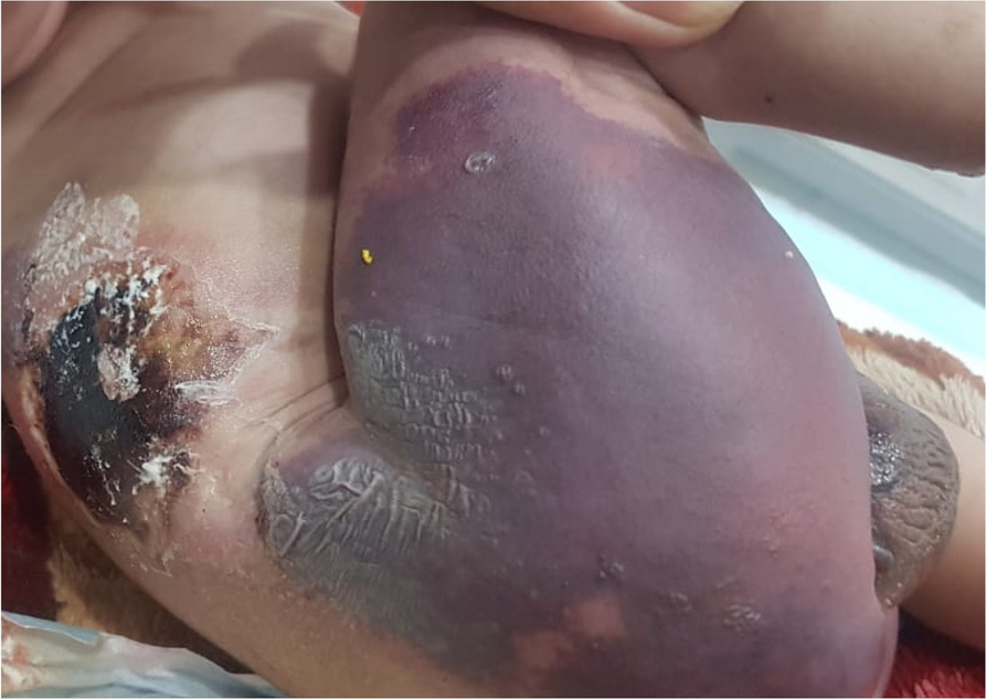


A 10-day-old male infant, delivered vaginally at full term following an uneventful pregnancy and uncomplicated delivery, was brought to the emergency room due to purplish patches on his right foot and buttocks that had been present for 1 day. Aside from these patches, the infant was in good health and was breastfeeding without any issues. There were no noteworthy details in the birth and prenatal history. He was the fifth child of parents in a consanguineous marriage (fourth-degree consanguinity). The mother had encountered three first-trimester miscarriages previously. However, obtaining the exact gestational age was not possible due to the mother’s inability to recall and the absence of documented proof. There was no family history of similar skin lesions. Notably, the newborn did not receive vitamin K prophylaxis or vaccination at birth. Initially, the patches manifested on the right ankle but progressed distally to cover approximately two-thirds of the right foot and extended proximally to the mid-thigh. Subsequently, a similar patch emerged on his right buttock, eventually involving the genital area (Fig. 1). Over time, these patches evolved from purplish lesions to necrosis, forming black eschars.
Fig. 1
Purpuric rash involving right hip and thigh
On examination, the patient’s vital signs indicated stability, with a heart rate of 140 beats per minute, a respiratory rate of 35 breaths per minute, and a temperature of 98.6 °F. Peripheral pulses in the left lower limb were detectable, while the right lower limb showed good blood flow in the femoral and popliteal arteries but weak flow in the tibialis anterior and posterior. Capillary refill time was less than 2 s in the upper limb and left lower limb, but it exceeded 2 s in the right lower limb. All extremities exhibited normal warmth.
The infant exhibited good tone, fair sucking, and an incomplete Moro’s reflex. Microcephaly and craniofacial abnormalities likely contributed to the infant’s presentation of an incomplete Moro reflex. The anterior fontanelle was open and flat, measuring approximately 0.5 cm, while the posterior fontanelle was closed. Fundus examination revealed normal findings: clear media, pink optic disc with sharp margins (cup-to-disc ratio ~ 0.35), normal vasculature, and a healthy retinal nerve fiber layer evidenced by a striated sheen. Additionally, clinical signs of microcephaly, a long philtrum, a small chin, and low-set ears were observed, possibly indicating craniofacial abnormalities. However, other systemic examinations yielded unremarkable findings.
With suspicions of a coagulopathy and an underlying syndrome, the neonate’s care plan involved essential interventions. These encompassed maintaining a nil per os (NPO) status with the insertion of a nasogastric (NG) tube, providing oxygen support, and ensuring the maintenance of an intravenous (IV) line. Antibiotic therapy included cefotaxime and amikacin, and transfusions of red blood cells, platelets, and fresh-frozen plasma (FFP) were administered as needed. Continuous monitoring was in place to assess for complications, with vital signs and blood sugar levels checked every 6 h. Comprehensive laboratory tests, including a thrombotic workup, were initiated to diagnose the underlying condition.
InvestigationLaboratory assessments at presentation revealed the following results: hemoglobin at 15 g/dl, TLC (total leukocyte count) of 15,000 cells/mm3, platelet count of 87,000 cells/mm3, prothrombin time (PT) extending to 61 s, activated partial thromboplastin time (APTT) slightly prolonged at 34 s, and an INR of 3.9, all indicative of coagulation abnormalities. Following management, laboratory tests were repeated at different intervals (Table 1).
Table 1 Blood parameters at admission, 48 h, and 120 hSpecific coagulation factor levels unveiled severe deficiencies: protein C at 10% (normal range: 72–106%), protein S at 41% (normal range: 60–110%), and antithrombin III at 55% (normal range: 80–120%). Blood and urine cultures were initially conducted to investigate infection upon presentation, yielding negative results. Subsequently, these cultures were repeated twice at different intervals of 1 week, and all results came back negative effectively eliminating the possibility of secondary protein C and S deficiency due to infection, and adequate hydration was ensured through the maintenance of IV lines in the infant, thereby ruling out the possibility of protein C and S deficiency caused by Z dehydration. A lumbar puncture was also conducted, yielding unremarkable findings. An ultrasound of the brain was conducted due to the neonate’s comparatively small anterior fontanelle, which revealed bilateral dilated lateral ventricles, measuring 3.4 cm on the right, displacing the choroid plexus in the depending portion with internal echoes. These findings indicated non-communicating hydrocephalus. However, further brain imaging modalities such as MRI and CT scan could not be performed due to the unstable condition of the baby. Limitation of resources prevented the conduction of genetic testing, serological assays, and molecular techniques, including PCR, which are essential for ruling out TORCH infections and cause of craniofacial malformations.
ManagementRecognizing the complexity of the case, a collaborative approach was adopted, with the involvement of a senior hematologist, vascular team, pediatric surgery team, and infectious diseases team for further evaluation.
Following the vascular team’s assessment, a Doppler ultrasound of the limbs indicated a monophasic spectrum with a normal peak velocity in the right posterior tibial artery at the site of an ulcerated wound. In contrast, the rest of the arteries displayed a triphasic spectrum with normal peak systolic velocities. In response to this assessment, the vascular team recommended the initiation of local glyceryl trinitrate (GTN) application at 8-h intervals.
The treatment plan included subcutaneous injections of enoxaparin at a dose of 2 mg/kg/day, administered twice daily to maintain anticoagulation. IV antibiotics and elevation of the legs were continued as part of the management. An echocardiogram, as suggested by the vascular team, was performed to rule out any cardiac causes, revealing a patent foramen ovale (PFO) with a left-to-right shunt. Subsequent Doppler ultrasounds showed gradual improvement in arterial insufficiency, allowing for the continuation of enoxaparin treatment. As the INR began to normalize, no further FFP transfusions were required. By the 7th day of admission, there was an improvement in the purpura of the right foot.
However, by the 10th day of admission, the neonate started to develop multiple bruises and eschar involving various areas, including the bilateral flank, perianal region (Fig. 2), and bilateral hands and feet. The infectious disease team was consulted, and they recommended discontinuing vancomycin after establishing a central line and starting clindamycin while continuing meropenem and colistin. Despite these efforts, the purpura worsened and took on a darker appearance. Due to the risk of developing necrotizing fasciitis, the LRINEC (laboratory risk indicator for necrotizing fasciitis) scoring system was applied, and the calculated score was 11, indicating a high risk. Pediatric surgery was consulted and advised debridement. However, due to the neonate’s hemodynamic instability and parental refusal to provide consent, the procedure was not performed.
Fig. 2
Multiple eschars involving bilateral flanks, genitalia, and perianal region
On the 19th day of admission, the neonate experienced apneic episodes and intermittent desaturation, with oxygen support including epinephrine. The infant went into apnea and bradycardia, and despite medical intervention, the parents declined cardiopulmonary resuscitation (CPR), resulting in the declaration of the neonate’s passing.
留言 (0)