
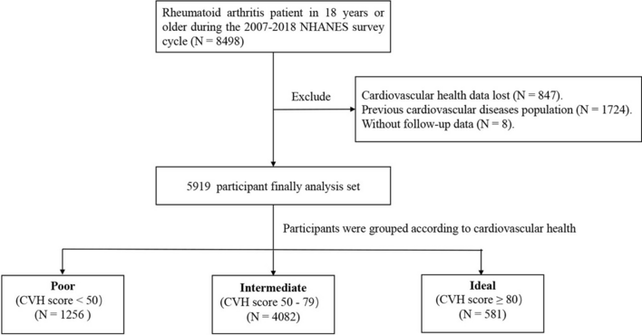
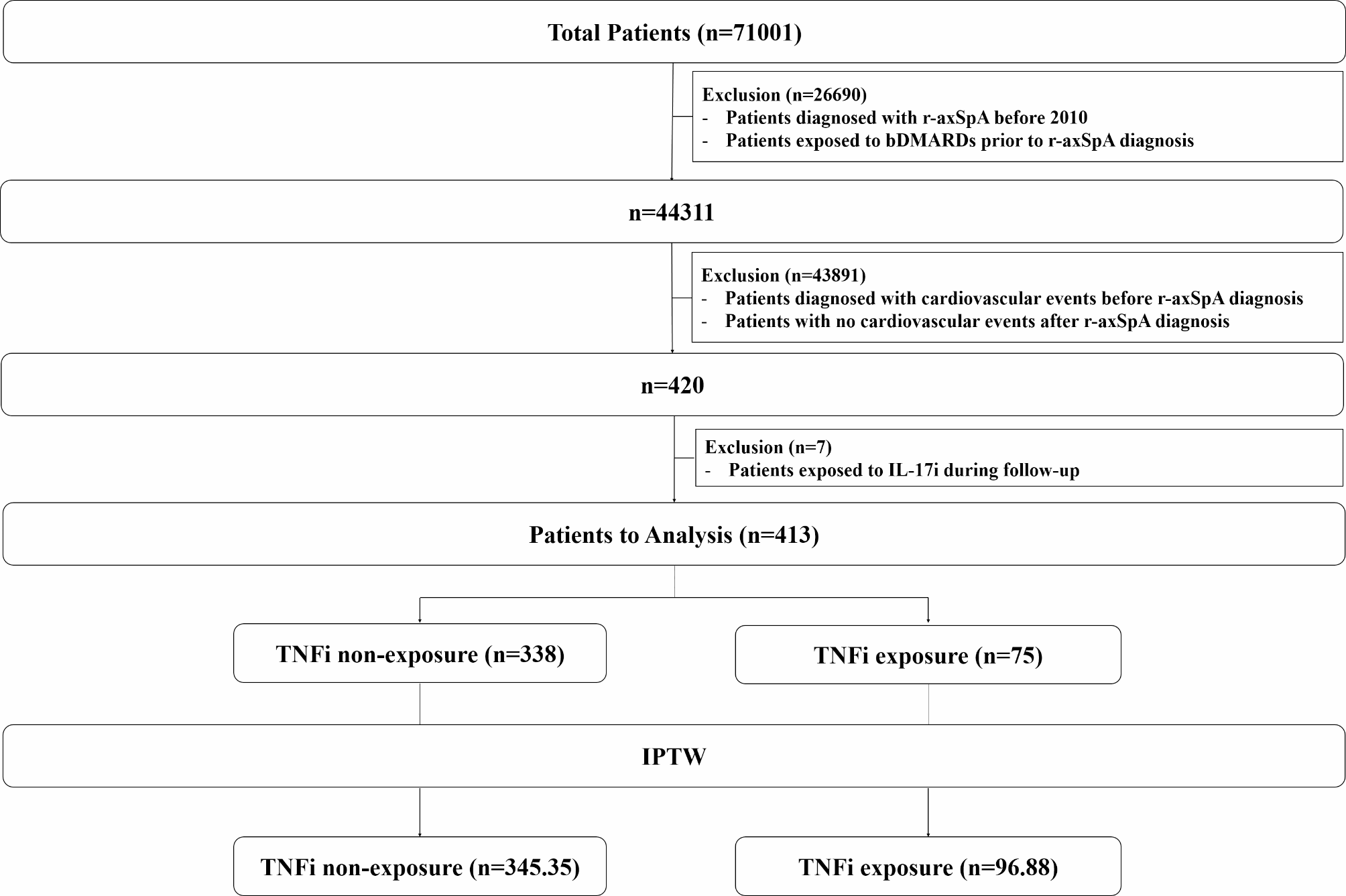
記住我


Firstly, we used the dimensionality reduction algorithm tSNE-CUDA to classify the different populations based on EPCs markers in PBMCs from patients with SLE. tSNE-CUDA is a graphics processing unit (GPU)-accelerated implementation of t-distributed Symmetric Neighbour Embedding (t-SNE) for visualizing datasets and models, allowing large scale visualizations of modern computer vision datasets. Compared to classical flow cytometry assays, tSNE-CUDA is able to characterize populations thorough the combination of markers used [28].
Using this approach, we identified 8 different population clusters in HC and patients with SLE (Fig. 1A). Most of these clusters did not show characteristics of EPCs; however, clusters 1 and 5 expressed markers of MACs (CD45 + , CD14 + and CD31 +), while cluster 6 showed phenotypic characteristics that define ECFCs, such as absence of CD45 and CD14 and expression of CD31 and CD309 (Fig. 1B and Supplementary Figure S1). The percentage of these clusters was similar in patients with SLE and HC except cluster 6, which was significantly higher in patients with SLE (Fig. 1C).
Fig. 1
Patients with SLE present EPCs clusters and subclusters that are involved in disease. A High dimensional characterization of EPCs clusters in patients with systemic lupus erythematosus (SLE) using tSNE-CUDA dimensionality reduction algorithm. B Characterization of EPCs clusters identified in (A) according to the expression of surface protein markers. C Percentage of cluster expression in HC and patients with SLE. D High dimensional characterization of EPCs subclusters in patients with SLE (yellow) and HC (purple) using tSNE-CUDA dimensionality reduction algorithm. E Characterization of EPCs clusters identified in (D) according to the expression of surface protein markers. F Percentage of subclusters expression in HC and SLE patients stratified according to the 2021 Definition of Remission in SLE (DORIS). Data are shown as percentage of cells. Bars show the median + interquartile range. **** p < 0.0001, using Mann-Whitney and Kruskal-Wallis tests
We next analyzed these clusters performing a stratification of group patients based on traditional CVE risk factors, such as cholesterol, triglycerides levels and carotid intima media thickness (CIMT), but also depending on specific factors involved in risk of CVE in SLE pathogenesis, such as interferon gene signature (IFNGS), disease status and current treatments [4]. Cluster 5 positively correlated with SLE activity (SLEDAS and SLEDAI), while clusters 3 and 7 were positively correlated with triglyceride levels. On the other hand, we found a significant increase in cluster 4 in patients not treated with glucocorticoids compared to those treated, and in patients in remission (DORIS +) compared to patients in active disease. We did not find any other association between cluster expression and traditional and SLE-specific CVE risk factors (Supplementary Figure S2 and S3).
We also identified and characterized different subclusters within the previous described clusters. These subclusters expressed CD34, CD45, CD144, but no expression of CD31 and CD309, and showed differential expression levels in patients with SLE compared to HC. Indeed, we found four specific subclusters that were expressed in 50% of patients with SLE, while only in 25% of healthy controls (Fig. 1D, E). Thus, patients with SLE showed an increased expression of these subclusters compared to HC, although differences were not significant due to the absence of the subclusters also in several patients (Supplementary Figure S4). These subclusters S2-S4 were highly correlated among them (Supplementary Figure S5) and patients in remission showed a higher expression of these subclusters compared to HC and patients with active disease (Fig. 1F). We did not find any association with any other CVE risk factors (data non shown).
Altogether, this flow cytometry approach identified specific cluster and subclusters in patients with SLE, which are associated with the activity of the disease.
SLE patients with organ damage showed specific EPCs populationsInterestingly, when we studied the possible associations of the 8 clusters previously identified with the clinical characteristics of the patients, we found 4 patients that behaved atypically in their cluster expression levels, independently of the parameters analyzed (Supplementary Figure S3). When we checked in detail the characteristics of these patients, we found that they were the patients with organ damage (SDI +), so we decided to study them in depth. The damage observed in these patients are detailed in Supplementary Table 1.
Clusters 5 and 8 were barely expressed by SDI- patients, while the expression of clusters 1, 2 and 4 was almost absent in SDI + patients (Fig. 2A, B). Since cluster 4 was slightly expressed in SDI- patients, clusters 1 and 2 were determined as specific to SDI- patients and clusters 5 and 8 as specific to SDI + patients (Fig. 2C). We did not observe differences between SDI + and SDI- patients in the expression of cluster 6, which was the only one with higher expression in patients compared to HC (Fig. 2C). Notably, despite the similar expression of this cluster between SDI + compared to SDI- patients, the expression of the angiogenic markers CD31, Tie2, CD105 and CD309 was significantly lower in the SDI + group (Fig. 2D), suggesting functional differences in this cell population between these groups of patients.
Fig. 2
Specific EPCs populations in SLE patients with organ damage. A High dimensional characterization of EPCs clusters in SLE patients positive (SDI+, green) or negative (SDI-, grey) for the Systemic Lupus International Collaborating Clinics Damage (SLICC) Index, using tSNE-CUDA dimensionality reduction algorithm. B, C Phenotypic characteristics (B) and percentage of expression (C) of specific clusters for SDI+ patients (5 and 8), SDI- patients (1 and 2) and shared clusters (3,4,6,7). D CD31, Tie2, CD105, CD309 protein expression cluster 6 in HC, SDI- and SDI+ patients. Data are shown as percentage of cells and Median Fluorescence Intensity (MFI). Bars show the median + interquartile range. * p < 0.05; ** p < 0.01; *** p < 0.001, using Kruskal-Wallis test
ECFCs pure populations are increased in patients with SLE and are associated with clinical activity and organ damageSince the new approach here performed does not represent pure cell populations, classical flow cytometry analysis of MACs and ECFCs populations was performed in order to validate our initial findings (Supplementary Figure S6). ECFCs were significantly increased in patients with SLE compared to HC. MACs population was also elevated in patients, but differences were not significant. Importantly, the expression of CD31 and CD105 was significantly lower in ECFCs from patients with SLE, while CD309 expression was reduced in both ECFCs and MACs (Fig. 3A, B).
Fig. 3
Endothelial cells forming colonies (ECFCs) expression is elevated in patients with SLE. A-B Percentage of ECFCs (A) and MACs (B) and CD31, Tie2, CD105, CD309 protein expression in HC and patients with SLE. Data are shown as percentage of cells and Median Fluorescence Intensity [MFI]. Bars show the median + interquartile range. * p < 0.05 and ** p < 0.01, using Mann-Whitney test. HC: healthy control; SLE: Systemic Lupus Erythematosus
Altogether, these data support our findings in the EPCs clusters, which suggest that EPCs from patients with SLE may have a functional angiogenic impairment.
ECFCs and MAC are associated with clinical activity and organ damageFinally, we sought associations between ECFCs, MACs and clinical activity. Then, correlation analysis between ECFCs, MACs and clinical parameters were performed. Firstly, EFCFs frequency and CD31 showed positive and negative correlations, respectively, with triglycerides levels, a CVE traditional factor [5]. Regarding clinical disease parameters, a positive correlation between ECFCs percentage and IFNGS, SLEDAS and SLEDAI scores was found. Interestingly, these scores showed a significant negative correlation with CD31 expression by ECFCs (Fig. 4A and Supplementary Figure S7).
Fig. 4
ECFCs are associated with clinical activity and organ damage. A Correlation between EPCs and IFN Gene Signature (IFNGS) score, Systemic Lupus Erythematosus Disease Activity Score (SLEDAS), Systemic Lupus Erythematosus Disease Activity Index (SLEDAI) and triglycerides levels (TGD). Spearman r correlation was used. B, C Percentage of ECFCs and CD31, Tie2, CD105, CD309 protein expression in HC and SLE patients stratified according to the 2021 Definition of Remission in SLE (DORIS, B) or the Systemic Lupus International Collaborating Clinics Damage (SLICC) Index (C). Data are shown as percentage of cells and Median Fluorescence Intensity (MFI). Bars show the median + interquartile range. using Mann-Whitney test. * p < 0.05 and ** p < 0.01, using Kruskal-Wallis test
ECFCs percentage was independent of clinical disease status, since the percentage was significantly elevated in both DORIS + and DORIS- patients. However, the expression of CD31, CD309 and Tie2 was lower in DORIS- compared to DORIS + patients, although differences were only significant in the case of CD31 (Fig. 4B).
When we stratified according to organ damage, ECFCs percentage was only elevated in SDI- patients. CD31, CD309, CD105 and Tie2 expression was reduced in SDI- and SDI + patients, but reduction was much more pronounced in the latter (Fig. 4C).
In the case of MACS, we did not find any relevant correlation (Supplementary Figure S8). But the frequency of MACS was elevated in DORIS + patients compared to HC and DORIS- patients. In addition, CD31 expression was significantly reduced in patients in remission, while the other angiogenic markers were not modulated (Fig. 5A).
Fig. 5
MACs are associated with clinical activity and organ damage. A, B Percentage of MACs and CD31, Tie2, CD105, CD309 protein expression in HC and SLE patients stratified according to the 2021 Definition of Remission in SLE (DORIS, A) or the Systemic Lupus International Collaborating Clinics Damage (SLICC) Index (B). Data are shown as percentage of cells and Median Fluorescence Intensity (MFI). Bars show the median + interquartile range. using Mann-Whitney test. * p < 0.05, using Kruskal-Wallis test
Finally, there were no differences in the percentage of MACS according to the organ damage, although the expression of CD309, CD105 and Tie2 was lower in SDI + patients compared to HC and SDI- patients (Fig. 5B).
In summary, our results show that ECFCs and MACs populations are associated with disease activity parameters and the reduced expression of angiogenic and endothelial stability markers observed in patients with SLE suggest an impaired function of these cell populations.
留言 (0)