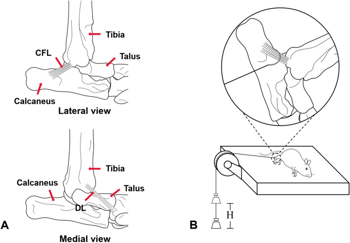
Ankle sprain (AS) is one of the most common injuries in the process of sports. A sprain can lead to the relaxation or fracture of ligaments around the ankle joint, which causes a change in the mechanical properties of the ankle joint or the adjacent joints and further causes instability of the ankle joint or adjacent joints [31,32,33]. The current mouse models of ankle instability are established by cutting the ligaments that maintain the stability of the ankle joint [10, 20, 34, 35]. However, in actual clinical practice, most ankle instability patients do not have broken ligaments around the ankle joint, but rather the ligaments are in a relaxed state [36]. Therefore, we established a mouse model of ankle instability with ankle osteoarthritis with the help of relaxation of CFL and DL caused by the rapid descent of weights. Balance assessments and footprint analysis were performed to evaluate whether the motor level and balance ability of mice were impaired. In contrast, micro-CT scanning and histomorphometry analysis were performed to evaluate whether long-term ankle instability causes PTOA.
In this study, it was found that ligament laxity would lead to the degeneration of articular cartilage and then develop into osteoarthritis, which was similar to the results of osteoarthritis caused by transverse ligament dissection. Compared with previous studies, it was found that within 8 weeks, the time of passing the balance beam was similar between the ligamentous relaxation group and the ligamentous transverse section group [10]. At the 12th week, the time for the ligament laxity group to pass the balance beam was shorter than that for the ligament transverse section group. However, different molding methods did not affect the number of slides [10, 34]. By comparing the OARSI score and the modified Mankin score of the ankle and subtalar joints after ligament transection and ligament relaxation, the score was higher after ligament transection, indicating that the degenerative changes of articular cartilage were more significant after ligament transection [10]. However, different molding methods had no significant effect on the expression of type II collagen. In addition, different molding methods have different effects on the bone volume fraction of ankle joint and subtalar joint. The results showed that the bone volume fraction of the transverse ligament group was similar to that of the CFL laxity relaxation group, but the bone volume fraction increased significantly compared with that of the DL ligament relaxation group [37]. In general, ligament relaxation can lead to ankle instability, and then develop osteoarthritis, but its severity is not as severe as the transverse ligament model.
Balance assessments
With regard to the time required to pass the balance beam, the CFL laxity group and DL laxity group required significantly longer time than the SHAM group at three days and one week after surgery. This phenomenon may be because of the obvious edema and pain in the right hind foot ankle joint of mice in the early stage after surgery. This resulted in the slowing down of the mouse crawling on the balance beam owing to pain feedback and relatively increased the time taken. This suggests that physical activity levels should be reduced early after an acute ankle injury, as the Hubbard-Turner study reported that rest after a severe ankle sprain is essential to restore physical activity levels throughout the life cycle [38]. At postoperative 4 weeks, the time difference between the CFL and DL laxity groups and the SHAM group for crossing the balance beam was minimal. At this time, the surgical incision at the right hind foot ankle joint was completely healed, and the soft tissue swelling had completely subsided. At the later stage of the study (8 weeks after surgery), mice in the CFL and DL laxity groups took longer to traverse the balance beam than those in the SHAM group, which is consistent with what was observed after the transection of the ligaments around the ankle [10, 30, 34, 39]. At this time, the right hind foot ankle joint of mice was significantly deformed, and the stiffness of the joint caused instability in the right hind foot, possibly reducing the speed of the mice while crossing the balance beam.
With regard to the number of slips from the balance beam, the number of slips of the right hind foot in the CFL laxity group and DL laxity group were significantly higher than those in the SHAM group at 3 days, 4 weeks, 8 weeks, and 12 weeks after surgery.However, there was no significant difference between the two groups at 1 week after surgery. The reason for this phenomenon may be that the ankle joint edema of mice decreased 1 week after surgery, and the number of slips decreased owing to the slow speed because of pain when crawling across the balance beam. At 12 weeks after surgery, the number of slips of the right hind foot in the CFL laxity group mice was 3.45 times that observed before surgery, and the number of slips of the right hind foot in the DL laxity group mice was 1.99 times that observed before surgery. The number of slips of the right hind foot in the CFL laxity group mice was 1.58 times that observed in the DL laxity group mice. These results are similar to the experimental results observed by the Hubbard Turner research group by cutting the ligaments around the ankle joint [30, 39]. At this point, the mouse may have progressed from long-term chronic ankle instability to PTOA. Previous studies have shown that individuals with PTOA have balance disorders and changes in movement patterns, which are consistent with our experimental results [40, 41]. In the whole postoperative balance beam experiment, the right hind foot of mice in the CFL laxity group slipped slightly more than that of mice in the DL laxity group, and the difference was statistically significant 12 weeks after surgery. This also reflected that CFL, as the only ligament connecting the tibiotalar joint and subtalar joint at the same time, plays an important role in maintaining the stability of both ankle and subtalar joints [10, 42, 43].
Footprint analysis
The stride length and stride width data measured at 12 weeks in the SHAM group were greater than those observed before surgery. During the study period, the mice continued to grow, leading to individual enlargement and corresponding increases in stride length and width. However, although the mice in the CFL laxity group and DL laxity group grew larger, the stride length and stride width did not show an increase but rather decreased compared with those before surgery. They were significantly shortened compared with the stride length and width in the SHAM group in the same period. The research results suggest that CFL and DL relaxation can cause ankle instability, thereby affecting the normal gait of mice, similar to the results reported by the Hubbard Turner research team wherein the gait of mice was damaged by cutting the lateral ligament of the ankle [39].
Micro-CT scanning
Under normal physiological conditions, osteoclast bone resorption and osteoblast bone formation maintain the dynamic balance of the bone state. Micro-CT quantitative analysis showed that the ankle bone volume fraction in the CFL laxity group and DL laxity group was 6.71% and 4.23% higher than that in the SHAM group, respectively. These results indicated that the relaxation of the ligament around the right hind foot ankle joint in mice broke the stable bone balance, resulting in the abnormal activity of osteoblasts, increased bone density, bone hyperplasia, subchondral bone hardening and thickening, and degenerative changes at the joint site. This result is similar to the CT analysis results of Chang et al. [20], who established the ankle osteoarthritis model through surgery. In addition, in the process of CT image reconstruction and analysis, degenerative changes also occurred in the subtalar joint of mice. The bone volume fractions of the subtalar joint in the CFL laxity group and DL laxity group were 9.39% and 4.37% higher than those of the subtalar joint in the SHAM group, respectively. This result is consistent with the results of previous studies showing that severe foot instability can lead to the degeneration of adjacent ankle joints, suggesting that ankle joint instability in the animal model in this study is more serious [10, 44].
Histomorphometry analysis
In typical H&E staining, SOFG staining, and TB staining, obvious discontinuity was observed on the ankle surface in the CFL laxity group and DL laxity group. The number of chondrocytes on the joint surface was lower or chondrocytes had completely disappeared, and there were empty nucleated chondrocytes, suggesting that ligament relaxation could lead to ankle instability. Ankle instability without effective treatment can lead to PTOA. The results are similar to those obtained by cutting the ligaments around the ankle in mice to construct animal models of ankle instability and ankle osteoarthritis [10, 20]. In addition, large defects were also observed on the surface of subtalar joints in the CFL laxity group. In contrast, damage to subtalar joints in the DL laxity group was less than that in the CFL laxity group. This phenomenon may be caused by the fact that CFL connects the tibial talar joint and the subtalar joint at the same time [10, 42, 43], and its relaxation not only causes ankle injury but also damages the subtalar joint to some extent. In fact, the ankle joint and the subtalar joint are two inseparable structures, which can be called the ankle-subtalar joint complex. Hence, the subtalar joint surface in the DL laxity group also had certain injuries, which was similar to the results reported by Liu et al. [10] In typical type II collagen immunohistochemical staining, the type II collagen-positive area in the ankle joint and subtalar articular cartilage layer in the CFL laxity group and DL laxity group was significantly reduced compared with that in the SHAM group. This difference may be caused by the destruction of some articular chondrocytes, which fail to normally produce type II collagen. Type II collagen is a type of high molecular weight protein produced by articular chondrocytes, which can produce certain mechanical strength to maintain the normal physiological function of articular cartilage. When articular chondrocytes are destroyed, the production of type II collagen also decreases. This result was consistent with the results of the abovementioned three staining methods and with the results of Liang et al. [45], who constructed a rat model of post-traumatic osteoarthritis through ankle fracture.
In this study, the effects of chronic ankle instability after grade I ankle sprain on post-traumatic osteoarthritis were described from macroscopic and microscopic levels. From a macro perspective, we found through behavioral experiments, including balance beam experiment and gait analysis, that the balance ability of mice decreased after ligament relaxation led to chronic ankle instability. On the micro level, we found that ankle sprain would lead to chronic ankle instability and further develop into degenerative changes of the joint, specifically manifested as the reduction of chondrocytes and the reduction of type II collagen positive area. In addition, Micro-CT results also indicated increased bone density and subtalar bone ossification in the ankle and subchondral joints. The macro instability of the mouse ankle joint leads to the abnormal stress stimulation of the mouse ankle, which results in the degeneration of the joint and the ossification of the cartilage at the micro level.
Limitations
There are some limitations to the study. First, the animal used in the experiment is mice. Although the structure of the hind foot and ankle joint of mice is similar to that of humans, it should be noted that mice are quadruped, while humans are biped. Thus, the consistency of movement patterns and behavior needs to be further verified and studied. Second, this study needs to further elucidate the signaling pathway and molecular mechanisms of joint degeneration at a cellular level. More in-depth studies and more reasonable animal models should be developed in future studies.


留言 (0)