Cell lines
Five NB cell lines used in the study, including SK-N-SH, SK-N-BE (2), SH-SY5Y, IMR32, and SK-N-AS, were purchased from COBIOER BIOSCIENCES (Nanjing, China). SK-N-BE (2) and SH-SY5Y were cultured in MEM/F12 (1:1) (Gibco, USA) supplemented with 10% fetal bovine serum (FBS, Gibco, USA); SK-N-SH and IMR32 were cultured in MEM (Gibco, USA) supplemented with 10% FBS (Gibco, USA); SK-N-AS was cultured in DMEM (Gibco, USA) supplemented with 10% FBS (Gibco, USA). All mediums were supplemented with 1% penicillin–streptomycin solution (Gibco, USA). All cell lines had been authenticated by STR and are free from mycoplasma.
Cell counting kit-8 (CCK-8) assay
NB cells were seeded in 96-well plates with three replicates for each sample. After the indicated treatment, 10% CCK-8 solution (APExBIO, USA) was added to the plate and continued to be incubated for 3 h at 37 °C in the cell incubator. Then, the absorbance was detected with the spectrophotometry at 450 nm.
Compounds and drug screening
The small molecule compound library containing 288 molecular drugs was purchased from Selleck (Shanghai, China). Two representative NB cell lines were selected for the high-throughput screening: SK-N-SH (MYCN non-amplified) and SK-N-BE (2) (MYCN amplified). Cells pre-inoculated into 96-well plates at a density of 5000 cells/well. The working concentration of the drug was 5 μM, with DMSO as the control. After 72 h, cell viability was detected by CCK-8 and the survival rate was calculated [52]. An effective drug is defined as the tumor survival rate lower than 50%. The drugs that showed co-efficacy in both two cell lines were screened out, and the natural antifungal product chaetocin, with a significant killing effect was identified.
Bioinformatics analysis
The combined cohort of TCGA, TARGET, and GTEx samples were obtained from UCSC Xena browser (TCGA TARGET GTEx cohort, https://xenabrowser.net/datapages/?cohort=TCGA%20TARGET%20GTEx&removeHub=https%3A%2F%2Fxena.treehouse.gi.ucsc.edu%3A443), and there were 162 NB samples and 1280 normal tissues (128 normal adrenal gland and 1152 normal brain tissues) [19]. The Wilcox method was used for the difference analysis, with R package used for the bioinformatics analysis of the SUV39H1 expression levels between NB samples and normal tissues. For the Kaplan–Meier survival analysis, overall survival (OS) and event-free survival (EFS) curves between NB patients expressing high/low levels of SUV39H1 or MCPIP1 were generated through the R2 online website platform (http://r2.amc.nl), followed by the log-rank test. The scanning method was used to determine the cut-off [53, 54]. For the Spearman correlation analysis based on the TARGET database, the mRNA expression levels of indicated genes were analyzed based on the RNA-seq results from 143 NB samples (https://www.cbioportal.org/).
Immunohistochemistry (IHC) assay
The 61 NB tissue specimens obtained from our center (all patients were <18 years old with the initial diagnosis as NB, and the follow-up data was complete) were fixed in formalin, embedded in paraffin blocks, and sectioned with a thickness of 4 μm. Then, IHC staining was performed following instructions as previously described [19]. Anti-SUV39H1 rabbit pAb (Abclonal, China, A3277) was used in this assay. We used a fluorescence microscope to capture images (Olympus BX61, Japan). The intensity was scored as follows: 0—negative; 1—weak; 2—moderate; 3—strong. The proportion of positive cells was scored as follows: 0—less than 25%; 1—25% to 50%; 2—50% to 75%; 3—75% to 100%. The product of the above two scores indicated the composite staining score. Two expression staining levels were defined: scores of 0–4 were classified as a low expression, whereas scores of 5–9 were classified as a high expression.
RNA isolation and quantitative real-time polymerase chain reaction (qRT-PCR) analysis
The total RNA from the cultured cells was extracted with the Trizol reagent (Life Technologies, USA). The RNA was reverse transcribed into cDNA using the Prime Script RT Master Mix (TaKaRa, Japan) following the manufacturer’s instructions. Then, qRT-PCR was conducted with a Power SYBR Green Master Mix (Dongsheng Biotech, China) using primers listed in Supplementary Table 1, with the β-actin as an endogenous control.
Western blotting (WB) analysis and antibodies
The Whole Cell Lysis Assay Kit (KeyGEN, China) was used to extract the whole-cell lysates. The BCA method (Thermo Fisher Scientific, USA) was applied to detect the protein concentration. β-actin (1: 1000, Proteintech, China, 66009) was used as the internal control. An equal amount of each protein sample was separated with SDS-PAGE and transferred onto a PVDF membrane. Then, the membrane was blocked in 5% skim milk and incubated with primary antibodies overnight, followed by incubating with indicated secondary antibody for 1 h at room temperature. Primary antibodies used this study were listed in Supplementary Table 2. The signal detection was performed with the ECL chemiluminescence detection system (Bio-Rad, USA).
Colony formation assay
Two thousand cells were seeded in a 6-well plate. After 10–14 days, the cell colonies were fixed in 4% polyformaldehyde and stained with 0.1% crystal violet for 15 min. After staining, the colonies were washed with tap water to remove excessive crystal violet. Images were taken by a camera, and the number of colonies was quantified by ImageJ (NIH).
Wound healing assay
NB cells were seeded in 6-well plates at a density of 5 × 10⁴ cells/well with scratch plug-in components. The cells were cultured with the culture medium containing 0.5% FBS in the incubator overnight. The scratch plug-in components were taken out the next day, and an even wound formed then. Images of samples were captured at appropriate time with the microscope (Leica, USA) at ×20 magnification. In order to exclude the effect of cell proliferation in wound healing assay, the CCK-8 assay for cell proliferation was performed among indicated cells with the same condition used in wound healing assay, and the results indicated that there was no significant statistical difference among indicated cells, confirming that changes in cell migration ability were independent of cell proliferation (Supplementary Fig. 1).
Transwell assay
A density at 1 × 10⁵ cells/well of NB cells was seeded in 24-well plates in 200 μL FBS-free culture medium, and was then plated into the upper chamber with 8 μm pores. Accordingly, 650 μL medium containing 10% FBS was added into the lower chamber. After continuously incubated for about 24 h, the migrated cells were fixed by 4% paraformaldehyde for 20 min and followed by stained with crystal violet for 10 min. Images of the migrated cells were captured with microscopy (Leica, USA). For each well, 5 randomized fields within the view were selected for imaging, and the numbers were counted.
Apoptosis analysis
Cell apoptosis of NB cells was analyzed by flow cytometry using the Annexin V-Alexa Fluor 647/7-AAD Kit (4A Biotech, China) according to the manufacturer’s instructions. In brief, 5 × 10⁵ of indicated NB cells were prepared, and incubated with 5 μL Annexin V-Alexa Fluor 647 for 15 min in the dark, then 10 μL of 7-AAD was added into each tube, immediately analyzed with the flow cytometry (SP6800, Sony, Japan) within 1 h.
Cell cycle analysis
The cell cycle phases of NB cells were analyzed by flow cytometry using the PI Cell Cycle Detection Kit (4A Biotech, China). Briefly, cells with the indicated treatment were fixed in 70% ethanol at 4 °C overnight. Then cells were washed and stained with 50 ug/mL propidium iodide (PI) in solution of 2 mg/mL RNase [55, 56]. Flow cytometry (SP6800, Sony, Japan) was performed on the cells. Modfit software was used for the cell cycle distribution analysis.
Immunofluorescence (IF) staining and imaging
NB cells were prepared in the glass-bottom cell culture dishes (NEST, Wuxi NEST Biotechnology Co., Ltd., China) and fixed with 4% paraformaldehyde for 20 min at room temperature. Then, cells were permeabilized with 0.1% Triton X-100 for 5 min following by blocked in 5% BSA for 1 h. The primary anti-SUV39H1 (Santa Cruz Biotechnology, USA) antibody was used and incubated with cells for 1 h at room temperature. NB cells were washed with PBS and incubated with Alexa Fluor 594-conjugated secondary antibodies (FuDe, China) for 1 h in the dark. DAPI (Beyotime, Shanghai, China) was added and incubated for 5 min to stain the cell nuclei. The confocal microscope (LSM880, Carl Zeiss, Oberkochen, Germany) was used for image acquisition.
Xenograft tumorigenesis in vivo
The NOD/SCID mice aged 3–4 weeks were purchased from Vital River Laboratory Animal Technology (Beijing, China). A mixture of SK-N-BE (2) cells (8 × 10⁶ cells/mice) and thawed Matrigel (Corning, USA) (at a ratio of 3:1) was subcutaneously injected into the right axilla area of the mice [55, 57]. When the tumor volume reached 100 mm3 (tumor volumes = short diameter*short diameter*long diameter/2), the mice were divided randomly into the vehicle group and the treatment group. The treatment group was given 100 uL of chaetocin (0.5 mg/kg) intraperitoneally every 2 days. Body weight and tumor volumes of mice were recorded every other day. Any mice with tumor reached the maximum permitted diameter of 2 cm was regarded as the humane endpoint. After euthanized, the subcutaneous tumors of the mice were isolated and weighed immediately. The in vivo experiments were performed with the approval of the Ethics Committee of Sun Yat-sen University. Investigators were not blinded in this experiment procedure.
RNA sequencing and data analysis
Biological triplicate RNA-seq analysis was performed on NB cells of SUV39H1 inhibition: siNC-NB cells (3 samples), siSUV39H1-NB cells (3 samples), DMSO-NB cells (3 samples), and chaetocin-NB cells (50 nM, 3 samples). Total RNA was extracted with TRIzol reagents (Ambion, Life Technologies, USA). Then, cDNA library building and RNA sequencing (RNA-seq) were carried out via a commercially available service [58] (Huada Gene, Wuhan, China). RNA-seq reads were aligned to the hg38 genome, using |log2FC| ≥ 0.5 and FDR ≤ 0.01 as the cutoff criteria to identify the DEGs. Data can be accessed via GSE254118.
siRNA transfection
Human siRNA oligos were purchased from Ribo (Shanghai, China). For transfection, NB cells were seeded onto 6-well plates and transfected with 20 nM SUV39H1(siSUV39H1) or scrambled control (siNC) using Lipofectamine 3000 (Invitrogen, USA) according to the manufacturer’s instructions. After 24 h, the RNA was harvested and analyzed by qRT-PCR to verify the transfect efficiency. The transfected cells were cultured for up to 48 h for further experiment studies.
Lentiviral transduction of NB cells
For the stably knockdown of SUV39H1, the SUV39H1 and control shRNA plasmids (Cat#:LPP-HSH062782-LVRU6GH-200) were purchased from GeneCopoeia (Guangzhou, China). For the stably overexpression of SUV39H1, MCPIP1 and AURKA, the pCDH-SUV39H1-3xflag-puro plasmid (Cat#:OENM_001282166), the pcDNA3.1-ZC3H12A-3xFlag (Cat#:OENM_025079) and the HY22766 pCDH-CMV-AURKA-EF1-Puro-3xFlag (Cat#:OENM_198433) plasmids were purchased from DaHong Biosciences (Guangzhou, China). The lentiviral transduction system construction was conducted as the previous description [19, 58], with the core plasmids: packaging plasmid psPAX2: envelope plasmid pMD2.G at a ratio of 10:7.5:3. HEK293T cells were used for the lentiviral particle production. The SK-N-SH and SK-N-BE (2) cells were infected with viral supernatant (diluted at 1:1 with fresh media), followed by puromycin selection at 1 μg/mL for 48 h.
Chromatin immunoprecipitation (ChIP) assays
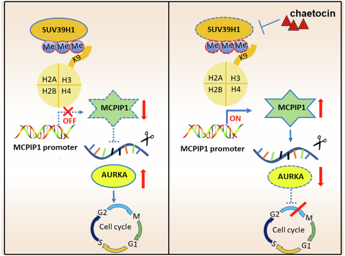
ChIP assay was performed using a SimpleChiP (R) Kit (Magnetic Beads) (Cell Signaling Technology, USA, 91820s) according to the manufacturer’s protocol. NB cells were seeded in 15 cm cell culture dish and were collected following by fixed with 1% formaldehyde at room temperature for 10 min. Subsequently, glycine was added to stop this reaction, and cells were sonicated to shear the DNA into fragments at 200–1000 bp. Then, sheared DNA was incubated with antibodies against H3K9me3 (Cell Signaling Technology, USA) or IgG control (Santa Cruz Biotechnology, USA, sc-2027). DNA-protein-antibody complexes were incubated with Protein A/G magnetic beads, and were washed sequentially with gradient salt buffer, eluted with 1% SDS/NaHCO3, and finally, enriched DNA fragments were subjected to qPCR analysis. IgG was used as the negative control. The primers used for the ChIP-qPCR assays are listed in Supplementary Table 3.
Statistical analysis
Statistical analyses were performed with SPSS 22.0 software (SPSS, Chicago, IL, USA) by Student’s t test, one or two-way ANOVA, or χ2 test. The variance analyzed in this study was assumed as similar with normal distribution. The Kaplan–Meier survival curves were generated based on a log-rank test to compare the survival rates between different groups. All functional assays were independently repeated at least three times, and the results were expressed as mean ± SEM. p < 0.05 was considered statistically significant.


留言 (0)