Ethics statement
All experimental procedures involving animals received approval from the Institutional Animal Care and Use Committee of Sichuan Agricultural University, China, under Permit Number SYXK(川)2019-187.
Cells, animals, and virus
BHK-21 cells were maintained in Dulbecco’s modified Eagle medium (DMEM, Gibco, USA) supplemented with 10% new bovine serum (NBS, Gibco, USA). Nine-day-old duck embryos were sourced from the duck industry in Ya’an, China. Duck embryo fibroblasts (DEFs) were cultured in a minimum essential medium (MEM, Gibco, USA) supplemented with 10% NBS (Gibco, USA). One-day-old Peking ducks were obtained from the duck industry in Chengdu, China. The 20-day-old chickens were kindly provided by Dr. Xia, Sichuan Agricultural University. The TMUV CQW1 strain (GenBank: KM233707.1) was propagated in DEFs as previously described51.
Bio-information analysis of duCD40L
Nine ORF sequences of CD40L retrieved from GenBank were compiled. Phylogenetic relationships among these sequences were assessed utilizing the neighbor-joining method within MEGA software version 7.0. Protein domains were identified using the Simple Modular Architecture Research Tool (SMART, http://smart.embl-heidelberg.de/). Alignment of the amino acid sequences of the dusCD40L from various species was performed either through DNAMAN or an online BLAST tool (https://blast.ncbi.nlm.nih.gov/Blast.cgi).
Plasmids construction
DusCD40L with codon optimization for duck protein expression was synthesized by Wuhan Jinkairui Bioengineering Co., China. Then, the fragment was integrated into the pVAX1 vector from Invitrogen, which includes the signal sequence of tissue plasminogen activator (TPA) (MDAMKRGLCCVLLLCGAVFVS), resulting in the construction of the dusCD40L eukaryotic expression plasmid designated as pVAX-TPA-dusCD40L. The fragments dusCD40L, P2A, ILZ, and Leu-Leu were introduced into the pVAX-prM-E plasmid as described by Huang et al.51 using specific primers (refer to Supplementary Table 1). A schematic representation of pVAX-prM-E-dusCD40L can be observed in Fig. 4a.
Western blot (WB) assay
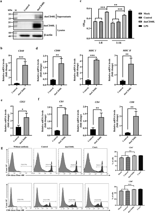
DEFs were seeded at a density of 2 × 10⁶ cells per well in a six-well plate and maintained at 37 °C in an atmosphere containing 5% CO2. The following plasmids, pVAX1, pVAX-TPA, pVAX-TPA-dusCD40L, pVAX-prM-E, or pVAX-prM-E-dusCD40L, were individually transfected into DEFs when the cells reached a confluence of 90%. Transfection was carried out using TransIntroTM EL Transfection Reagent (TransGen Biotech, China) following the manufacturer’s instructions. After 48 h post-transfection, both cell supernatants and cell pellets were harvested. The pellets were lysed using 100 μL of radioimmunoprecipitation assay (RIPA) lysis buffer (Beyotime, China) containing 1% phenylmethanesulfonyl fluoride (PMSF, Beyotime, China). Protein separation was accomplished by performing sodium dodecyl sulfate-polyacrylamide gel electrophoresis (SDS-PAGE) using a 15% acrylamide gel for resolving cell supernatants and lysates. The samples were separately loaded into the gels, 20 μL/lane. Subsequently, proteins were transferred onto a PVDF membrane (Bio-Rad, USA) at a current of 220 mA for 80 minutes. The membrane was blocked with 5% nonfat milk for 2 h at room temperature. Primary antibodies used included rabbit anti-E polyclonal antibody (prepared in this study, 1:1000) and rabbit anti-duCD40L polyclonal antibody (prepared in this study, 1:500), both diluted accordingly. Horseradish peroxidase (HRP)-conjugated goat anti-rabbit IgG (H + L) (Cat# RS0002, Immunoway, China) at a dilution of 1:5000 served as the secondary antibody for the detection of E protein and dusCD40L expression. Membrane imaging was achieved using Clarity™ Western ECL Substrate (Bio-Rad, USA). Uncropped and unprocessed scans of the blots are in the Supplementary Figs. 4 and 5. All blots derive from the same experiment and that they were processed in parallel.
Indirect immunofluorescence assay (IFA)
When DEFs reached approximately 90% confluence, they were individually transfected with the following plasmids, pVAX1, pVAX-prM-E, and pVAX-prM-E-dusCD40L. Subsequently, the cells were fixed by incubating them with 0.5% paraformaldehyde for an overnight period at 4 °C at 48 h post-transfection. To block nonspecific binding sites, a 5% nonfat milk solution was applied to the cells for 30 min at room temperature. Following this, the cells were subjected to incubation with rabbit anti-duCD40L polyclonal antibody (prepared in this study, 1:250) or rabbit anti-E polyclonal antibody (prepared in this study, 1:500) at 4 °C. After overnight incubation, a secondary antibody, PE-conjugated goat anti-rabbit IgG (Cat# HS121-01, TransGen, China) at a dilution of 1:1000, was utilized. The visualization of DEFs was accomplished using a fluorescence microscope (Nikon, Japan).
qRT-PCR assay
DEFs were subjected to transfection with either pVAX-TPA (control) or pVAX-TPA-dusCD40L (dusCD40L) using TransIntroTM EL Transfection Reagent (TransGen Biotech, China) as per the manufacturer’s instructions. At 48 h post-transfection, cell culture supernatants were collected for the following assays.
Ducks around 30-day-old were used for blood collection. DuPBMCs were isolated using a Peripheral Blood Mononuclear Cell Separation Kit (Tianjin Hao Yang Biological Products Technology Co., Ltd., Tianjin, China) following the manufacturer’s protocols. DuPBMCs were then treated with the collected supernatants at dilutions of 1:8 or 1:16 for a 48-hour incubation period. Total RNA was extracted from the treated duPBMCs using RNAiso (Lablead, China), and this RNA was reverse-transcribed into cDNA utilizing a PrimeScript RT Reagent kit with gDNA Eraser (TaKaRa, Japan).
QRT-PCR was employed to analyze the relative mRNA expression levels of various genes, including MHCI, MHCII, CD3, CD4, CD8, CD21, CD40, CD80, Il-6, Il-10, Il-12, Ifnγ, BAFF, and GAPDH. The primers for qRT-PCR are listed in Supplementary Table 1. The qRT-PCR was conducted following the manufacturer’s instructions for TB Green® Premix Ex TaqTM (Tli RNaseH Plus) (TaKaRa, Japan), comprising an initial denaturation step at 95 °C for 30 s, followed by 40 cycles of denaturation at 95 °C for 5 s and annealing at 60 °C for 30 s. The relative gene expression levels were calculated using the 2-ΔΔCt method.
Flow cytometry
DuPBMCs were isolated as mentioned above. CD4+ and CD8+ T cells were measured by the flow cytometry assay. It was carried out as described previously52. Briefly, duPBMCs were adjusted to 1 × 10⁷ cells per mL and seeded into 96 well plates, 100 μL/well. They were stimulated with dusCD40L (1:8), control (1:8), ConA (5 μg/mL), or DMEM (mock) for 48 h. Then, the suspended cells were collected. The anti-duck CD4 monoclonal antibody (Cat# MCA2478, Bio-rad, USA) at 1:200 dilution and the anti-duck CD8 monoclonal antibody (Cat# MCA2479, Bio-rad, USA) at 1:200 dilution were added into the cells, respectively. The cells were separately incubated with the antibodies at 4 °C, 30 min. After washing with PBS, Alexa Flour 488-labeled goat anti-mouse IgG (Cat# A32723, ThermoFisher, USA) as the secondary antibody at 1:500 dilution was added and the cells were incubated in the dark. Flow cytometry was performed on an CytoFLEX™ (Beckman counlter, USA). Data was analyzed using CytExpert software.
Immunization and challenge
A total of forty 1-day-old ducklings were reared for 1 w and subsequently randomly allocated into four groups, each consisting of ten ducks. Vaccination was administered to the ducks when they were 7 days old. One group of ducks received an intramuscular injection of pVAX1 at a dose of 200 μg in 0.5 mL of PBS. Two other groups of ducks were intramuscularly injected with either the vaccine pVAX-prM-E or pVAX-prM-E-dusCD40L at a dose of 200 μg in 0.5 mL of PBS. The final group of ducks received an intramuscular injection of 0.5 mL of inactivated TMUV vaccine (Yangzhou Ubang Biopharmaceutical Co., China) in the lateral thigh. Ducks in the prM-E-based groups were subjected to intramuscular injections in the lateral thigh three times at 3-week intervals, while ducks in the inactivated vaccine group received two intramuscular injections in the lateral thigh at 3-week intervals. At 7 days after the first immunization, three ducks from DNA-vaccinated groups were randomly selected and euthanized for spleen collection. At 5 w, 6 w, 7 w, 8 w, and 10 w post the first immunization, three ducks from each group were randomly selected for blood collection. Subsequently, ducks from each group were challenged with 108 TCID50 TMUV CQW1 strain per duck via intramuscular injection at 5 w after the third immunization. The clinical symptoms were recorded for continuous 7 days. At 7 days post-challenge, three ducks from each group were randomly selected and euthanized for serum, spleen, and brain sampling.
Immunohistochemical analysis
In each DNA-vaccinated group, ducks were euthanized at 7 days after the first immunization (n = 3). They were anesthetized by sodium pentobarbital (100 mg/kg) and were cut open for bloodletting bled. Then the spleens were collected. The presence of the E protein was confirmed through immunohistochemistry, following established protocols51. Briefly, the collected tissues were sectioned at a thickness of 4 μm using a Leica RM2128 microtome (Germany). These sections were initially fixed with 4% paraformaldehyde and then embedded in paraffin. Subsequently, the sections underwent dewaxing with xylene, rehydration with a gradient of ethanol, and were subsequently treated with 0.3% hydrogen peroxide (H2O2). Antigen retrieval was performed using a citrate buffer solution (CBS, 0.01 M, pH 6.0). The sections were incubated with a 10% normal goat serum to block nonspecific antigens for 1 h at 37 °C. Afterward, the sections were stained with a rabbit polyclonal antibody targeting TMUV E protein (prepared in this study) at a 1:200 dilution, followed by an incubation overnight at 4 °C. An HRP-conjugated goat anti-rabbit antibody (Cat# HS101-01, Transgen Biotech, China) at a 1:1000 dilution was used for a 2 h incubation at room temperature. The visualization of E protein expression was achieved using DAB (Solarbio, China), and the images were captured using microscopy (Olympus, Japan).
Cell counting kit-8 (CCK-8) assay
DuPBMCs were obtained from 30-day-old ducks or ducks that had been vaccinated at 5 w post the initial vaccination using the peripheral blood mononuclear cell separation kit (Tianjin Hao Yang Biological Products Technology Co., Ltd., Tianjin, China) following the manufacturer’s protocols. ChPBMCs were isolated from 20-day-old chickens by the same method as duPBMCs. The freshly isolated duPBMCs and chPBMCs were adjusted to a concentration of 5 × 10⁶ cells/mL and were then seeded into 96-well plates at a volume of 100 μL per well, respectively. Subsequently, they were stimulated with dusCD40L at dilutions of 1:8 or 1:16 (prepared in this study), LPS (5 μg/mL, Solarbio, China), ConA (5 μg/mL, Solarbio, China), or 100 µg/mL of inactivated TMUV (prepared in this study) for a duration of 48 h at 37 °C in an atmosphere containing 5% CO2. Following stimulation, lymphocyte proliferation was assessed by measuring the optical density at 450 nm (OD450) using a CCK-8 kit (Solarbio, China), following the manufacturer’s instructions.
ELISA assay
For the detection of antibodies against inactivated TMUV, a 96-well ELISA plate was coated with inactivated TMUV (3.2 μg/well) in a coating buffer (pH 9.6) overnight at 4 °C. Following this, the plates were washed three times with PBS (pH 7.4) containing 0.1% Tween-20 (PBS-T). After blocking with PBS-T containing 5% nonfat milk, the plates were incubated with serum samples (diluted 1:400, 200 μL/well) collected earlier for 2 h at 37 °C. Subsequently, the plates were washed five times with PBS-T. HRP-conjugated goat anti-duck IgG (Cat# 5220-0296, KPL, USA), diluted at 1:2000, was added to the plates (100 μL/well) for 1 h at 37 °C. Following this incubation, the plates were washed, and 3, 3′, 5, 5′-tetramethylbenzidine (TMB) was added (100 μL/well) for 10 min at room temperature in the dark. The reaction was halted by adding 2 M H2SO4 (50 μL/well), and the OD450 was measured using a spectrophotometer (Bio-Rad, USA).
To determine IFNγ titers, a 96-well ELISA plate was coated with rabbit anti-duIFNγ polyclonal antibody (prepared in this study, 1:80 dilution) in coating buffer (pH 9.6) overnight at 4 °C. The plates were then blocked with PBS-T containing 5% nonfat milk at 37 °C for 1 h. DuPBMCs were seeded into 96-well plates with 1 × 10⁷ cell/well in 100 μL. They were obtained and treated as mentioned above. The supernatants of the duPBMCs were separately collected at 48 h post dusCD40L stimulation. They and serum samples (1:30 dilution) from vaccinated ducks collected at 5 w and 8 w post-primary immunization were added to the plates and incubated at 37 °C for 1.5 h. Mouse anti-duIFN-γ polyclonal antibody (prepared in this study), at a 1:160 dilution, was subsequently added to the plates, and the plates were incubated at 37 °C for 2 h. HRP-conjugated goat anti-mouse IgG antibody (Cat# HS201-01, TransGen, China) at a 1:8000 dilution was added to the plates and incubated at 37 °C for 1 h. After the incubation, the plates were washed, and TMB (100 μL/well) was added for 10 min at room temperature in the dark. The reaction was stopped with 2 M H2SO4 (50 μL/well), and the OD450 values were measured using a spectrophotometer (Bio-Rad, USA).
Neutralizing antibodies measurement
Sera were collected at 5 w and 8 w post the initial immunization with TMUV vaccines, as previously described. The collected sera were subjected to inactivation at 56 °C for 30 min and then were serially diluted in twofold increments (2−1–2−8) using DMEM. Each dilution was mixed with an equal volume of TMUV CQW1 at a concentration of 200 TCID50. The resulting mixture was added to BHK-21 cells cultured in 96-well plates with approximately 90% confluence, at a volume of 100 μL per well. After 2 h, the supernatant was removed, and the plates were washed three times with PBS. Following the washes, the cells were cultured in DMEM supplemented with 2% NBS, at a volume of 100 μL per well, and maintained at 37 °C in an atmosphere containing 5% CO2 for 5 days. Each dilution was replicated in 10 wells. The cytopathic effect (CPE) was observed and recorded during this period. Neutralizing activity was subsequently analyzed using the Reed-Muench method.
TCID50 assay for viral titration
Ducks from each group were anesthetized and euthanized as mentioned above at 7 days post-challenge (n = 3). The sera, spleens and brains were collected at 7 dpi. A total of 100 mg/spleen or 100 mg/brain was added to 1 mL DMEM with 1% penicillin and 1 μg/mL streptomycin for homogenization. The homogenized samples were centrifuged at 12,000 r/min for 10 min at 4 °C. Then the supernatants were collected. They and the sera were diluted in serial 10-fold increments (10−1–10−9) in DMEM containing 2% FBS. When the BHK-21 cells grown in 96-well plates reached approximately 90% confluence, the samples were added to the plates individually at 100 µL/well. The plates were incubated at 37 °C with 5% CO2 for 5 days. Then, the CPE was recorded and TCID50 was calculated by the Reed-Muench method.
Statistical analysis
Data were analyzed by GraphPad Prism (version 8.0) and presented as the means ± standard deviation (s.d.). Statistical significance was assessed using an unpaired Student’s t-test. The significant differences are annotated as *P < 0.05, **P < 0.01, ***P < 0.001.
Reporting summary
Further information on research design is available in the Nature Research Reporting Summary linked to this article.


留言 (0)