Cells and viruses
BHK-21 cells and BSR cells were cultured in Dulbecco’s Modified Eagle Medium (DMEM, Gibco, USA) and supplemented with 10% fetal bovine serum (FBS, Gibco, Australia) at 37 °C in a 5% CO2 humidified incubator. Rabies virus CVS-11 strain (GenBank No. GQ918139.1) and street virus BD06 strain were propagated in BHK-21 cells and used throughout the experiments. Rabies virus SC16, GD1, NM3, QH2, LY, YN3, and XZ17 strains belonged to China I–VII clades, respectively. The background information of representative strains are shown in Table 1.
Table 1 Rabies virus background informationVaccine
The development and characterization of mRNA vaccine (LVRNA001) were previously described23. Briefly, the mRNA encoding the rabies virus glycoprotein (RABV-G) of the CTN-1 strain (GenBank No. ACR39382.1) was produced by in vitro transcription (IVT) and encapsulated using lipid nanoparticle (LNP) technology. Inactivated rabies virus vaccines were purchased from Liaoning Chengda Biotechnology Co., Ltd. (Lot number 4150466A human use, freeze-dried, 1 dose ≥2.5 IU).
Animal ethics statement
This study was carried out in strict adherence to recommendations described in the Guide for the Care and Use of Laboratory Animals, the Office of Animal Welfare, and the United States Department of Agriculture. The procedures used for anesthesia and euthanasia of study animals followed tenets of the ARRIVE reporting guidelines39. Dogs and cynomolgus macaques were anesthetized with intramuscular injection of ketamine (10 mg/kg, 50 mg/mL) followed by intramuscular injection of xylazine hydrochloride (2 mg/kg, 20 mg/mL). Then, the animals were euthanized through exsanguination via the femoral artery. Mice were euthanized using carbon dioxide inhalation. For Sprague-Dawley rats, F0-generation male rats were anesthetized with isoflurane inhalation and then euthanized through exsanguination via the abdominal aorta and/or posterior vena cava. F0-generation female rats and F1-generation rats were euthanized using carbon dioxide inhalation. All procedures were carried out under trained personnel and under the supervision of veterinary staff.
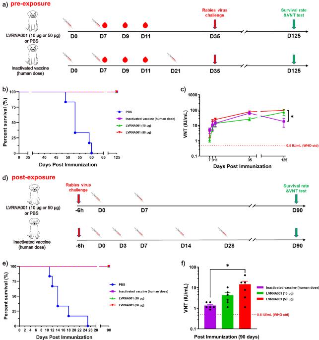
Pre-exposure vaccination against RABV infection in dogs
Dogs (4–6 month-old beagles) were obtained from the Institute of Military Veterinary Medicine, Academy of Military Medical Sciences (Changchun, China). All experiments were approved by the Research Ethics Committee of the College of the Institute of Military Veterinary Medicine, Academy of Military Medical Sciences (IACUC of IAC22W002). Dogs were fed and watered regularly twice a day under normal conditions before being challenged with the virus. The challenged dogs were observed and raised in the designated animal safety laboratory. Dogs were randomized by gender, age, and weight into four experimental groups, and the study was performed in a double-blind randomized fashion. Six dogs per group were vaccinated i.m. with LVRNA001 (10 or 50 μg, 0d/7d), inactivated vaccine (human use, 1 dose ≥2.5 IU, 0d/7d/21d) in 500 μL, or phosphate-buffered saline (PBS) (500 μL, 0d/7d). Dogs were challenged with 50-fold LD50 of street virus BD06 strain in the skeletal muscle of opposite hind legs on D35 (35 days post vaccination). Blood samples were collected on D7, D9, D11, D35, and D125 for neutralization antibody tests. Observation of animal survival continued through 125 days after vaccination. The experimental endpoint of this study was 125 days after the first vaccination.
Post-exposure vaccination against RABV infection in dogs
Dogs (4–6-month-old beagles) were injected i.m. with 50-fold LD50 of virulent RABV-BD06 strain in the biceps femoris of the hindlimb. Six hours after infection, dogs were i.m. vaccinated with LVRNA001 (10 or 50 μg, 0d/7d), inactivated vaccine (human use, 1 dose ≥2.5 IU, 0d/3d/7d/14d/28d) in 500 μL, or PBS (500 μL, 0d/7d). Animal survival was evaluated until 3 months after the first dose. Blood samples were collected on D90 and used for neutralization antibody tests. The experimental endpoint of this study was 90 days after the first vaccination.
Pre-exposure and post-exposure vaccination against different rabies strains in mice
Mice (BALB/c, female, 6 weeks old) were obtained from Huafukang Biotechnology Co., Ltd. (Beijing). The IACUC approval number was 2023-CCDC-IACUC-009. The animals were housed under specific pathogen-free conditions and a standard light cycle (12 h light/dark cycle).
In the pre-exposure prophylaxis murine model, ten mice per group were vaccinated i.m. with LVRNA001 (5 μg/high-dose group, 0d or 0d/7d), LVRNA001 (1.67 μg/low-dose group, 0d or 0d/7d), or inactivated vaccine (1/10 or 1/30 human use, 0d/7d). On Day 14 after the first vaccination, mice were challenged with 50-fold LD50 of RABVs (SC16, GD1, NM3, QH2, LY, YN3, and XZ17, belonging to China I–VII clades, respectively) delivered i.m. Observation of animal survival continued through 42 days after immunization. The experimental endpoint of this study was 42 days after the first vaccination.
In the post-exposure prophylaxis murine model, mice were injected i.m. with 50-fold LD50 of RABVs (SC16, GD1, NM3, QH2, LY, YN3, and XZ17, belonging to China I–VII clades, respectively). Two hours after infection, ten mice per group were vaccinated with LVRNA001 (5 μg/high-dose group, 0d/7d or 0d/14d), LVRNA001 (1.67 μg/low-dose group, 0d/7d or 0d/14d) or inactivated vaccine (1/10 or 1/30 human use, 0d/3d/7d/14d/28d). Mice were monitored for 35 days. Animal survival was evaluated 28 days after immunization. The experimental endpoint of this study was 35 days after the first vaccination.
Immune generation period and immunodynamic testing in mice
Groups of six mice (BALB/c, female, 6 weeks old) were vaccinated i.m. with LVRNA001 (5 μg, 0d or 0d/7d or 0d/14d) or inactivated vaccine (1/10 human use, 0d/3d/7d/14d/28d). Blood samples were collected on D3, D5, D7, D10, D14, D21, D28, and D42 after the first vaccination. Neutralizing antibody titers were detected in sera by rapid fluorescent focus inhibition test (RFFIT). The experimental endpoint of this study was 42 days after immunization.
Rapid fluorescent focus inhibition test (RFFIT)
Sera was inactivated by heating at 56 °C for 30 min, then serially diluted in a 96-well plate with human rabies immunoglobulin standards. CVS-11, at a dose that caused 80% infection of BSR cells in each well, was incubated with the serum for 1 h at 37 °C. Then BSR cells were added to each well and incubated for 24 h at 37 °C in 5% CO2. Finally, the cells were fixed and stained with a fluorescein isothiocyanate-conjugated antibody (Fujirebio, Cat.800-092) at a 1:50 dilution. The percentage of infected cells at each serum dilution was detected in each well using a fluorescence microscope. The RVNA titer (50% neutralization, ED50) of a test sample was mathematically interpolated using the Reed and Muench method40 and was calibrated and converted into IU/mL against the WHO SRIG (standard rabies immune globulins). Sera with a neutralizing antibody titer ≥0.5 IU/mL were considered positive.
Single-dose acute toxicity study in CD-1 mice
A total of 30 CD-1 mice (half male and half female) were randomly divided into 3 groups (n = 10, half male and half female): negative (Saline), LVRNA001 (10 μg, low-dose group), and LVRNA001 (50 μg, high-dose group), which were injected into the thigh of the right hindlimb on D0. Then, the mice were continuously monitored for 14 days. Body weight was measured and weight gain rate was calculated on D-1 (1 day before administration), D3, D5, D7, D9, D11, and D14. The IACUC approval number was S-ACU22-0994. The experimental endpoint of this study was 14 days after immunization.
Vaccination of cynomolgus macaques
According to the ICH Guideline S6 (R1) issued in 2011, cynomolgus monkey is recognized as an appropriate animal model for toxicological studies of biological products that have been or are intended to be used in humans. A total of 40 cynomolgus macaques aged 2.8 to 4.4 years, weighing from 2.16 to 4.48 kg, half male and half female, were purchased from Xiongsen Primate Laboratory Animal Breeding Development Co. Ltd. (Guangxi, China). Animals were included in the study after quarantine (Laboratory Animal Production License No: SCXK (Gui) 2021-0004, Laboratory Animal Quality certificate No: 0002990). The IACUC approval number was S-ACU22-1084. The animals were housed in stainless steel cages, with males and females separated into different cages. Each cage did not exceed five animals per gender per group. The temperature in the animal room was controlled within the range of 18–26 °C, with a relative humidity maintained between 40 and 70%. The vivarium light cycle was set at 12:12 h of light:dark. All animals were fed a non-human primate diet, which is nutritionally complete. The diet was supplemented with a variety of fruits and vegetables at a minimum of three times each week.
Cynomolgus macaques were randomly divided into 4 groups (n = 10, half male and half female): Saline group (negative control), Empty LNP group, LVRNA001 (50 μg) (low-dose group), and LVRNA001 (150 μg) (high-dose group), which were injected into the thigh of the right hindlimb on D0 (0-day post vaccination), D7, D21, and D35. The administration volume of the test product was 0.5 mL per animal. Cynomolgus macaques experienced repeated administration for 5 weeks, followed by a 4-week convalescence period. The cynomolgus macaques were observed for a total of 9 weeks. Cynomolgus macaques were clinically evaluated throughout the duration of the experiment, and body weight and temperature were monitored. The first six animals in each group were euthanized and used for histopathological examination on D39 (4 days after the administration of the last dose), whereas the remaining animals continued to be monitored during the convalescence period. Blood samples were collected on D-3 (3 days before administration), 4 h, 24 h, D4, D7, D11, D21, D25, D35, D37, D39, and D64, and used to measure neutralization antibodies, lymphocyte subsets, and biochemical indexes. The experimental endpoint of this study was 64 days after the first vaccination.
Fluorescent antibody virus neutralization test (FAVN)
The titers of rabies virus-neutralizing antibodies (RVNAs) were determined using FAVN assays according to standards GB/T 34739-2017. Briefly, 3-fold dilutions of heat-inactivated sera were incubated with 50 µl suspension containing 50% tissue culture infectious dose (TCID50) of rabies virus CVS-11 strain for 1 h at 37 °C in 96-well plates. Then, 50 µl BHK-21 cells (4 × 10⁵/mL) were added to each well and cultured in a 5% CO2 incubator at 37 °C for 48 h. The cells were fixed with 80% ice-cold acetone, and stained with a RABV-specific monoclonal antibody (homemade) at a 1:200 dilution followed by FITC-conjugated goat anti-mouse IgG. Fluorescence in each well was detected with a fluorescence microscope, and the titers of RVNAs were calculated as IU/mL in order to be compared against the WHO standard (0.5 IU/mL).
Biochemical evaluation of cynomolgus macaques
Venous blood from the hind limbs of cynomolgus macaques was collected into a heparinized tube. Plasma was separated at 1500 g and assayed to quantify the levels of fibrinogen (FIB) using a coagulation analyser (CS-5100, Sysmex Europe), aspartate aminotransferase (AST), alanine aminotransferase (ALT), alkaline phosphatase (ALP), albumin/globulin ratio (A/G), and C-reactive protein (CRP) using a Clinical Chemistry Analyzer (TBA-120FR, Canon).
Analysis of lymphocyte subsets
To investigate lymphocyte subsets in cynomolgus macaques, blood was collected for peripheral blood mononuclear cells (PBMC) preparation. The PBMCs were stained with 10 µl of different combinations of PerCP mouse anti-human CD3 (BD Biosciences, Cat. 552851, 1/5 final dilution), FITC anti-human CD4 (Biolegend, Cat. 317408, 1/20 final dilution), APC anti-human CD8a (Biolegend, Cat. 301049, 1/20 final dilution), and PE anti-human CD20 (Biolegend, Cat. 302306, 1/20 final dilution) antibodies and incubated for 15 min at room temperature. Then, cells were detected on BD FACSCantoTMII (BD Bioscience) and data were analyzed using FlowJo software (Tree Star).
IFN-γ enzyme-linked immunospot (ELISpot) assays
PBMCs collected from the immunized cynomolgus macaques on D39 were prepared in 96-well ELISpot plates. The PBMCs were stimulated with RABV-G protein peptide pools (101 overlapping 15-mer peptides with 11-amino acids overlap, 2 µg/peptide/mL) for 21 h at 37 °C. IFN-γ secreting cells were visualized using an ELISpot assay kit (Cat. 3421M-4APW-10, MabTech, Sweden) according to the manufacturer’s instructions. The resulting spot-forming cells (SFCs) were counted with an ELISpot reader (ImmunoSpot S6, CTL).
Histopathological examination
For histopathological examination, 54 tissues were collected, including brain, heart, liver, spleen, lung, kidney, injected muscle, eyeball, optic nerve, and testis. The eyeball, optic nerve, and testis were fixed in Davidson’s fixative, and all other tissues were fixed in a 10% neutral formalin solution. Tissues were embedded in paraffin, sectioned, and stained with hematoxylin and eosin (HE). The sections were examined and photographed by a pathologist. The data were analyzed by the Provantis system (SAS 9.4 statistical software with mean ± standard deviation).
Reproductive toxicity study in Sprague-Dawley rats
Sprague-Dawley rats were group housed (up to 4 per cage) in single-sex groups until paired for mating, at which time females were housed 1:1 with a nontreated breeding male. The female rats were individually housed through gestation and lactation following evidence of mating. Rats were provided with a complete rodent breeding diet and locally sourced water. Environmental conditions throughout the studies were set to maintain a relative humidity of 40–60% and temperature of 22–25 °C, along with the room lighting set to provide a 12 h light/dark cycle. All animal care and experimental procedures were conducted in compliance with guidelines for the care and use of laboratory animals and the relevant regulations of the Institutional Animal Care and Use Committee (IACUC) and approved by the IACUC (approval number: S-ACU22-1431).
A total of 336 Sprague-Dawley rats (224/female, 112/male) were randomly divided into four groups according to body weight, with 28 males and 56 females in each group. The four groups were the Saline group (negative control, 0.5 mL/ rat), empty LNP group (0.5 mL/ rat), LVRNA001 (25 μg) (low-dose group, 0.25 mL/ rat), and LVRNA001 (50 μg) (high-dose group, 0.5 mL/ rat), which were injected into the left hind limb muscle. The study was split into two phases: an EFD phase and a littering phase. EFD phase F0 females (n = 28/group; the number of pregnant rats in groups 1–4 were 28, 28, 27, and 25, respectively) were vaccinated i.m. with LVRNA001 (25 or 50 μg), Empty LNP, or PBS on D14 (28 days prior to pairing for mating), D21, D35, and GD6. Rats were terminally anesthetized and dissected for fetal examination on GD20. After mating, littering phase F0 females (n = 28/group; the number of pregnant rats of groups 1–4 were 27, 25, 25, and 21, respectively) were additionally dosed on postnatal day PND7. Rats were used for normal delivery until the end of PND21. F0 males (n = 28/group) were administrated on D0, D7, D21, and D35. Mating behavior was facilitated on D42. In the EFD phase, the number of pregnancies, uterine and placenta weight, number of live fetuses, and rate of dead fetuses were monitored. In the littering phase, the number of pregnancies, total pups on PND0, the number of pups that survived on PND0, live pups retained on PND4, birth index (the proportion of pups who were born alive), and lactation index (the proportion of pups alive on PND4 who were alive on PND21) were monitored and recorded. The IACUC approval number was S-ACU22-1431.
Statistical analysis
The data were statistically analyzed by GraphPad Prism (8.0) and presented as the means ± standard deviation. Comparisons among groups were performed using an ordinary one-way or two-way analysis of variance (ANOVA) test followed by Tukey’s test. A p value less than 0.05 was considered significant.


留言 (0)