
記住我
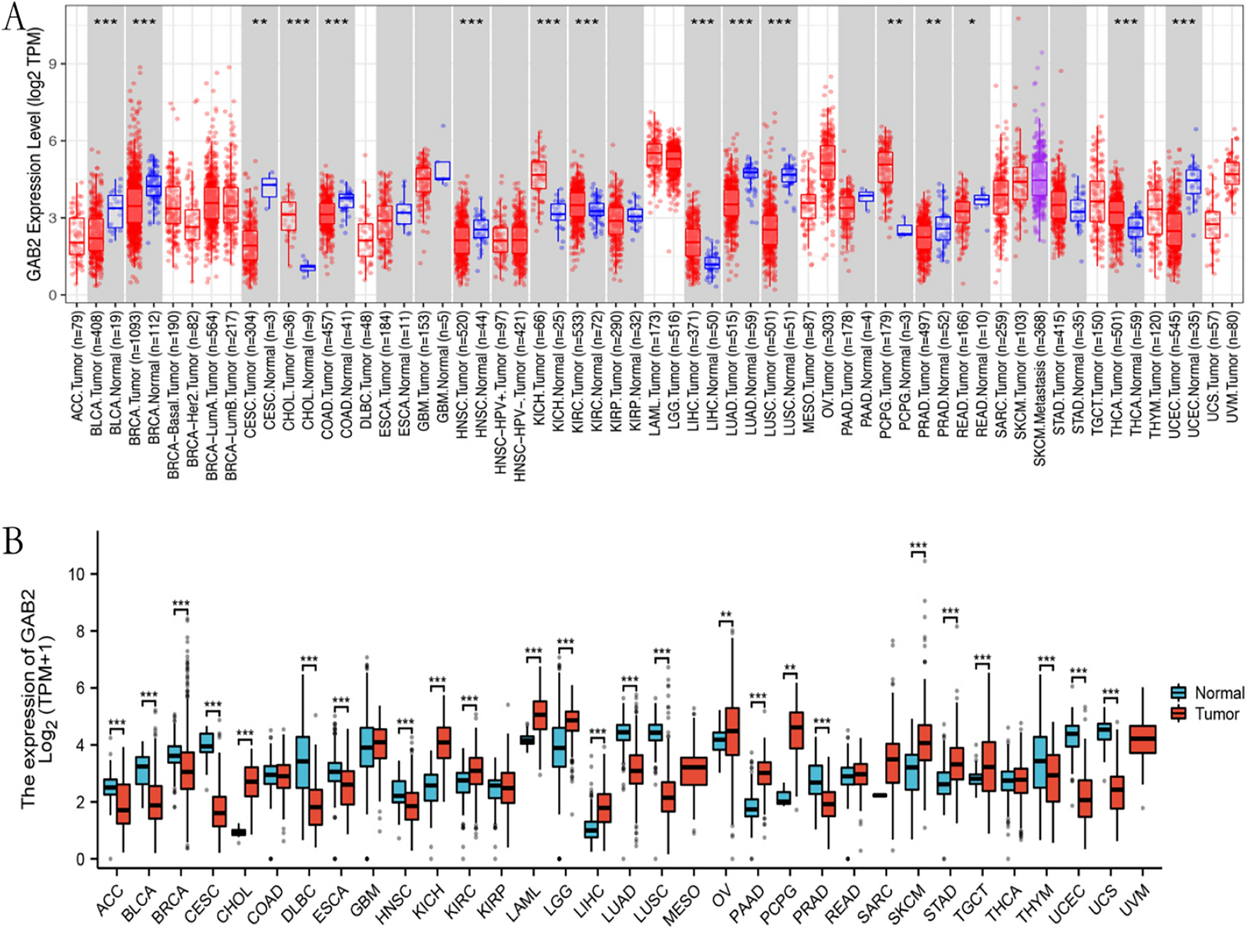
We utilized the TIMER 2.0 online tool to explore the expression of GAB2 mRNA in tumor tissues and corresponding normal tissues in the TCGA database. As shown in Fig. 1A, the expression level of GAB2 mRNA increased in cancer in CHOL, KICH, KIRC, LIHC, PCPG, and THCA, while decreased in cancer in BLCA, BRCA, CESC, COAD, HNSC, LUAD, LUSC, PRAD, READ, and UCEC.
Fig. 1
GAB2 mRNA expression levels in cancers. A GAB2 mRNA expression levels in pan-cancer from TCGA data in TIMER 2.0. B GAB2 mRNA expression levels in pan-cancer from TCGA and GTEx data. *P < 0.05, **P < 0.01, ***P < 0.001
Due to the little or no normal samples of some tumors in the TCGA database, we conducted further analysis in TCGA and GTEx database. As shown in Fig. 1B, the expression level of GAB2 mRNA increased in some tumors, such as CHOL, KICH, KIRC, LAML, LGG, LIHC, OV, PAAD, PCPG, SKCM, STAD, and TGCT; and the expression was reduced in cancer in ACC, BLCA, BRCA, CESC, DLBC, ESCA, HNSC, LUAD, LUSC, PRAD, THYM, UCEC, and UCS.
3.2 The relationship between GAB2 and prognosis in cancerTo further clarify the impact of GAB2 expression on the prognosis of patients. Based on the TCGA database, we used Kaplan–Meier Plotter to analyze the relationship between GAB2 expression and OS and RFS in pan-cancer. The result was shown in Fig. 2A, in BLCA, LUSC, and UCEC, high levels of GAB2 indicated shorter OS. However, we still found the opposite phenomenon, patients with high GAB2 expression had significantly longer OS in BRCA, KIRC, LUAD, READ, SARC, THYM and THCA. Subsequently, we further analyzed the predictive significance of GAB2 for RFS (Fig. 2B). It was found that high level of GAB2 was associated with shorter RFS in certain tumors, such as CESC and HNSC. In BRCA, TGCT and UCEC, the high level of GAB2 was associated with longer RFS.
Fig. 2
Survival analysis of GAB2 in pan-cancer. A)Correlation of GAB2 expression with OS in patients. B Correlation of GAB2 expression with RFS in patients
3.3 Phosphorylation analysis of GAB2 in pan-cancerAs shown in Fig. 3, in breast cancer, the GAB2 phosphorylation of T508, S185 and Y373 sites is higher in normal tissue. GAB2 phosphorylation of S330, S505, and T353 shows higher expression in normal tissue in Lung adenocarcinoma. GAB2 phosphorylation of S505, S172, and T353 in normal tissue is higher than that in primary tumor tissue in UCEC. GAB2 phosphorylation of T353 shows higher expression in normal tissue in colon cancer. However, in ovarian cancer, GAB2 phosphorylation of T353 and S172 in tumor tissues were significantly higher than those in normal tissues. Meanwhile, GAB2 phosphorylation of T353 was also higher in the tumor in renal clear cell carcinoma.
Fig. 3
Phosphorylation analysis of GAB2 protein in different tumors. Based on the CPTAC dataset, we analyzed the expression level of GAB2 phosphoprotein between normal tissue and tumor tissue. We supply the box plots for different cancers, including A breast cancer, B Lung adenocarcinoma, C UCEC, D Ovarian cancer, E Colon cancer, F clear cancer RCC. Product-limit method was used
3.4 Analysis of GAB2 mutation in pan-cancerWe used cBioPortal and COSMIC to explore GAB2 mutative status. As shown in Fig. 4A, the highest GAB2 alteration frequency occurred in melanoma, followed by OV, NSCLC, BRCA and BLCA. A variety of cancers were discovered to have GAB2 amplification alteration. From the GAB2 mutation sites (Fig. 4D) and corresponding 3D structures (Fig. 4E) W108Sfs*20/Pfs*20 was the most frequent mutation site. GAB2 mutation sites and their corresponding 3D structures were shown in Fig. 4D. Missense substitutions were the major mutation type and took 19.56% of 1145 GAB2 mutation (Fig. 4B). Moreover, GAB2 substitution mutations mostly happened on C>T (45.65%) and G>A (23.10%) (Fig. 4C). In addition, we utilized cBioPortal to examine the impact of GAB2 alterations on OS across multiple cancer types. The results showed that GAB2 alterations could significantly shorten OS (p = 0.0109, Fig. 4F). Then, we explored the distribution of different types of GAB2’s CNV (Fig. 4G) and the correlations between GAB2 expression and CNV (Fig. 4H). We found that The significant correlations between GAB2 expression and CNV could be found in COAD, KIRC, CESC, UCS, GBM, LIHC, LUSC, BLCA, KIRP, LUAD, ESCA, LGG, SARC, KICH, PCPG, HNSC, STAD, SKCM, BRCA and OV. Furthermore, the CNV of GAB2 showed a strong association with unfavorable OS in various cancer including KIRP, UCEC, ACC, KIRC, LGG, MESO, OV, and PAAD as well as poor PFS in 10 cancer types (UCEC, ACC, KIRC, LGG,MESO, PAAD, LUAD, SARC THCA and UCS) (Fig 4I).
Fig. 4
Mutation features of GAB2 in human cancers. Overview of the mutation status of GAB2 across TCGA cancers by A cBioPortal and B COSMIC. C Summary of GAB2 substitutional mutation types by COSMIC. D Mutation sites and E corresponding 3D structures of GAB2 displayed by cBioPortal. The correlations between pan-cancer GAB2 mutation status and F OS. G Summary of GAB2’s CNV across TCGA cancers. H The correlation between GAB2 expression and CNV in different cancers by GSCA. I The difference of OS and PFS between GAB2’s CNV and wide type in different cancers by GSCA
3.5 Molecular dockingUse GSCA database to screen out molecular compounds acting on GAB2, and the results are shown in the Fig. 5A. In the screening results, we found that Docetaxel can act on GAB2, and the correlation is relatively large (Fig. 5B). Docetaxel is also a commonly used clinical anti-tumor drug, so we chose Docetaxel for further analysis. We docked Docetaxel with GAB2 and analyzed the binding energy obtained. The binding energy is negative. The larger the result, the lower the energy, and the more stable the binding between target and molecule. The final binding energy of Docetaxel and GAB2 was − 4.875. This shows that Docetaxel and GAB2 combined stably.
Fig. 5
Molecular docking. A Main molecular compounds acting on GAB2. B Molecular docking results of docetaxel and GAB2
3.6 Performing correlation analysis between GAB2 expression and the presence of infiltrating immune cellsTo explore the correlation between GAB2 and the degree of immune cell infiltration. We used the TIMER database to analyze the relationship between GAB2 and the degree of infiltration of B cells, CD4+ T cells, CD8+ T cells, neutrophils, macrophages, and dendritic cells in pan-cancer. We found immune cell infiltration in various tumors, such as ACC, BRCA, COAD, HNSC, KIRC, KIRP, LGG, LIHC, LUAD, LUSC, PAAD, PRAD, READ, STAD, TGCT, THCA, and THYM (Fig. 6).
Fig. 6
Correlation of GAB2 expression with immune infiltration
留言 (0)