
記住我
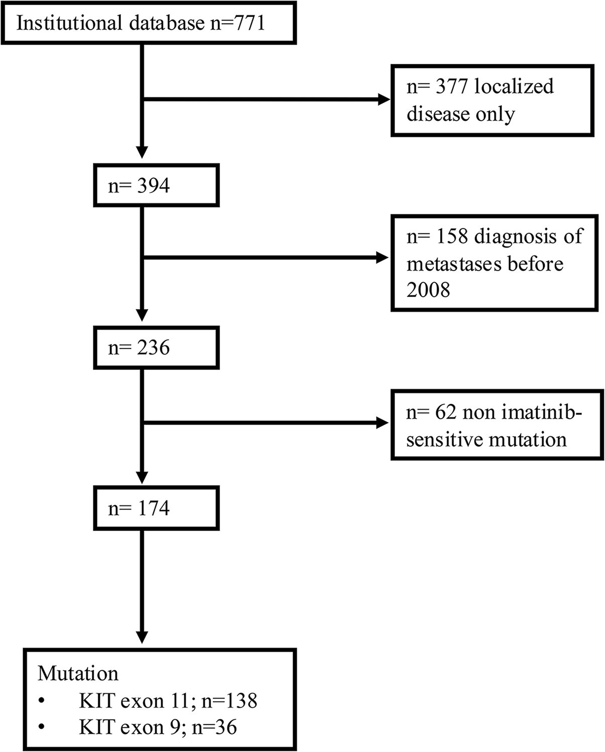


Routine blood markers were available for n = 164 patients. The median age of the patients was 64 years, and 58.5% were male. Radiological progression was recorded in n = 139 patients. Serum tumour markers were available for n = 70 patients (43%), showing similar demographics: a median age of 64 years and 63% being male. The dataset for routine blood markers consisted of 3923 longitudinal routine blood tests, with a median of 21 blood tests (each blood test containing the measurements of multiple markers) collected from each patient at a median interval of approximately 14 days. A complete list of the utilised 31 routine blood markers and their abbreviations are reported in Supplementary Table 1. The subset of tumour markers dataset consisted of 1509 longitudinal blood tests, with each patient receiving a median of 20 tests at approximately 25-day median intervals. A total of five serum tumour markers were included: cytokeratin 19 fragment antigen (CYFRA 21.1), carcinoembryonic antigen (CEA), neuron-specific enolase (NSE), cancer antigen 125 (CA125), and squamous cell carcinoma antigen (SCC), with the first three being the most prevalent in our dataset. Validation details for each endpoint in each MCCV fold are reported in the supplements.
Association between blood markers and PFSTime-varying regression analysis was employed to study longitudinal links between blood markers and progression (Supplementary Tables 2 and 3). In routine blood values, increases in the levels of C-reactive protein (CRP) and alkaline phosphatase (ALP) were significantly associated with progression events (HR = 3.68, and 7.18; p = 0.018, and 0.018 respectively; Table 2). When combined with tumour markers, these blood values kept the same positive trends, but only CRP maintained its statistical significance (p = 0.011). Furthermore, CYFRA 21.1 also showed statistical significance (HR = 22.26, p = 0.008). The hazard ratios of the variables significantly associated with PFS are described in Table 2.
Table 2 Results of the time-varying Cox regression analysisTo quantify the predictive value of blood markers to identify concurrent radiological progression, we used machine learning approaches. From now on, we refer to the results of the RF classifier. The results of SVMs and logistic regressions can be found in Supplementary Table 4, with comparable trends. Using only routine blood markers, we observed a moderate predictive value of 0.67 AUC (CI: 0.60–0.74). A similar result was observed when employing tumour markers, with an AUC of 0.67 (CI: 0.54–0.79). Combining routine blood and tumour markers increased the AUC to 0.69 (CI: 0.55–0.82). The AUC-based performance of the RF machine learning models is detailed in Table 3. Supporting metrics are provided in Supplementary Table 6. Using SHAP to uncover the most important markers for the machine learning models based on RFs (see Fig. 2a and Table 4), we observed ALP and CRP to be among the top most predictive markers for both models based on routine blood markers alone and on combined routine blood and tumour markers, and CYFRA 21.1 to follow in importance in the combined routine blood and tumour markers, suggesting an association of these markers with progression. SHAP summary plots for all prediction endpoints are included in the supplements (Supplementary Figs. 1–12).
Table 3 PFS and OS prediction performance using routine blood markers with/out combining tumour markers, with/out including death-defined progression casesFig. 2
a Venn diagrams showing blood markers found related to RECIST progression across different analyses, namely the top predictive values for the SHAP analysis and the significant values for the time varying model analysis b Frequency of missing values in the dataset
Table 4 Most predictive blood markers (in order of importance) for RECIST-based PFS prediction according to SHAP using routine blood data alone and combined with serum tumour markersRelation between predictive blood markers and PFSGiven the frequency of ALP and CRP emerging as important markers across the results of different types of RF analysis, we investigated further their relationship with PFS. We stratified the population of lab exams into subgroups based on the cutoff values used in our clinical workflow to define normal/abnormal ALP and CRP values: i.e. ALP > = 98 U/l for females, 115 U/l for males, and CRP > = 8 mg/l. This split created four subgroups: (1) normal-ALP normal-CRP, (2) normal-CRP abnormal-ALP, (3) abnormal-CRP normal-ALP, and (4) abnormal-ALP normal-CRP. Moreover, we split the analysis into the different stages of the treatment timeline, looking separately at the relationship between PFS and blood values collected pre-treatment (maximum one month), early on during the treatment (between 4 and 6 weeks), and at a later stage during treatment (between 10 and 12 weeks). Figure 3 shows the survival curves.
Fig. 3
Survival KM curves of normal/abnormal CRP and ALP groups, as measured at a before start of treatment, b early treatment and c late during the treatment
The group with abnormal CRP and ALP values showed shorter progression time, compared to the other subgroups, ranging from a median of 52 days for blood samples collected early during treatment to 123 days for blood samples collected later on during treatment (Supplementary Table 7). Interestingly, the normal CRP and ALP group showed lower median PFS pre-treatment (143 days; Fig. 3) than the groups with at most one abnormal value. During treatment, however, normalcy in both values was characteristic of longer PFS, reaching a median of 493 days, compared to 286 days of abnormal ALP group, 190 days of abnormal CRP group, and 52 days of abnormal CRP and ALP. Overall, no clear pattern of behaviour is visible among these groups, suggesting a complex relationship between CRP, ALP, and progression during treatment.
Predicting future progressionTo investigate whether blood and tumour markers can predict future progression, we set up the machine learning pipeline to predict whether progression will happen within a given time frame from the moment of the blood sampling. The timeframes investigated were one, three, six, nine and twelve months. To predict progression within one month from the moment of blood withdrawal, the model reached an AUC of 0.74 for both routine markers, as well as tumour markers. The combination of routine markers and tumour markers yielded a higher performance, with an AUC of 0.83. To predict progression within three months from the moment of blood withdrawal, the model reached the highest performance in all three combinations with 0.86 AUC, observed in the combination of routine and tumour markers, being the highest. The performance of the model is then observed to decrease from three months on: from 0.75 to 0.69 AUC to predict progression within the next 12 months in blood markers-only models, from 0.79 to 0.71 AUC in tumour markers-only models, and from 0.86 to 0.72 AUC for combined tumour and blood markers models. Results are displayed in Fig. 4 and Table 3. ALP and CRP were also among the most important markers for predicting future progression, as seen in Table 4. Additional evaluation metrics are provided in Supplementary Table 5. The numbers of progressive patients in all validation sets for all studied endpoints and data modalities is reported in the supplements.
Fig. 4
Comparative analysis of the performance with respect to a its inputs, and b its outputs
Relationship between PFS and OSWe also investigated the relationship between PFS and OS in our cohort. In order to do that, we quantified the performance of our models, trained to predict PFS (PFS models), in the prediction of OS: if PFS and OS were to be independent variables, the models would not show any predictive value for the OS endpoint. Surprisingly, the models were observed to perform, on average, 3–4% AUC higher in the prediction of OS within three months from the moment of blood withdrawal in all modalities, up to 11% better performance for routine blood markers only models within one month from blood withdrawal (Table 3). Similar performances between PFS and OS were observed for the rest of the timeline.
These findings suggest a significant overlap of progression and the occurrence of death. Because progression in PFS is defined as either RECIST progression or death of the patient, these were analysed separately. We analysed the performance of the PFS models to predict RECIST progression only, and removed from the analysis the patients whose progression was defined by death. The models were retrained to ensure that no influence of death events can be traced into the models. We observed an average drop of 2–3% AUC in predictive performance of the models, up to 5% AUC drop for combined tumour and blood markers models within three months from the blood withdrawal. Similar performances between PFS and OS were observed for the rest of the timeline. Figure 4b shows the change in performances over time.
留言 (0)