The 6MHP (Lot AC0626) (Table 1), and 12MP (Lot AC0558) (Table 1) vaccines were synthesized, purified, and vialed, and lyophilized under GMP conditions by Clinalfa-Merck as previously described (Chianese-Bullock et al. 2009). No stabilizers were used. The peptides were originally solubilized with some electrolytes to approach serum osmolarity. Some of the peptides required slightly acidic or basic conditions to support solubilization: dilute acetic acid or sodium bicarbonate were used for those purposes. For 12MP, the peptides were vialed at 200 mcg/ml in 80% Lactated Ringer’s solution + 20% sterile water, and < 1% NaHCO3 and < 0.1% acetic Lactated Ringer’s solution, USP, contains NaCl (6 mg/ml), Na lactate 3.1 mg/ml, KCl 0.3 mg/ml, calcium chloride dihydrate 0.2 mg/ml.
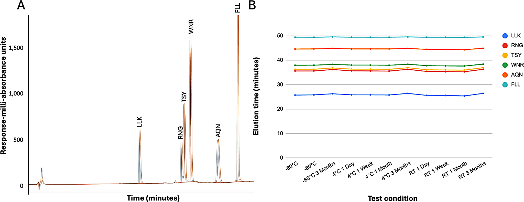
These lyophilized samples were stored in a light-protected, -80 °C environment until moved to their assigned testing condition. Conditions included storage at -80 °C, +4 °C, and room temperature (approximately 20–22 °C), light-protected, for one day, one week, or one month. Samples were then assessed using high-performance liquid chromatography (HPLC) and mass spectrometry by the University of Virginia Biomolecular Facility Core Laboratory.
Table 1 Twelve melanoma peptide (12MP) and Six melanoma helper peptide (6MHP) vaccinesHigh-Performance Liquid Chromatography
Each vial of lyophilized peptides was reconstituted with sterile water. A 3-mcg sample of each peptide was diluted to 200 mcL with 0.1% trifluoroacetic acid (TFA) before injection. Reverse phase chromatography was performed in University of Virginia’s Biomolecular Analysis Facility Core on a Phenomenex Jupiter C18 (catalog 00 F-4053-B0), 2 mm × 150 mm. Solvent A was 0.1% TFA, solvent B was 0.09% TFA in acetonitrile. The column equilibrated in 5% B (95% A). The elution gradient was 5% B for 5 min, 5–29% B in 36 min, and 29–70% B in 9 min. The Agilent 1100 pump (Agilent Technologies, Inc.; Santa Clara, CA) was used. Flow rate was 200 mcL/min, column temperature was 40 °C, and the effluent was measured by absorbance at 215 nm. Fractions were collected manually.
Mass Spectrometry
The HPLC fractions were immediately vacuum-dried and stored at -80 °C. These samples were reconstituted with 1 mL 0.1% formic acid (FA), and 100 mcL of a 1:10 sample dilution in 0.1% FA was added to autosampler vials. The LC-MS system had a Thermo nanoEASY-LC 1200 coupled to a Thermo Orbitrap Exploris 480 mass spectrometer with an Easy Spray ion source. Samples were injected into the mass spectrometer through an analytical PepMap RSLC C-18 Easy Spray column (Thermo Scientific − 3 mcm particle size, 100 Å pore size, 150 mm column length; 75 mcm internal diameter. Catalog ES900) with a pre-column Acclaim PepMap 100 C-18 (Thermo Scientific − 3 mcm particle size, 100 Å pore size, 20 mm column length; 75 mcm internal diameter. Catalog 164,946). 1 mcL of each sample was injected. Peptides were eluted from the column using an acetonitrile gradient in 0.1% FA (5–60% B in 20 min, 60–95% B in 4 min, and held at 95% for 6 min. Mobile phases: Solvent A 0.1% FA, solvent B 0.1% FA in 80% acetonitrile) at a 0.3 mcL/min flow rate. The mass spectrometer was operated in positive, data-dependent mode, in which one full MS scan was acquired in the m/z range of 375–1500 (repeat count = 3, exclusion duration = 20s, threshold = 1E + 06) followed by MS/MS acquisition using higher energy collisional dissociation of the ten most intense ions from the MS scan using window width of 2.0 m/z. The MS scans were manually examined for non-background ions. Observed ions were manually sequenced. For each fraction, the following data were presented for each species detected: selected ion chromatogram (SIC), MS spectrum to determine the (M + H)+, MS/MS spectrum to confirm the sequence. For fractions that contained more than one species, the approximate percentage of each species was calculated using the area under the SIC curves.


留言 (0)