
記住我


Microfluidic sperm selection techniques have emerged as advanced methods for isolating and sorting motile spermatozoa based on their functionality and morphology. These techniques utilize microfluidic devices that are fabricated using materials such as polydimethylsiloxane (PDMS), a silicon-based organic polymer. The devices consist of microchannels with specific dimensions tailored to the size of sperm cells. Creating flow conditions within the microchannels allows for the separation and collection of motile and morphologically normal spermatozoa, while non-motile spermatozoa and debris exit through a separate outlet(Fig. 1) [1,2,3, 17].
Fig. 1
Microfluidics Sperm Sorting Pathway: A Visual Overview
During the sorting process of sperm selection through microfluidic techniques, semen samples are introduced into the microfluidic devices. To isolate motile spermatozoa, different strategies are employed depending on the device design. These strategies may involve the use of parallel streams with varying widths, allowing motile spermatozoa to deviate into one stream while non-motile spermatozoa and debris continue along their initial streamlines. Other devices may utilize microporous membranes or precise flow control to separate motile spermatozoa from other components of the semen sample.
Microfluidic sperm selection techniques offer several advantages over conventional methods such as density gradient centrifugation and swim-up techniques (Table 1). These microfluidic techniques provide high selectivity and specificity in isolating motile and morphologically normal spermatozoa, resulting in improved sperm selection processes. Additionally, microfluidic devices enable real-time monitoring and analysis of sperm cells, allowing for precise selection based on parameters such as motility, morphology, and DNA integrity. The small-scale nature of microfluidic devices also allows for reduced sample volumes which improv handling of individual sperm cells.
However, it is important to note that microfluidic sperm selection techniques also possess limitations, including manipulation requirements, the potential of device clogging, and the cost of the device. One limitation is the complexity of device fabrication and the requirement for specialized equipment and expertise. The fabrication process involves precise control over microchannel dimensions and the integration of microfluidic components, thereby posing challenges in terms of scalability and accessibility. Another limitation is the potential for device clogging or blockage due to the presence of debris or non-motile spermatozoa in the semen sample. While microfluidic devices aim to separate motile spermatozoa from other components, the presence of debris or non-motile spermatozoa can adversely affect the sorting efficiency and accuracy of the technique. Furthermore, the cost associated with microfluidic sperm selection techniques may be higher compared to conventional methods. The fabrication of microfluidic devices along with the requirement for specialized equipment and materials contribute to the overall cost, which may limit their widespread adoption in certain settings or regions with limited resources.
In summary, microfluidic sperm selection techniques offer advanced capabilities for sperm selection in assisted reproductive technology. These techniques afford high selectivity and real-time analysis of sperm parameters, thereby leading to the enhanced quality of sperm. However, limitations such as the complexity of device fabrication, potential for device clogging, and high cost should be considered. The intricacy involved in the fabrication of devices for microfluidic sperm selection arises from the need for precise engineering and manufacturing processes. Fabricating microchannels with specific dimensions and integrating microfluidic components necessitate specialized expertise and equipment, often requiring cleanroom facilities. The intricate design and assembly process can be time-consuming and challenging, which may hinder widespread adoption, particularly in settings with limited resources where access to advanced fabrication techniques is limited.
One of the critical limitations of microfluidic sperm selection techniques is the potential for device clogging or blockage during the processing of semen samples. The microfluidic devices depend on the controlled fluid flow through microchannels to sort and isolate spermatozoa based on their motility and morphology. However, the presence of debris, non-motile spermatozoa, or other particulate matter in the semen sample may obstruct the microchannels, thereby compromising the accuracy and efficiency of the sperm sorting process. To mitigate this issue, researchers have been actively working on innovative solutions to prevent device clogging and enhance the performance of microfluidic platforms. For instance, Venugopal et al. [18] introduced a microfluidic platform with an array of uniquely designed multifunctional microposts to achieve higher capture efficiency and flow rates, while effectively avoiding clogging issues. They employed an alternative carry-forward path that allowed particles to bypass congested areas, mitigating the detrimental effects of surge pressure build-up and shear stress on cell viability.
Another approach to addressing clogging concerns is the bioinspired lobe filter system developed by Clark and San-Miguel [19]. Inspired by the filtration mechanism of manta rays, their microfluidic lobe filters enable efficient filtration of particles in the range of 10–30 μm with precise control and high throughput. The filtration efficiency increased with fluid flow rate, thereby highlighting the role of particle inertial effects in lobe filter separation. These innovations promise to significantly improve the reliability and performance of microfluidic sperm selection techniques, making them more suitable for high-throughput and continuous applications in assisted reproductive technology. In summary, while clogging remains a limitation in microfluidic sperm selection, ongoing research and the development of novel microfluidic designs, such as those inspired by nature and incorporating alternative carry-forward paths, demonstrate significant potential in overcoming this challenge and enhancing the effectiveness of microfluidic sperm sorting for ART.
The exorbitant price associated with microfluidic sperm selection techniques can pose financial challenges for healthcare facilities and patients. The fabrication of microfluidic devices requires specialized materials and equipment, thus contributing to their initial expenses. Additionally, the need for skilled personnel and proper training escalates the overall cost. While the potential benefits of improved sperm quality and ART outcomes are significant, cost considerations may hinder the widespread adoption of these techniques, particularly in regions with limited resources.
To surmount these limitations and fully connect the potential of microfluidic sperm selection techniques in assisted reproductive technology, a synergistic approach involving researchers, clinicians, and industry partners is imperative. Such collaborative endeavors can drive progress in multiple key domains, enhancing the accessibility, efficacy, and cost-effectiveness of these nascent technologies. Researchers could concentrate on simplifying device fabrication processes and refining device designs to mitigate the complexity and cost concerns. Additionally, explorations into novel sample preparation methods can ameliorate the risk of device clogging, ensuring more reliable and consistent results. Clinicians occupy a pivotal role in orchestrating extensive clinical trials to validate the long-term efficacy and impact of microfluidic sperm selection techniques on ART success rates. Collaboration with industry partners, strides in manufacturing technologies and materials can be actualized, thereby potentially diminishing the overall financial burden associated with microfluidic devices and facilitating their affordability for a broader range of healthcare facilities and patients. Through such collaborative endeavors, the field of assisted reproductive technology stands to make substantial progress, thereby extending personalized and effective fertility treatments to couples on a global scale.
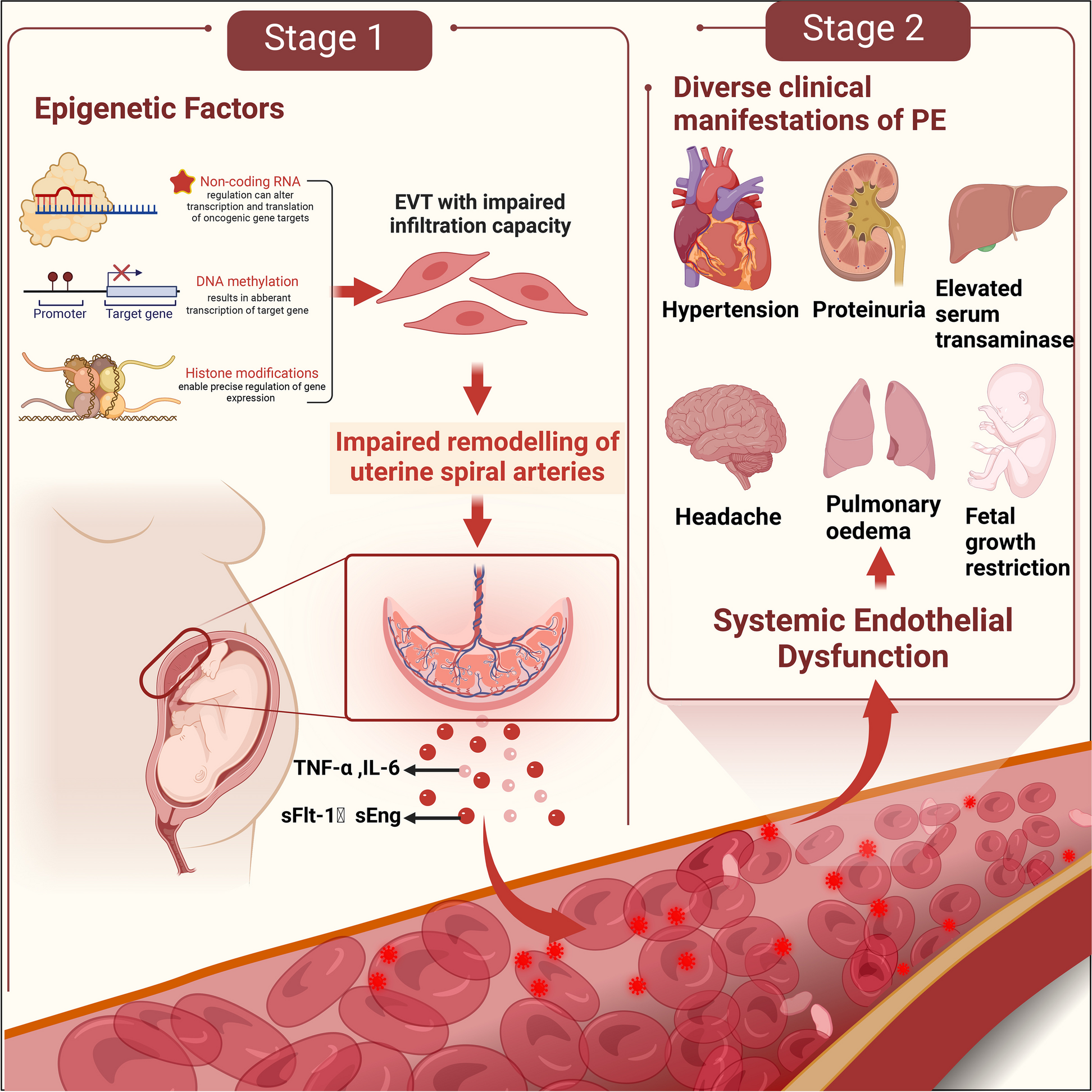
Magnetic-activated cell sorting (MACS)Magnetic-Activated Cell Sorting (MACS) has emerged as a leading technology with significant potential in sperm sorting for ART. This technology functions by utilizing magnetic microbeads coated with specific antibodies to isolate target cells based on their surface markers. In the context of sperm selection, Annexin-V, a calcium-dependent phospholipid-binding protein, is frequently used as the specific antibody to identify apoptotic spermatozoa [4,5,6,7, 20,21,22,23,24]. Annexin-V binds to phosphatidylserine (PS), a phospholipid typically confined to the inner leaflet of the plasma membrane in viable cells(Fig. 2). Nonetheless, during the process of apoptosis, PS is externalized to the outer leaflet of the plasma membrane, thus allowing its detection and binding by Annexin-V. Comprehensive research on this technique has yielded valuable insights into its effectiveness and potential clinical applications across varied patient demographics(statistics that describe populations and their characteristics).
Fig. 2
Magnetic-Activated Cell Sorting (MACS): A Visual Overview
The employment of MACS in sperm preparation for ART offers notable advantages, including the selective isolation of viable sperm characterized by reduced DNA fragmentation and enhanced genetic integrity. Such attributes have the potential to augment both fertilization and pregnancy rates (Table 1). Studies have demonstrated the efficiency of MACS in ameliorating sperm parameters, ranging from motility and morphology to chromatin integrity, not only in normozoospermic patients but also in those presenting with suboptimal semen parameters [7, 22, 24]. The synergistic use of MACS with density gradient centrifugation (DGC) has shown particularly promising results, leading to a substantial decline in apoptotic spermatozoa and an increase in sperm quality [6, 22, 24]. Furthermore, the integration of DGC-MACS has been associated with superior ART cycle parameters, encompassing diminished sperm DNA fragmentation rates and curtailed oxidative stress, potentially leading to increased success rates in Intracytoplasmic Sperm Injection (ICSI) cycles [20].
However, certain limitations should be acknowledged (Table 1). While MACS has demonstrated encouraging outcomes within specific patient groups, its integration into ART remains nascent. Further research is required to corroborate its efficacy and to buttress the evidence advocating its inclusion in routine sperm selection protocols [20]. Additionally, the technical intricacy inherent to MACS systems could pose obstacles to its ubiquitous acceptance in clinical environments. Such technical complexity of MACS systems can be attributed to the intricate process involved in isolation of target cells based on their surface markers, utilizing magnetic microbeads and designated antibodies. This protocol mandates exacting calibration and refinement to guarantee precise and efficacious spermatozoa sorting. Moreover, the apparatus and reagents used in MACS procedures necessitate rigorous maintenance and quality assurance, amplifying the overall intricacy of assimilating this technology within clinical laboratories. Consequently, dedicated training and proficiency became indispensable for laboratory personnel to adeptly conduct MACS, potentially constraining its pervasive acceptance in routine clinical practices.
The utilization of MACS in sperm preparation for ART presents a promising approach to augment sperm quality and thereby ameliorate fertility outcomes across a spectrum of patient populations. When amalgamated with DGC, MACS has evinced notable efficacy in the exclusion of apoptotic spermatozoa, favoring the selection of viable sperm characterized by reduced DNA fragmentation, which subsequently bolsters the probability of successful fertilization and elevated pregnancy rates. Such pioneering revelations fortify the expanding corpus of evidence advocating the integration of MACS in tailored sperm selection techniques addressing male factor infertility challenges. An exhaustive appraisal of sperm quality, undertaken via various assays, inclusive of evaluations pertaining to viability, motility, chromatin integrity, and the acrosome reaction, offers an encompassing perspective on sperm functionality and genetic integrity. This rigorous analysis is pivotal for precision-driven sperm selection and the ensuing success of ART, more so in cases marked by abnormal semen parameters or idiopathic infertility. The incorporation of MACS under such circumstances underscores its potential in revolutionizing fertility treatment strategies, thereby offering hope to couples facing challenges in conception.
With the relentless progression of reproductive medicine, further research and validation studies are warranted to not only solidify these previous findings but also to probe the potential expended applications of MACS in ART. An augmented, multi-centric study encompassing a broad spectrum of patient cohorts is pivotal to bolster the empirical evidence supporting MACS’ efficacy, thereby facilitating its integration into routine sperm selection protocols in clinical settings. The adoption of innovative approaches like MACS in ART has the potential to transform the landscape of fertility treatment, exemplifying personalized care and amplifying reproductive success for a on a global scale.
Electrophoretic sperm selectionElectrophoretic sperm selection emerges as a revolutionary modality within the realm of assisted reproductive technologies, capitalizing on the inherent electrical attributes of spermatozoa to segregate functionally adept cells [8, 25]. This technique exploits the distinctive electrical charge acquired by sperm throughout their maturation process - a characteristic that is instrumental in preventing aggregation, circumventing nonspecific binding, and mitigating undesired storage within the female reproductive tract [8]. Over time, considerable progress has been achieved in this field, leading to the refinement of methodologies that aim to ensure sample integrity, reduce DNA damage, and augment overall fertility potential [13, 23, 24]. This section delves into the advantages, limitations, addressed concerns, and future prospects of electrophoretic sperm selection.
The advent of electrophoretic sperm selection heralds a series of noteworthy advantages in the realm of assisted reproduction (Table 1). The approach introduces a non-invasive and rapid means of isolating spermatozoa with optimal fertilization potential. The CS-10 device pioneered this technique by capitalizing on the negatively charged attribute of mature sperm, resulting in the birth of viable offspring devoid of embryonic development problems [13]. The advancement embodied by the Felix™ apparatus further accentuates this advantage by incorporating a filtration system that effectively segregates contaminating cells. This results in the heightened purity and superior quality of isolated sperm populations [23]. Nevertheless, like any technological advancement, electrophoretic sperm selection presents certain limitations (Table 1). A primary challenge resides in maintaining a nuanced equilibrium between sperm membrane charge and functional attributes. Although the exclusion of negatively charged sperm through DGC may enhance DNA damage levels, the intricate dynamics of this process warrant further exploration. Moreover, the formulation of an optimized electrophoretic buffer should be a focal consideration during isolation procedure [14]. The composition and properties of the buffer critically influence the migration of spermatozoa, dictated by their charge, size, and various other determinants. Suboptimal buffer conditions could potentially lead to inconsistent results and compromised sperm quality during the separation process. The conductivity, pH, and ionic strength of the buffer necessitate rigorous optimization to ensure the precise and effective migration of sperm cells. Moreover, the constituents of the buffer should be carefully selected to mitigate potential detrimental impacts on sperm viability and function. Consequently, a comprehensive investigation of buffer formulations is essential to address this potential limitation, ensuring enhanced consistency, reliability, and reproducibility.
Additionally, the robustness of the technique across a diverse spectrum of semen samples, particularly from pathological donors, requires validation [24]. Thus, while electrophoretic sperm selection offers considerable potential, it is crucial to address these limitations to ascertain its clinical applicability.
The evolution of electrophoretic sperm selection epitomizes the dynamic nature of scientific progress in addressing pertinent concerns. The CS-10 device introduced the concept of charge-based sperm selection, revolutionizing the field and laying the foundation for subsequent advancements [13]. The Felix™ device, with its amalgamation of electrophoretic separation and a sophisticated filtration system, stands as a testament to the continual refinement of the technique. This innovation notably addresses the pivotal issue of sample purity by effectively eliminating contaminating cells [23]. Additionally, Simon et al. (2016) deepened our understanding of sperm membrane charge dynamics, highlighting the intricate relationship between charge and DNA integrity [26]. Furthermore, Ainsworth et al.‘s (2005) electrophoretic system presents a novel approach to sperm isolation, demonstrating its potential to mitigate DNA damage and enhance functional attributes [27]. The collective progress made in addressing these concerns underscores the trajectory of electrophoretic sperm selection.
The trajectory of advancements in electrophoretic sperm selection has been significant, showing great potential for the field of assisted reproduction. The transition from CS-10 to the Felix™ device exemplifies a proactive approach to addressing challenges and enhancing the efficacy of the technique [13, 23]. The studies by Simon et al. (2016) and Ainsworth et al. (2005) offer a comprehensive insight into the complex dynamics of sperm membrane charge and its implications for fertility potential [26, 27]. Additionally, these studies emphasize the importance of rigorous validation across a variety of semen samples to confirm the clinical applicability of this technique. The development of electrophoretic sperm selection is noteworthy, highlighting the capability of innovative approaches to redefine existing paradigms. As progress ensues, the field stands on the cusp of revolutionizing assisted reproduction by leveraging the electrical attributes of sperm for enhanced selection. Nevertheless, the path forward necessitates meticulous validation and comprehensive research to ensure that these advancements lead to discernible enhancements in clinical outcomes.
In the field of assisted reproductive technologies, the innovative approach of electrophoretic sperm selection presents a promising avenue for improving fertility outcomes. This technique capitalizes on the inherent electrical properties of spermatozoa, leveraging the distinct charge they acquire during maturation to facilitate selective isolation. The progression of this technique from its preliminary stages to the introduction of sophisticated devices like the Felix™ exemplifies the commitment of the field to addressing concerns and refining methodologies. Electrophoretic sperm selection offers a range of advantages, from rapid and non-invasive sperm isolation to augmented sample purity and heightened DNA integrity. Both the CS-10 and Felix™ devices, supplemented by additional pertinent studies, underscore the potential of this technique to revolutionize sperm selection, culminating in elevated success rates for assisted reproduction. However, as is the case with any emergent technology, certain challenges must be meticulously addressed. The nuanced balance between sperm membrane charge and functional attributes, coupled with the imperative for comprehensive validation across diverse semen samples, accentuates the intricacy of this technique. While electrophoretic sperm selection offers considerable potential, ongoing research and meticulous refinement are essential to guarantee its successful integration into clinical practice. In conclusion, the path of electrophoretic sperm selection is a continuous and evolving technology stream, remaining subject to further advancement and exploration. The concerted efforts of both researchers and clinicians have yielded significant progress in addressing concerns and unlocking the potential of this technique. As the field progresses, it is imperative to embrace these advancements while concurrently ensuring rigorous validation and the pursuit of improved fertility outcomes.
Intracytoplasmic morphologically selected sperm injection (IMSI)Intracytoplasmic Morphologically Selected Sperm Injection (IMSI) stands as a pioneering technique in assisted reproduction, with the primary objective of optimizing the selection of high-quality sperm for fertilization [10]. This advanced procedure employs high-magnification microscopy, typically around 6000x magnification, to meticulously assess sperm morphology. Through this rigorous assessment of parameters such as nuclear vacuoles, acrosomal integrity, and overall structure characteristics, IMSI seeks to pinpoint sperm that possess the optimal genetic and structural attributes [10, 28].
This approach offers a range of potential advantages [9, 11, 28](Table 1). A foremost advantage of IMSI lies in its capacity to enhance the selection of morphologically normal sperm. By leveraging higher magnification, even minute abnormalities become detectable abnormalities that might be overlooked at lower magnifications [28]. This enhanced precision is instrumental in singling out sperm with optimal genetic integrity. Moreover, the meticulous selection process of IMSI could mitigate the likelihood of transmitting genetic abnormalities or DNA damage to the developing embryo, potentially leading to improved pregnancy rates [11, 28].
However, despite its potential benefits, IMSI presents certain limitations. The exhaustive examination procedure requires considerable time, which could extend treatment durations and elevate potential patient stress [28]. Furthermore, while promising, compelling evidence delineating clear and significant improvements in clinical outcomes compared to conventional methods like ICSI remains somewhat scarce [12, 28]. Additionally, the requisites for specialized equipment and the expertise essential for high-magnification microscopy could contribute to increased costs and intricate procedures, potentially restricting accessibility for certain patients [12, 28, 29]. Firstly, the acquisition and maintenance of such advanced microscopy systems can significantly increase the overall financial strain associated with assisted reproductive treatments. The initial acquisition costs, in tandem with expenses related to routine maintenance, calibration, and prospective upgrades, amplify the financial implications of IMSI procedures compared to conventional methods. These elevated financial requirements could pose challenges for patients with limited financial means, potentially restricting their access to this advanced technique. Furthermore, the complexities associated with high-magnification microscopy necessitate an advanced degree of technical proficiency and specialized training for embryologists and laboratory personnel [29]. To attain accurate and consistent results, an in-depth grasp of the equipment is imperative, along with meticulous sample preparation and adapt interpretation of the complex morphological details shown by the high-magnification imagery. As a result, clinics offering IMSI must allocate resources towards training programs and continuous professional development for their staff members. This emphasis on specialized training and expertise adds an additional layer of complexity to the overall procedure, potentially necessitating an extended learning curve for embryologists transitioning to this technique. The combination of increased financial expenditures and heightened technical demands collectively contributes to the procedural complexity of IMSI. Although the prospective advantages of IMSI are substantial, the requisites for specialized apparatus, continual maintenance, and advanced technical expertise might render its adoption more challenging for certain clinics and patients. As the field of assisted reproduction endeavors towards inclusivity and egalitarian access to advanced treatments, devising strategies to surmount these challenges emerges as a critical priority. Efforts to enhance training programs, explore cost-effective equipment alternatives, and foster collaborations among clinics could pave the way toward broader accessibility of IMSI. This would ensure that patients from various socio-economic backgrounds have the opportunity to benefit from this advanced sperm selection technique [12, 28, 29].
The subjective element intrinsic to embryologists’ assessment of sperm morphology during the selection process carries the risk of introducing selection bias and result in variability of outcomes [12, 28]. This significant concern emphasizes the necessity for standardized criteria and comprehensive methodologies in sperm selection techniques, such as IMSI. Any deviations can markedly impact the success rates and overall efficiency of assisted reproductive procedures. Efforts toward standardization encompass the establishment of comprehensive guidelines for sperm selection criteria and morphological assessments in IMSI. The primary objective is to reduce observer variability. Ongoing work is carefully creating consistent rules to accurately define the parameters for selecting sperm and to enhance the accuracy of morphological assessment within the IMSI process [28]. This systematic approach aims to effectively diminish the impact of observer variability, ensuring a consistent and objective methodology for embryologists in the process of sperm selection. By minimizing inherent subjectivity through these guidelines, the overall reliability of IMSI outcomes could be markedly augmented, consequently translating into elevated success rates for assisted reproductive procedures. Furthermore, instituting standardized criteria might facilitate the development of exhaustive training initiatives for embryologists, ensuring their proficiency in accurately identifying and selecting morphologically normal sperm. This pedagogical aspect is crucial in ensuring uniformity and competence across different clinical settings. Furthermore, the adoption of standardized guidelines might foster collaboration within the reproductive medicine community, facilitating the exchange of best practices and continuous refinement of the IMSI technique.
As the path to standardization continues, it is imperative that these efforts remain adaptive to new research findings and technological advancements. By directly confronting the issue of observer variability, the field of assisted reproduction may advance towards elevated precision, ultimately benefiting couples seeking fertility treatments. Further exploration delves into the long-term health and developmental outcomes of embryos derived from IMSI-selected sperm [9, 28]. Researchers are actively exploring the combined use of IMSI with other advanced sperm selection techniques to leverage their potential synergistic benefits [11, 12,
留言 (0)