Chemicals and materials
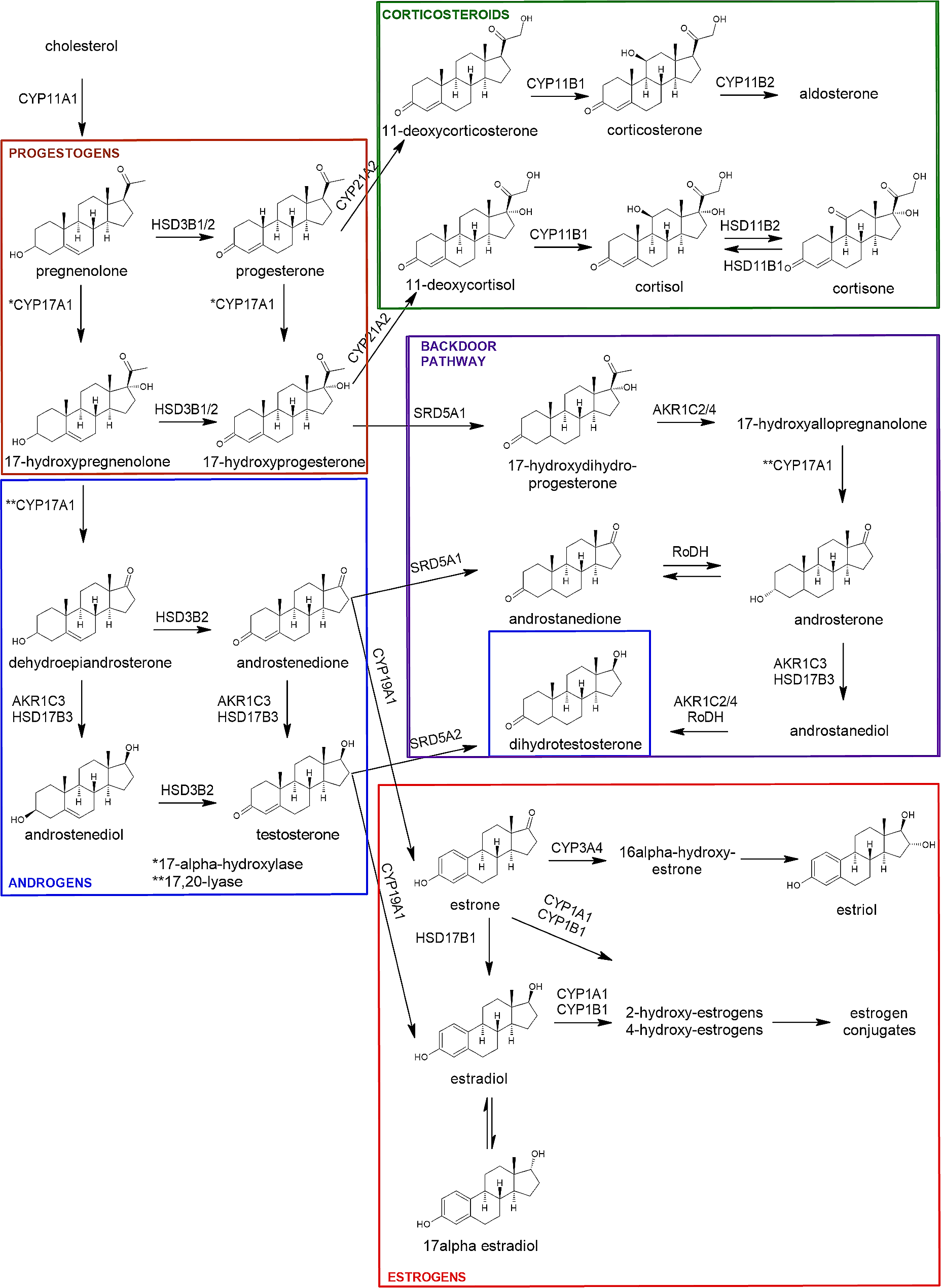
Cortisol, cortisone, corticosterone, 11-deoxycortisol, 11-deoxycorticosterone, androstenedione, dehydroepiandrosterone, testosterone, dihydrotestosterone, 5α-androsterone, 17α-hydroxyprogesterone, progesterone, pregnenolone, 17α-hydroxypregnenolone, 17β-estradiol, 17α-estradiol, estrone, estriol were purchased from Sigma-Aldrich. 5α-Androstenediol was acquired from Steraloids and 5α-Androstanedione from Eurisotop. 17α-hydroxydihydroprogesterone was purchased from Santa-Cruz. All native steroids had more than 99% purity. In addition, internal standards cortisol-9,11,12,12-D4, corticosterone-9,11,12,12-D4, 11-deoxycortisol-2,2,4,6,6-D5, progesterone-2,3,4-13C3, androstene-3,17-dione-2,3,4-13C3, 5α-dihydrotestosterone-16,16,17-D3, estrone-2,3,4-13C3, 17β-estradiol-2,3,4-13C3 were purchased from Sigma-Aldrich. Moreover, the internal standards pregnenolone-20,21-13C2-16,16-D2, 5α-androstanedione-2,3,4-13C3, dehydroepiandrosterone-2,2,3,4,4,6-D6, testosterone-2,3,4-13C3, estriol-13C3 were purchased from IsoSciences. Cortisone-2,2,4,6,6,9,12,12-D8, 17α-hydroxyprogesterone-2,3,4-13C3, 5α-androsterone-2,2,4,4-D4 from Eurisotop and 5α-androstenediol-16,16,17-D3 from Steraloids. Purity of all internal standards were more than 98%. Steroid names, abbreviations and CAS numbers are provided in Supplementary Table S1 and Table S2.
Solvents and reagents used for the steroid analysis were methanol (Biosolve BV), Milli-Q water (Millipak, Merck), acetone (Biosolve BV), formic acid (99%, Biosolve Chimie), sodium bicarbonate and sodium carbonate (Sigma-Aldrich), dansyl chloride (Sigma-Aldrich) and ammonium fluoride (Sigma-Aldrich). Cell media used for the validation of the analytical method consisted of DMEM/F-12 medium w/o phenol red (REF no. 11039-021), ITS + premix (cat. 354,352), Serum Charcoal dextran treated Hyclone (SH.30068.03), 1% Penicillin Streptomycin (REF no. 15140-122) and 0.1% dimethylsulfoxide (DMSO).
Human fetal ovarian cultures
For steroid quantification, 8 fetal ovaries were collected from first trimester fetuses between GW 10 (10 weeks of gestation) and GW 14 + 1 (14 weeks and 1 day of gestation) and another 7 fetal ovarian tissues from second trimester fetuses between GW 15 + 2 and 19 + 5 (Table S3). Fetal material was obtained as a part of the Scottish Advanced Fetal Research (SAFeR) Study as approved by NHS Grampian Research Ethics Committees (REC 15/NS/0123). Women over 16 years of age and between 7 and 20 gestational weeks (GW) seeking elective terminations of normally progressing pregnancy (determined at ultrasound scan prior to termination) were recruited with full written, informed consent by NHS Grampian research nurses working independently of the study. The aim of the study was to study the effects of EDCs on fetal ovaries. Fetuses were collected following termination by RU486 (mifepristone) treatment (200 mg) and misoprostol-induced delivery as previously described [21]. Fetal tissues were transported to the laboratory within 30 min of delivery, weighed, sexed and the crown-rump length as well as the foot length were recorded for further confirmation of gestational age. Ovaries separated from mesonephroi were dissected, weighed, and immediately placed into ice-cold phosphate-buffered saline. Fetal ovaries were processed as previously described [10]. Briefly, each ovary was cut into 1 mm3 size explants to ensure accessibility to nutrients and viability of the tissue. The same number of ovarian explants randomly picked from each ovary of the same fetus (2 per ovary, 4 in total) was used for each culture condition. Explants were placed into cell culture inserts (0.4 μm pores, Millipore #PICM01250) in wells filled with 400 µl of culture media containing 0.1% v/v dimethyl sulfoxide (DMSO). Culture media included phenol red-free Medium 199 (Invitrogen Life Technologies, Cergy-Pontoise, France) supplemented with 50 µg/mL gentamycin, 2.5 µg/mL fungizone (Sigma Aldrich Chemicals, Saint-Quentin Fallavier, France), and 1 g/L insulin, 0.55 g/L transferrin, and 0.67 mg/lsodium selenite (ITS, Corning, Fisher Scientific). Cultures were incubated for 7 days at 37 °C under 5% CO2 and humidity. Viability of tissue explants in culture was confirmed with immunohistochemistry using anti-cleaved caspase 3 antibody. Media were completely changed at first after 24 h and then every 48 h. Finally, culture media were collected, snap-frozen and stored at − 80 °C until steroid hormone analysis. Clinical metadata regarding the maternal age, BMI and smoking status according to questionnaire are given in Supplementary Table S3.
Human adult ovarian cultures
For this study, 13 human adult ovary culture-conditioned media samples were collected for steroid quantification. Human adult ovaries were collected from women undergoing Caesarean section (C-section), as described by Li et al. and part of the steroid hormone analysis data for these samples is re-used in the present study [22]. All women were younger than 35 years and gave consent to participate in the research project investigating the effects of EDCs on ovaries at Karolinska University Hospital Huddinge in Stockholm, Sweden, in accordance with the Declaration of Helsinki. Briefly, a superficial piece of cortical tissue (5 mm * 5 mm * 2 mm depth) was taken, placed in Dulbecco’s phosphate buffered saline with calcium, magnesium, glucose, sodium pyruvate (Life Technologies, Paisley, UK) and transferred to the laboratory within 10 min. Upon arrival in the laboratory, ovarian cortical tissues were cut into 1 mm * 1 mm pieces using scalpels and each piece was placed on a laminin-221 coated Millicell cell culture plate insert (Merck Millipore, Darmstadt, Germany) in 24-well plates (Sarstedt, Nümbrecht, Germany). The tissues were cultured in an incubator at 37 °C under 5% CO2. Full media change (350 µL) with 0.1% DMSO was performed every other day. Full culture media contained phenol red high glucose DMEM with glutamax (Life Technologies Grand Island, NY, USA) supplemented with 10% human serum albumin solution (Vitrolife, Göteborg, Sweden), 1% antibiotic-antimycotic (Life Technologies, Grand Island, NY, USA), 1% insulin-transferrin-selenium (Life Technologies, Grand Island, NY, USA), and 0.5 IU/ml human recombinant follicle-stimulating hormone (FSH, Fostimon, Italy). After 6 days of culture, media from 2 technical replicates were pooled and stored at -80 °C until steroid hormone analysis. LDH-Glo cytotoxicity assay kit (Promega, USA) was used to assess cytotoxicity for the ovarian tissue in culture. Clinical metadata regarding the women’s age, BMI, smoking status, disease and androgen treatment are given in Supplementary Table S4.
Human adult follicular fluid
Follicular fluids were collected from women undergoing infertility treatment in Estonia, as described by Bellavia et al. [15]. The full cohort encompassed 148 women (age: 23–43) undergoing fertility treatment at Nova Vita Clinic in Tallinn, and 14 follicular fluid samples were randomly selected for the present study. All women were informed regarding the study, and they signed an informed written consent form complying with the Declaration of Helsinki. Briefly, the follicular fluid samples were collected from the leading follicles avoiding blood contamination, centrifuged for 10 min at 300 g and then for another 10 min at 2000 g [15]. To prevent dilution of the samples, the flushing medium was eliminated from the needle and the hose prior to collection of follicular fluid. The cell-free follicular fluid samples after centrifugation were aliquoted, and within 2 h delivered on ice to the university where the samples were kept frozen at -80 °C till steroid analysis. Clinical metadata of the women in this study are given in Supplementary Table S5.
Quantification of steroid hormones
The samples were thawed at room temperature and extracted with offline solid phase extraction (SPE). 100 µl of each sample and 50 µl internal standard were added to 1 ml Milli-Q (Millipak, Merck) with 2% formic acid (Sigma-Aldrich), which made 1.15 ml loading solution for each sample ready for the SPE. The SPE was conducted with Agilent Versaplate-Plexa 96 well-plate preassembled with Bond Elut Plexa 30 mg cartridges. First, the cartridges were pre-conditioned with 0.5% formic acid in 1 ml methanol (Biosolve BV). Then, the cartridges were equilibrated with 0.5% formic acid in 1 ml Milli-Q and each sample was loaded afterwards. After this, the cartridges were pre-washed with 1 ml Milli-Q and washed by 1 ml methanol/Milli-Q (30:70). Vacuum was applied for 10 min. Finally, the steroid hormones were eluted with 0.7 ml methanol and collected to a 96 well-plate and vacuum was briefly applied to complete elution. Subsequently, the extracts were evaporated to dryness at 40 °C using CentriVap Concentrator (LABCONCO), were reconstituted with 150 µl methanol/Milli-Q (50:50) and were filtrated with Agilent Captiva 96 well filter plate (0.2 μm). Then the samples were analysed using LC-MS/MS for quantifying underivatised free steroids.
After analysing the underivatised steroid hormones, the samples were then used for quantifying low amounts of estrogens after dansylation. The samples were evaporated to dryness at 40° C using CentriVap Concentrator (LABCONCO) and subsequently 40 µl bicarbonate buffer (pH 10.5) and 40 µl dansyl chloride (Sigma-Aldrich) (1 mg powder in 1 ml acetone (Biosolve BV)) were added. The reaction was completed in the oven for 5 min at 60 °C. Then the samples were ready for analysis of the dansylated estrogens.
Steroid levels (pg/ml) found in procedural blanks (SPE extracted blank media) were subtracted. Steroid levels (pg/ml) found in blank media samples were subtracted from adult ovarian tissue cultures. No blank media samples were available from follicular fluid samples. While, only cortisol (CO-SOL) was quantified in higher level in the blank media than the levels in the fetal ovarian tissue samples, which was excluded from the measurements of fetal samples as explained in Results.
Both derivatised and underivatised samples were analysed with LC-MS/MS using AB Sciex 6500 + triple quadrupole mass spectrometer coupled to an AB Sciex Exion LC system. Ionisation was achieved with electrospray ionisation (ESI) in positive mode and only for underivatized estrogens in negative mode. The column used was Phenomenex Kinetex C18 column (150 × 3 mm, 2.6 μm particle size, pore-size 100 Å) with mobile phases A: Milli-Q water with 0.2 mM ammonium fluoride (Sigma-Aldrich) and B: methanol. The flow rate was 0.6 ml/min and the injection volume was 10 µl. The analytical method was validated in cell media spiked with steroid standards.
Steroid hormones validation
The method was validated for underivatized steroid hormones based on one level (6 ng/ml) for inter-day repeatability and based on two levels (300 pg/ml and 3 ng/ml) with spiked cell media for assessing intra-day repeatability. For the inter-day repeatability at 6 ng/ml, the accuracy was on average 90% (64-109%) and precision was 15% (4.2-57%) for all underivatised steroids. Similarly, dansylated estrogens were assessed for inter-day repeatability at 10 pg/ml. Accuracy and precision for all dansylated estrogens were on average 84% (79-92%) and 18% (14-22%) respectively. More information about the intra-day and inter-day repeatability assessment, limit of detection, transitions and calibration range is provided in Supplementary Tables S6-S8.
Statistical analysis
Steroid hormone peak data were processed with Sciex Analyst 1.7.2 software and further calculations were completed with Excel (Version 2208 Build 16.0.15601.20540). Steroid hormone concentrations for fetal and adult ovarian tissue cultures were normalised based on the volume of the culture media collected, size and number of tissue explants and time of incubation, based on which the corresponding steroid hormone release rates were calculated.
Graphs with steroid hormone release rates or concentrations and corresponding ratios were made with GraphPad Prism 9.5.0, and principal component analysis (PCA) was performed in R version 4.3.0 using base and attached packages: ggrepel_0.9.3, factoextra_1.0.7 ggplot2_3.4.2, FactoMineR_2.8 in R studio (version 2023.03.1 + 446) after data normalization in MetaboAnalyst 5.0. For the PCA plot, probabilistic quotient normalization, logarithmic transformation, and pareto scaling were used. Missing values were replaced by 1/5 of lowest release rate and steroid hormones with more than 55% missing values were eliminated, leading to a final dataset of only 13% predicted missing values. Logarithmic transformation of steroid hormone release rates or ratios were normally distributed, based on Shapiro-Wilk test and quantile-quantile plots. Unpaired multiple t-test assuming equal standard deviation using Holm-Šídák method and p value threshold set to 0.05 was applied for each steroid hormone release rate or ratio coming from two different sample types: first compared to second trimester fetal ovarian tissue cultures; first trimester fetal ovarian tissue cultures compared to adult ovarian tissue cultures; second trimester fetal compared to adult ovarian tissue cultures; adult ovarian tissue cultures compared to follicular fluid. Comparisons were performed between means of steroid hormone release rates and ratios. The units for steroid hormones were different in adult ovarian tissue cultures and follicular fluid and thus only steroid hormone ratios were compared in this case. Outlier detection for adult ovarian tissue cultures was performed with Rout and with Grubb’s tests and was confirmed visually with principal component analysis.


留言 (0)